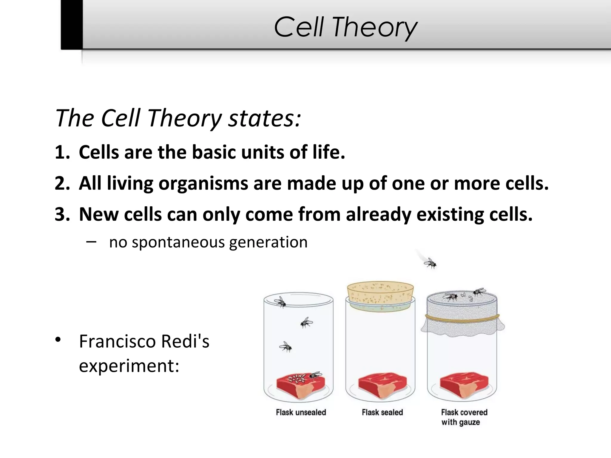
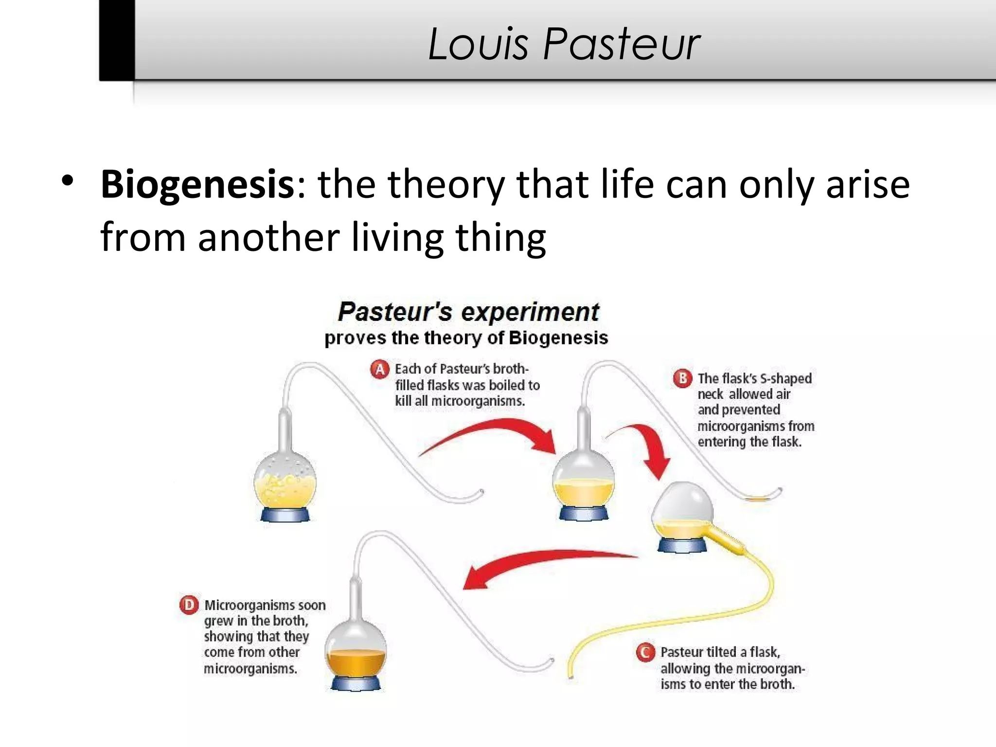
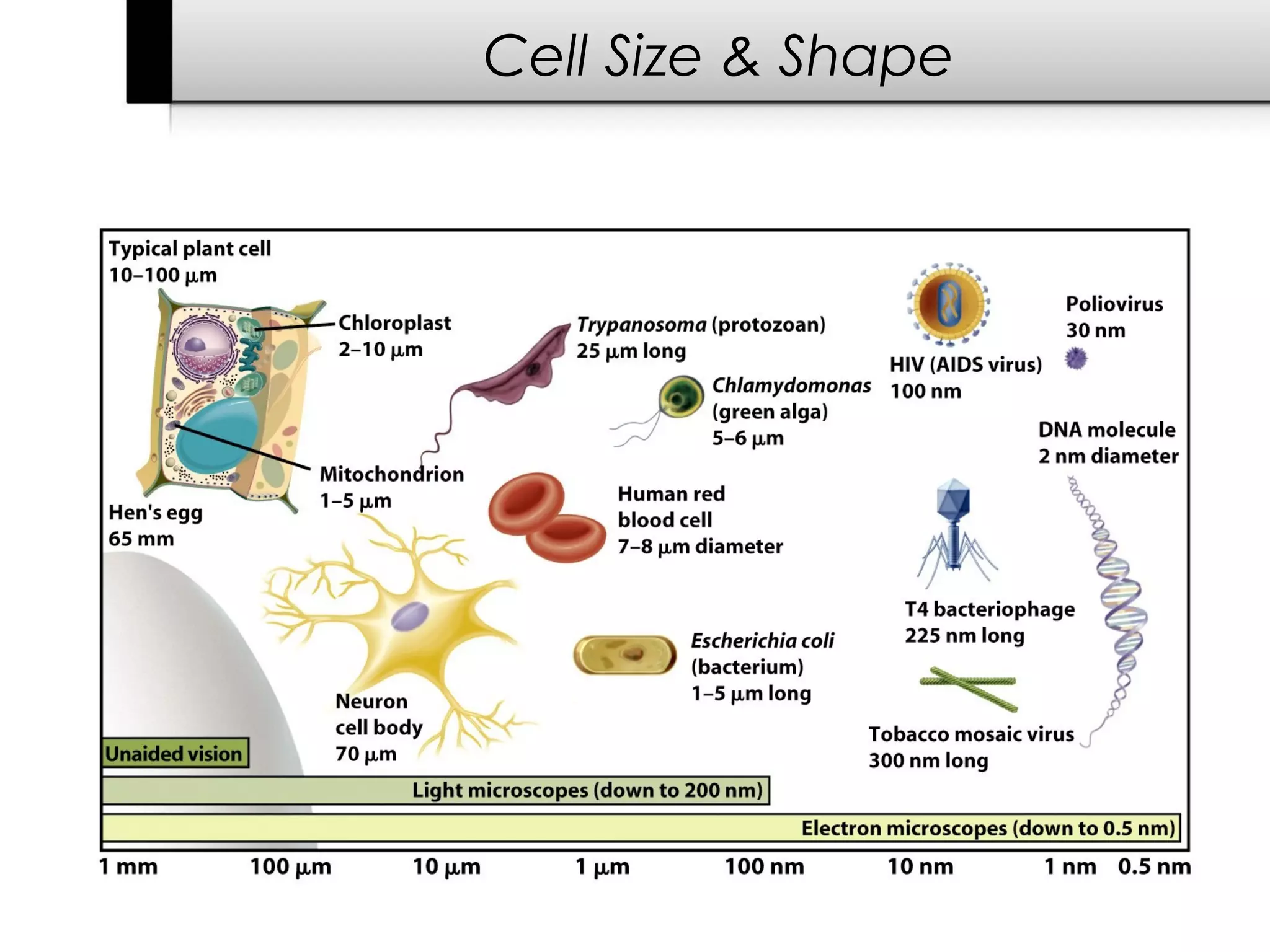
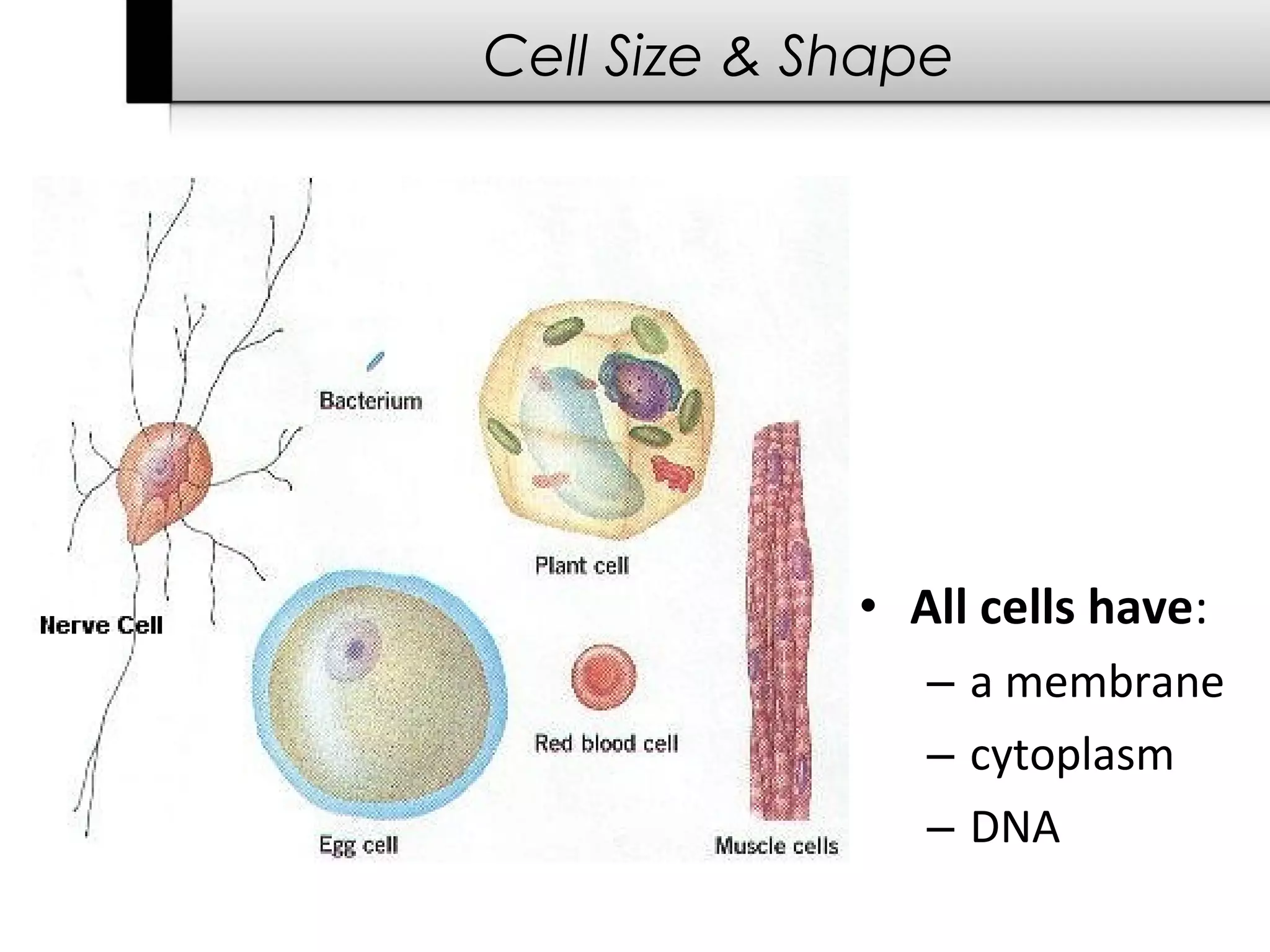
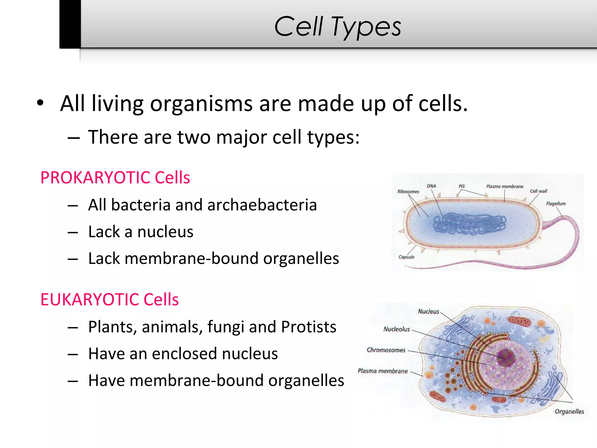
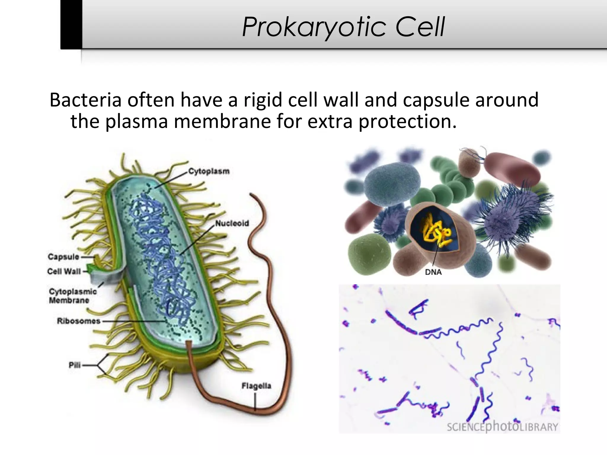
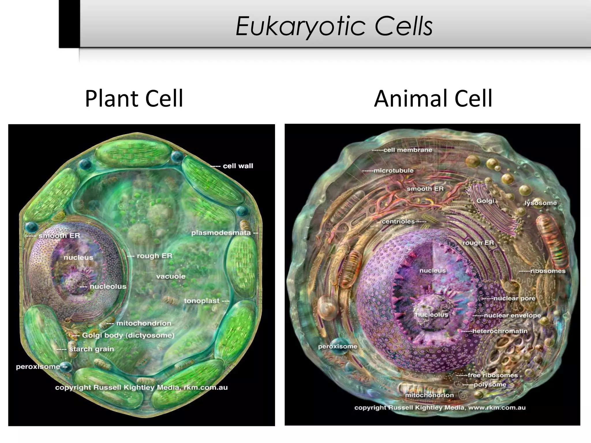
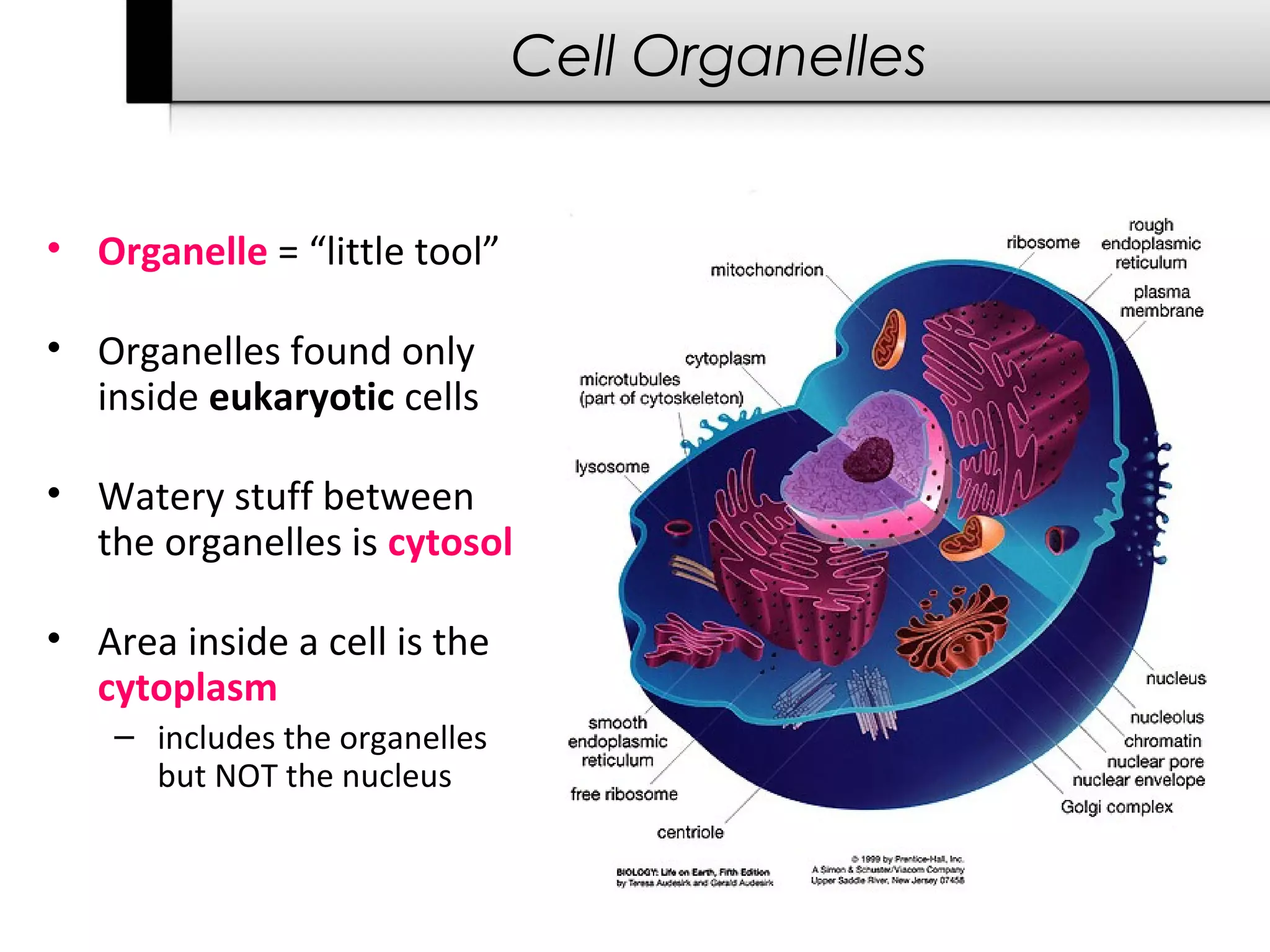
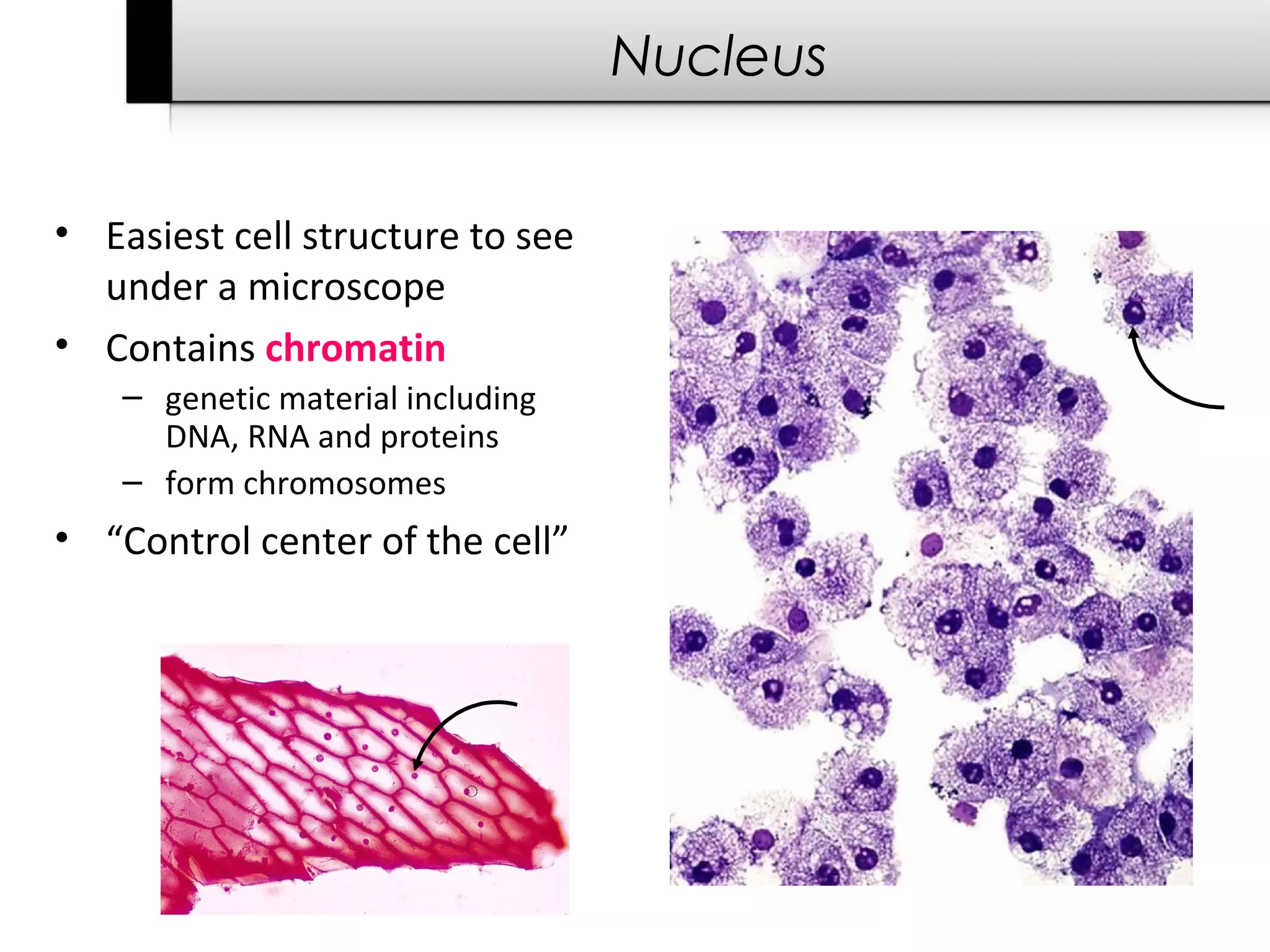
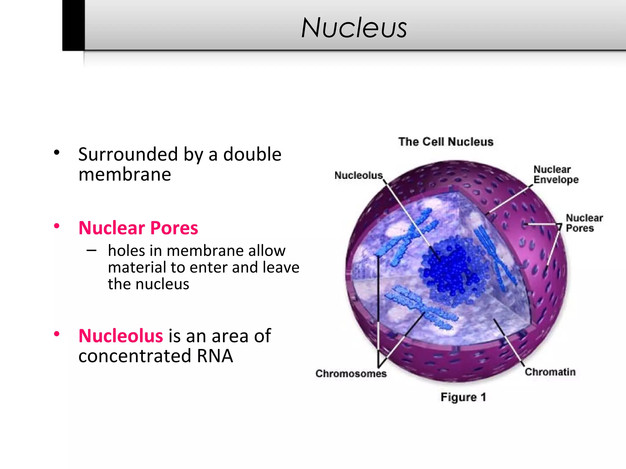
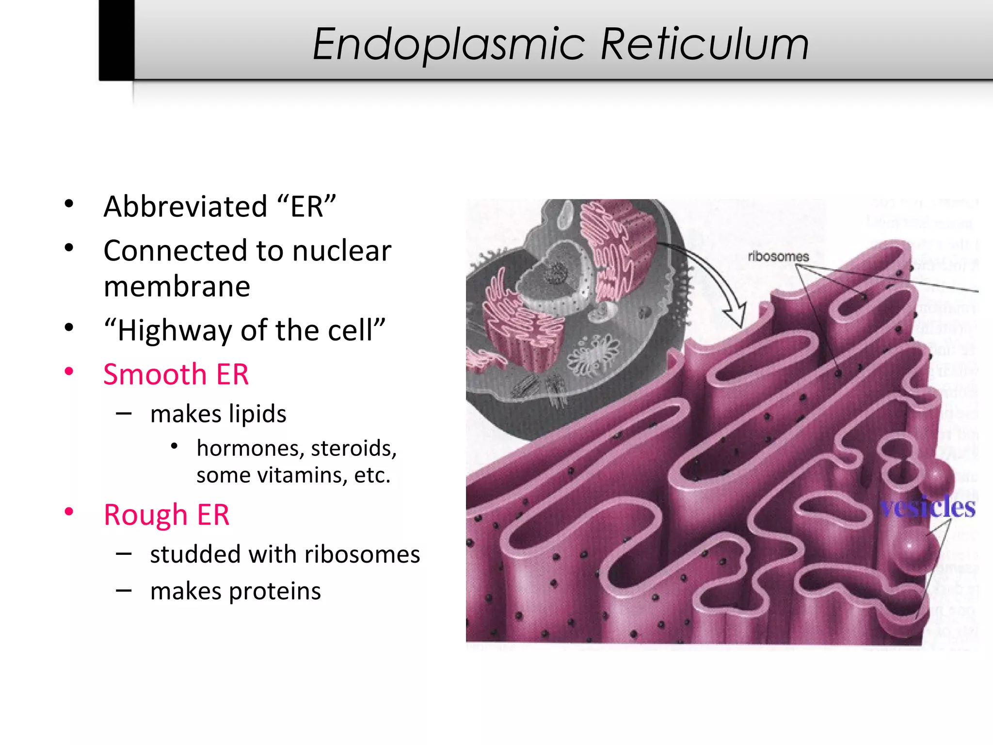
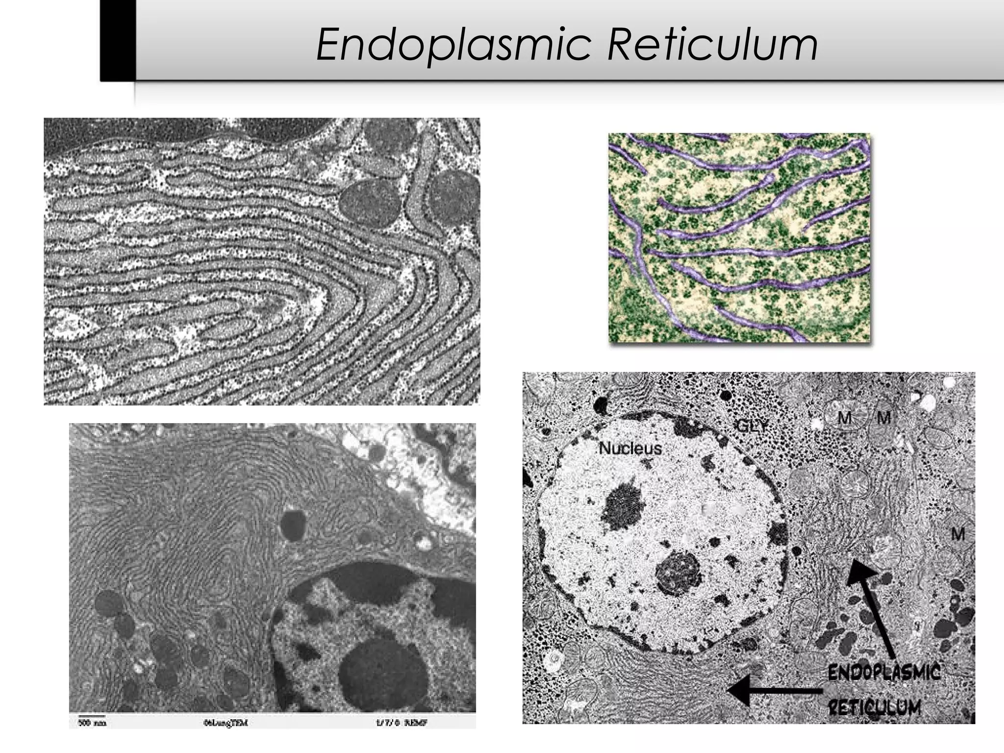
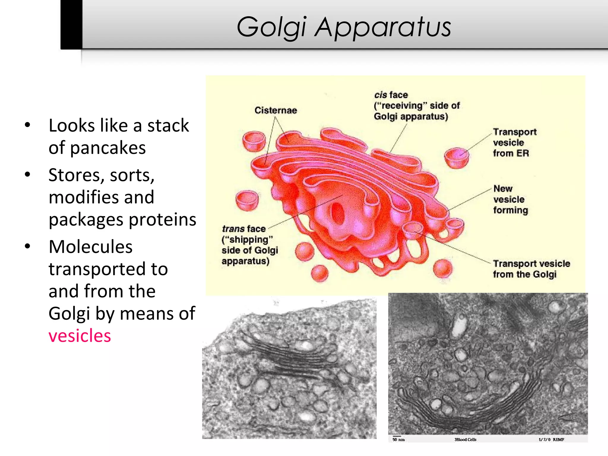
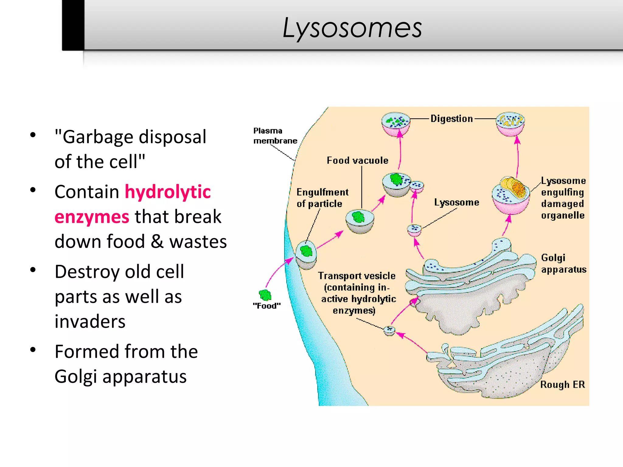
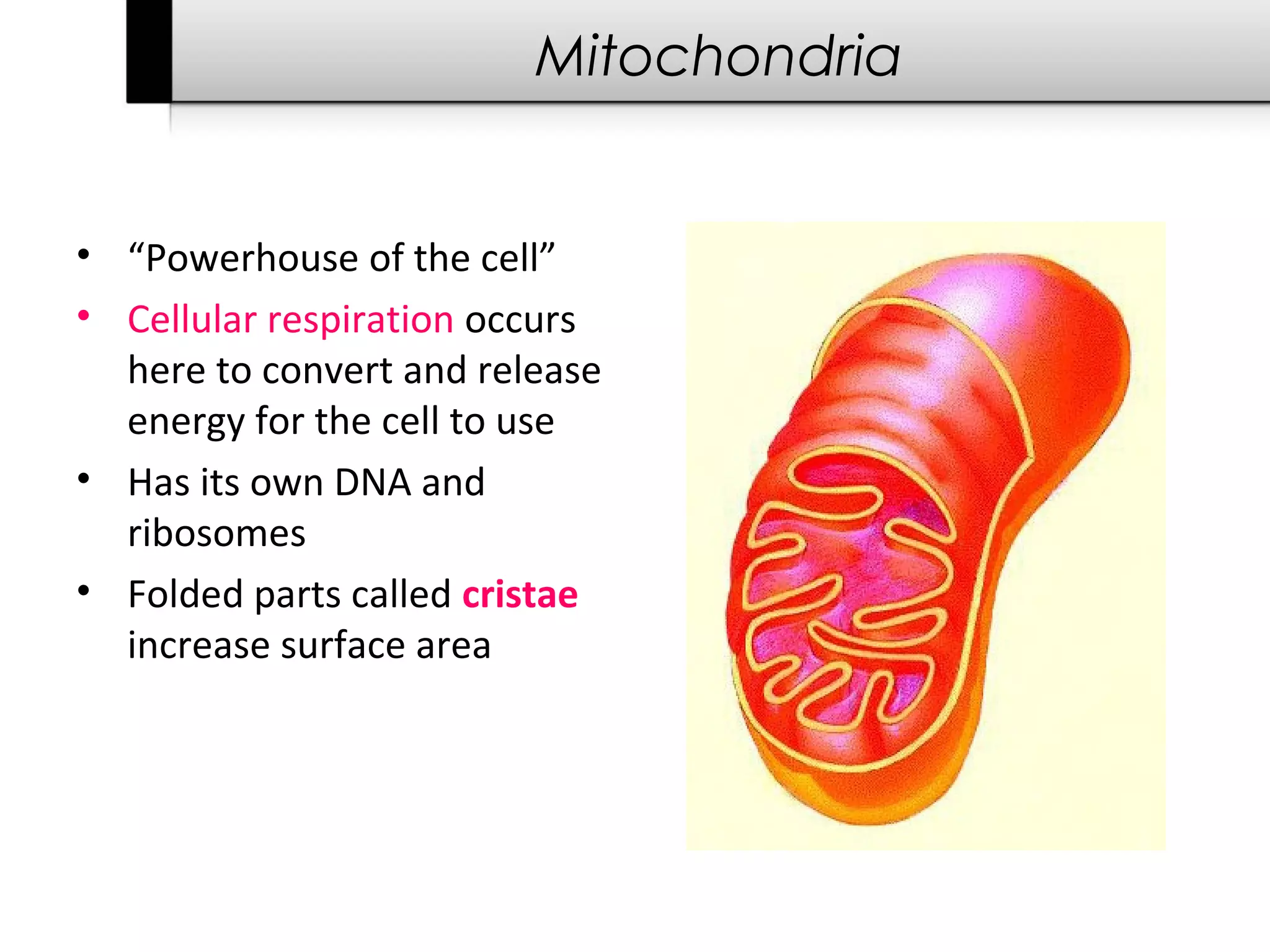
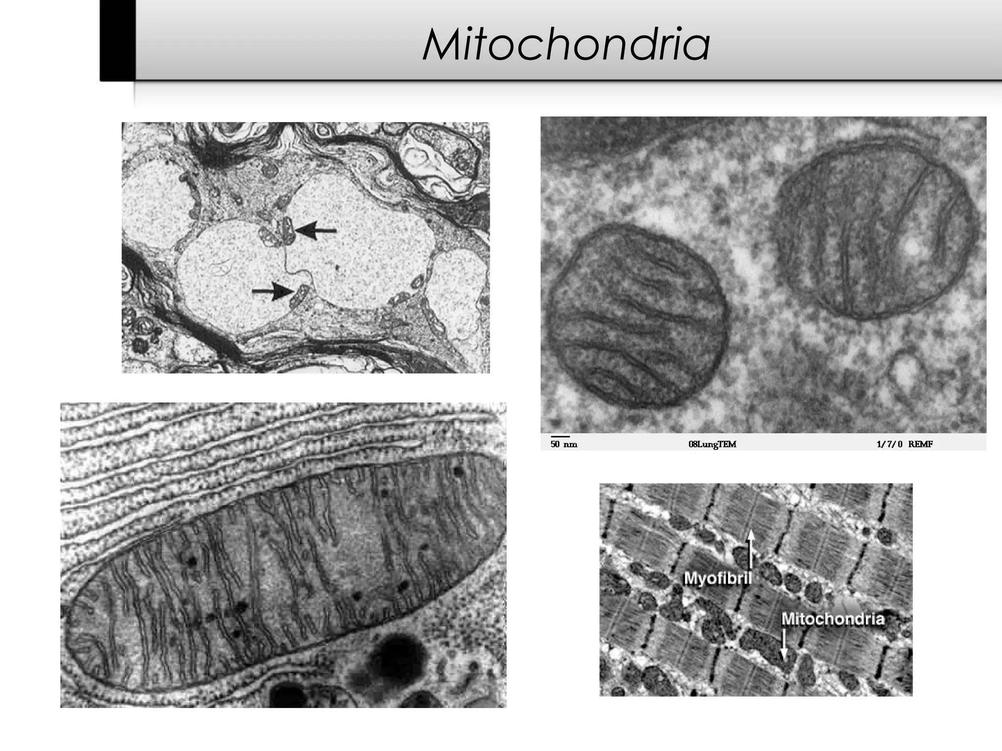
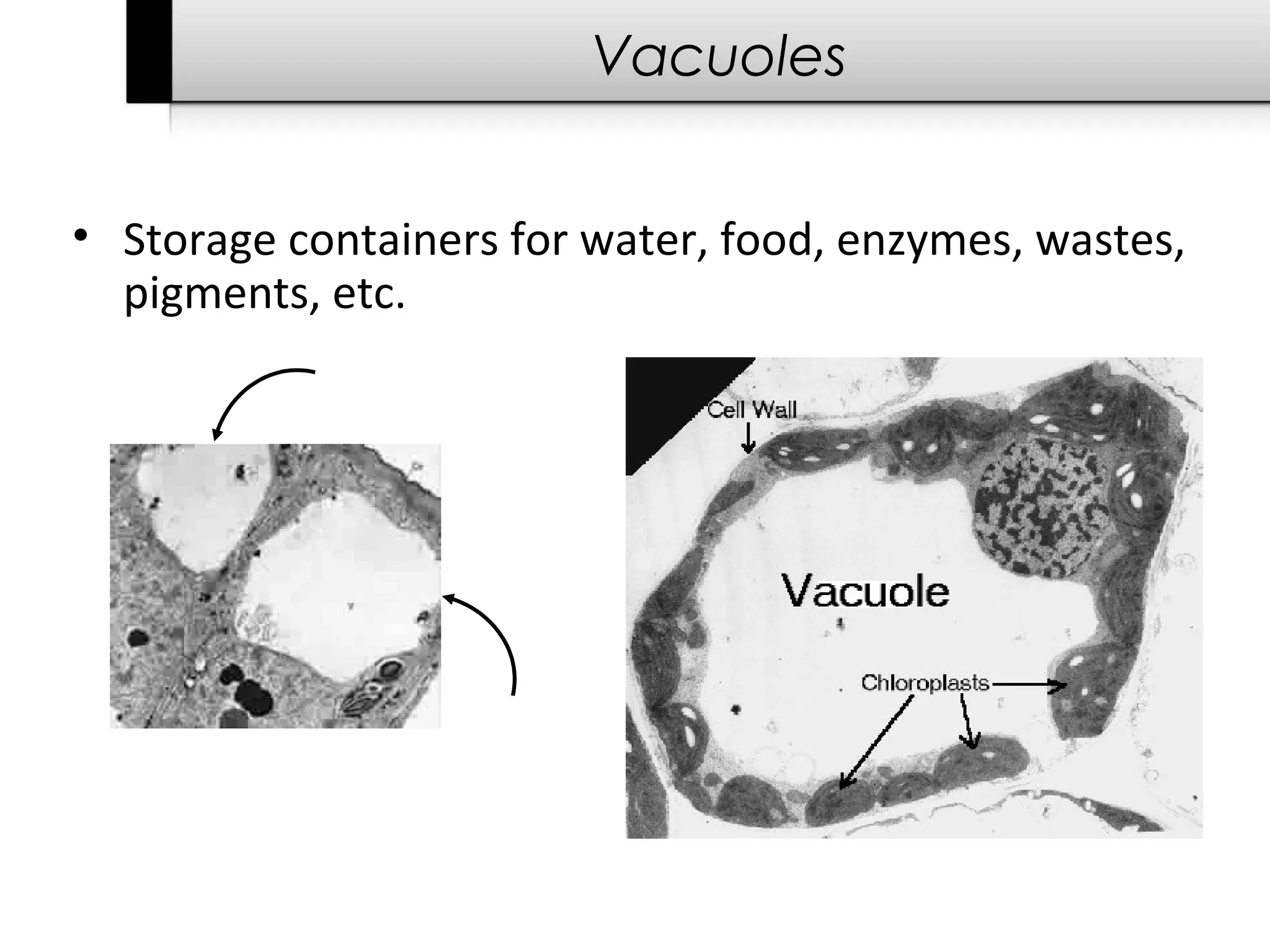
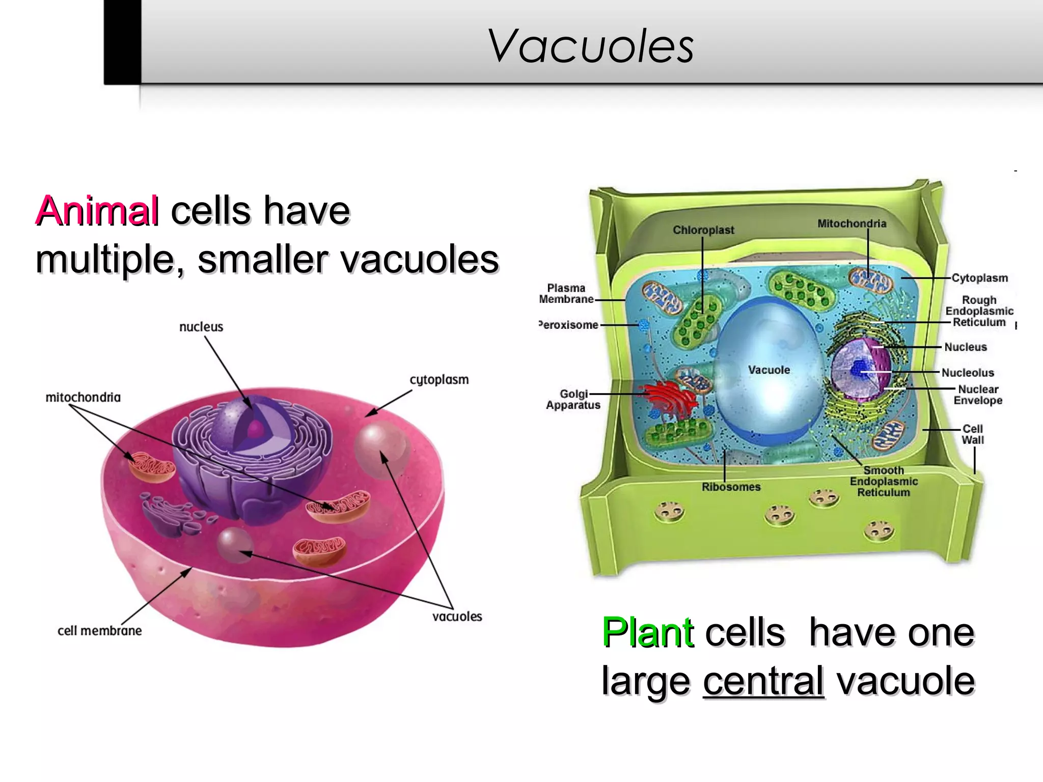
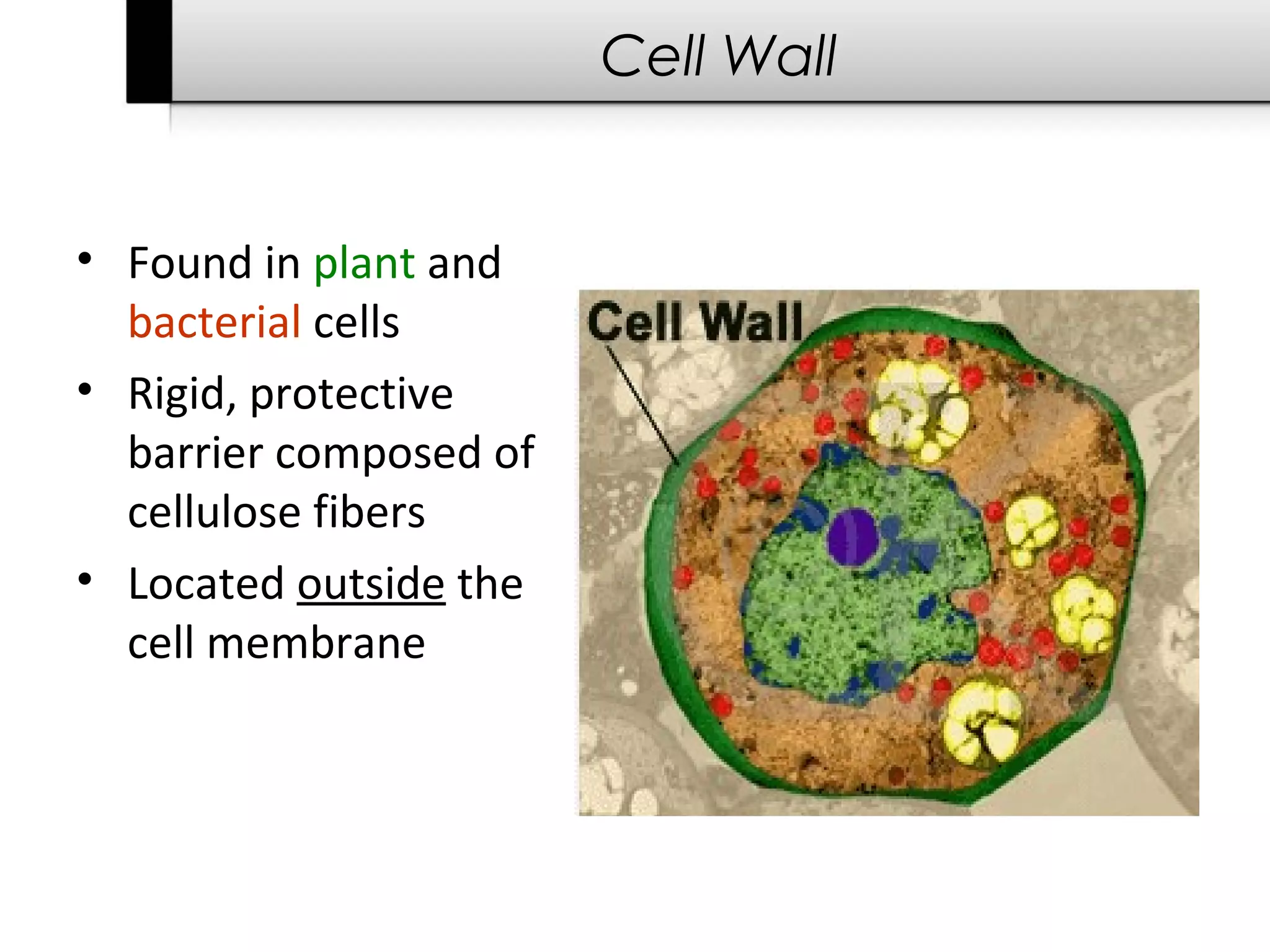
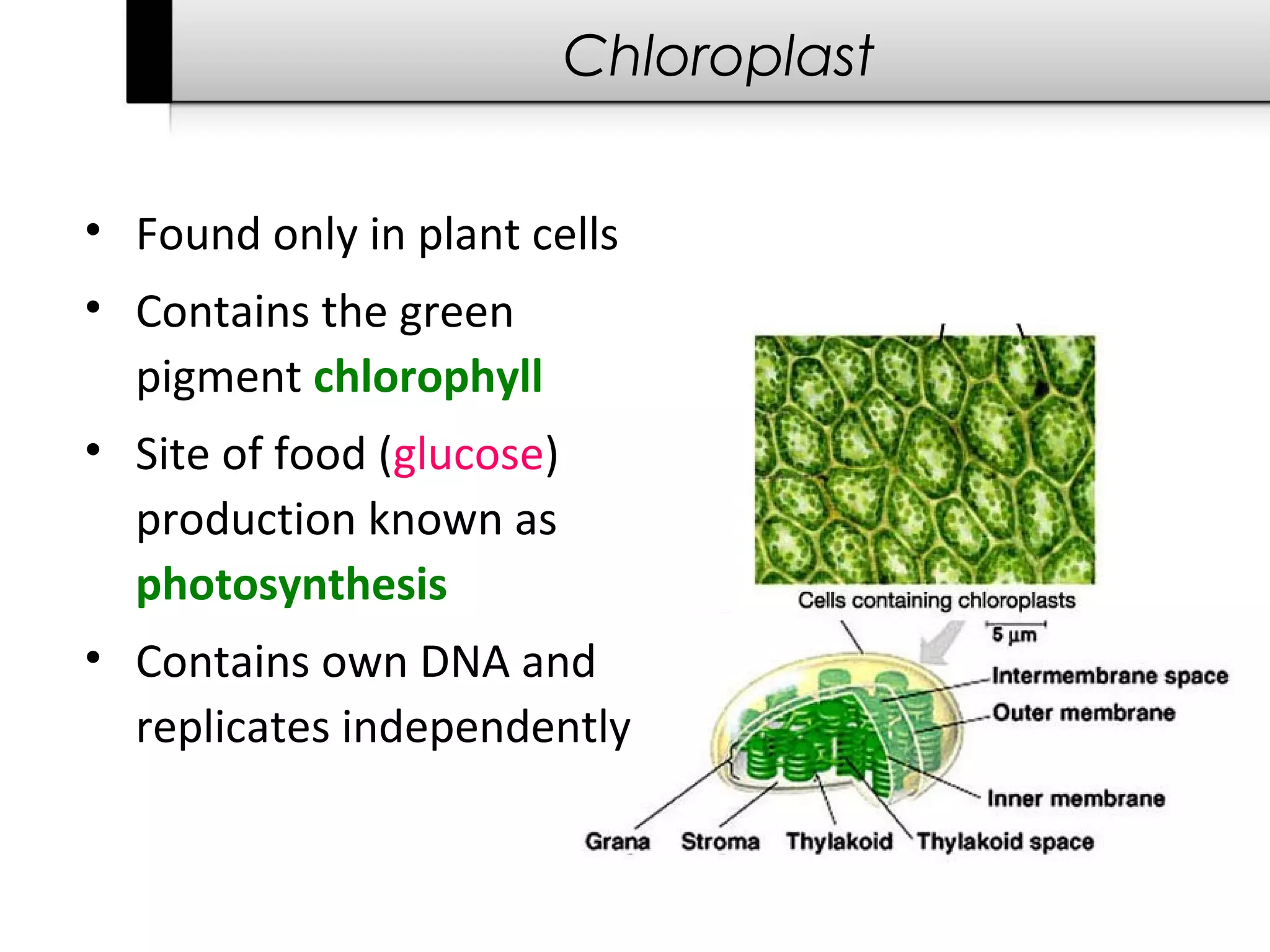
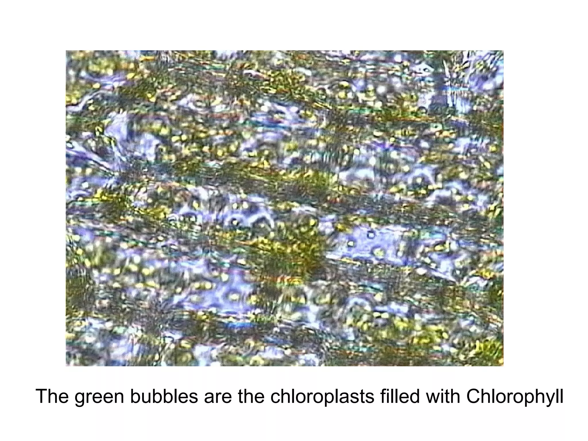
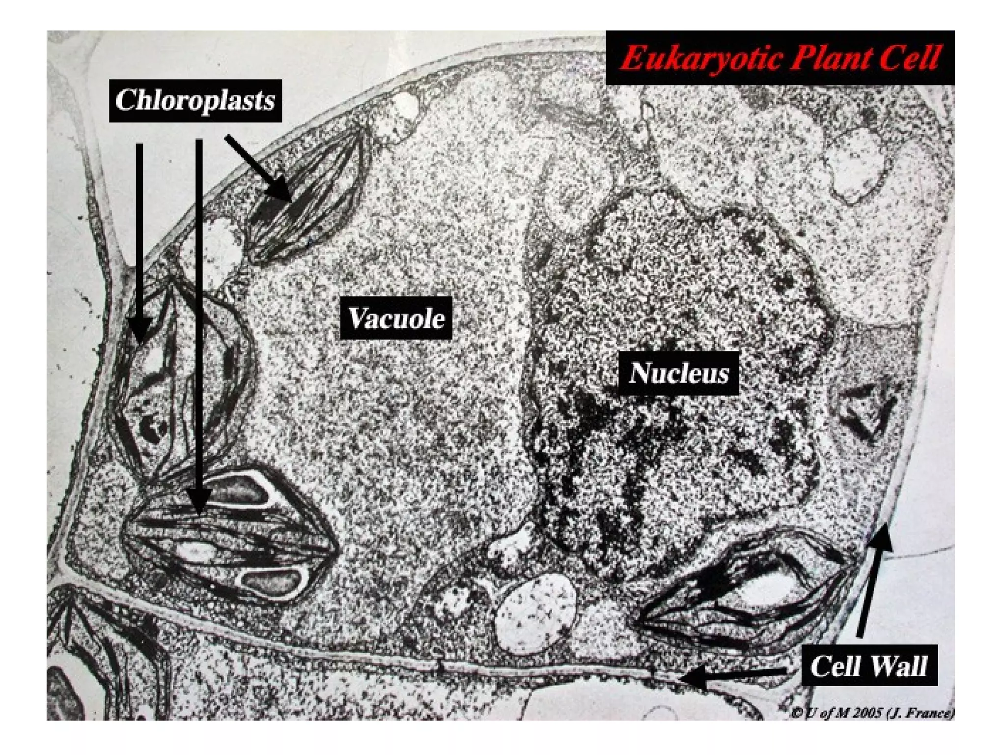
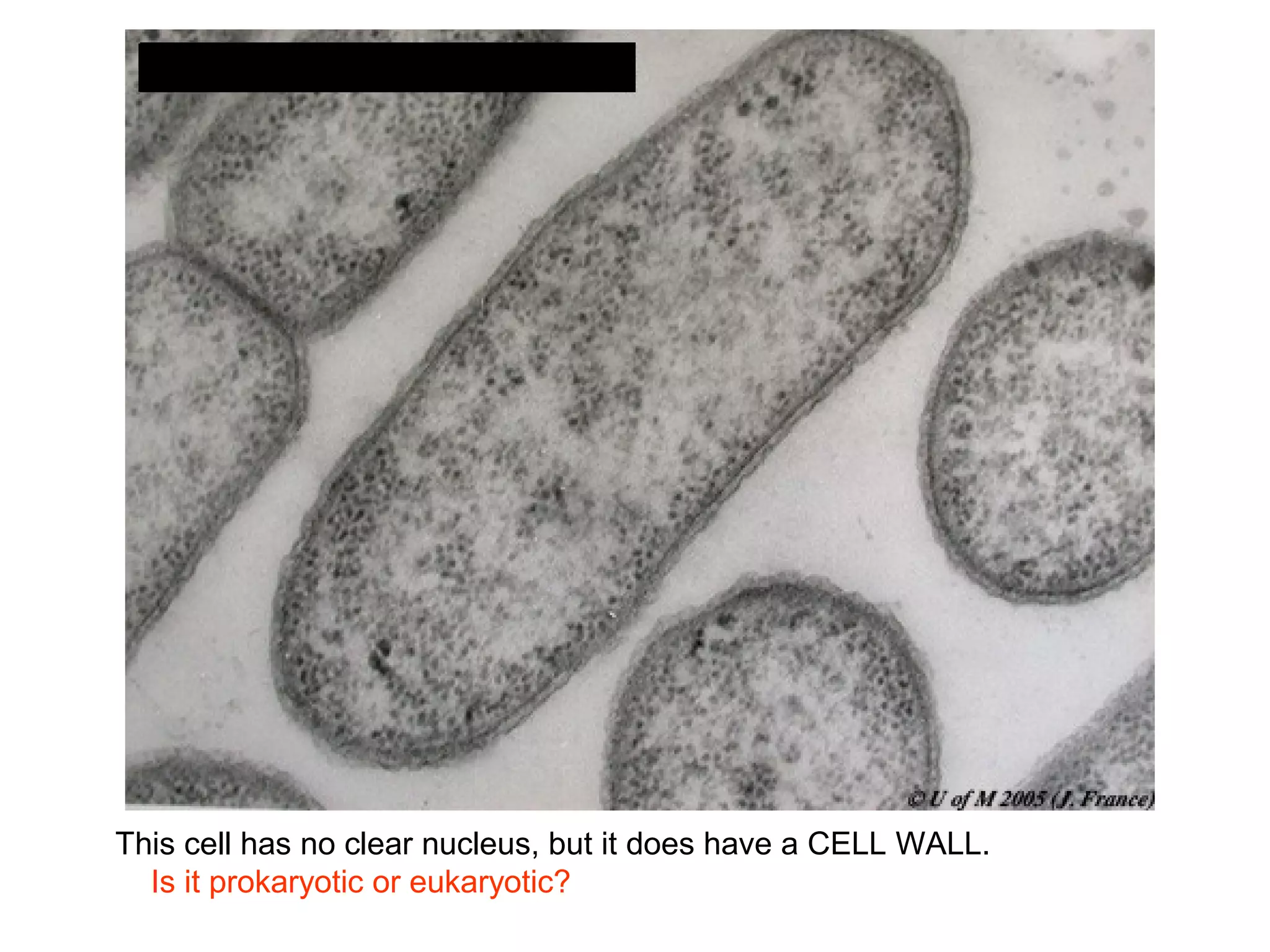
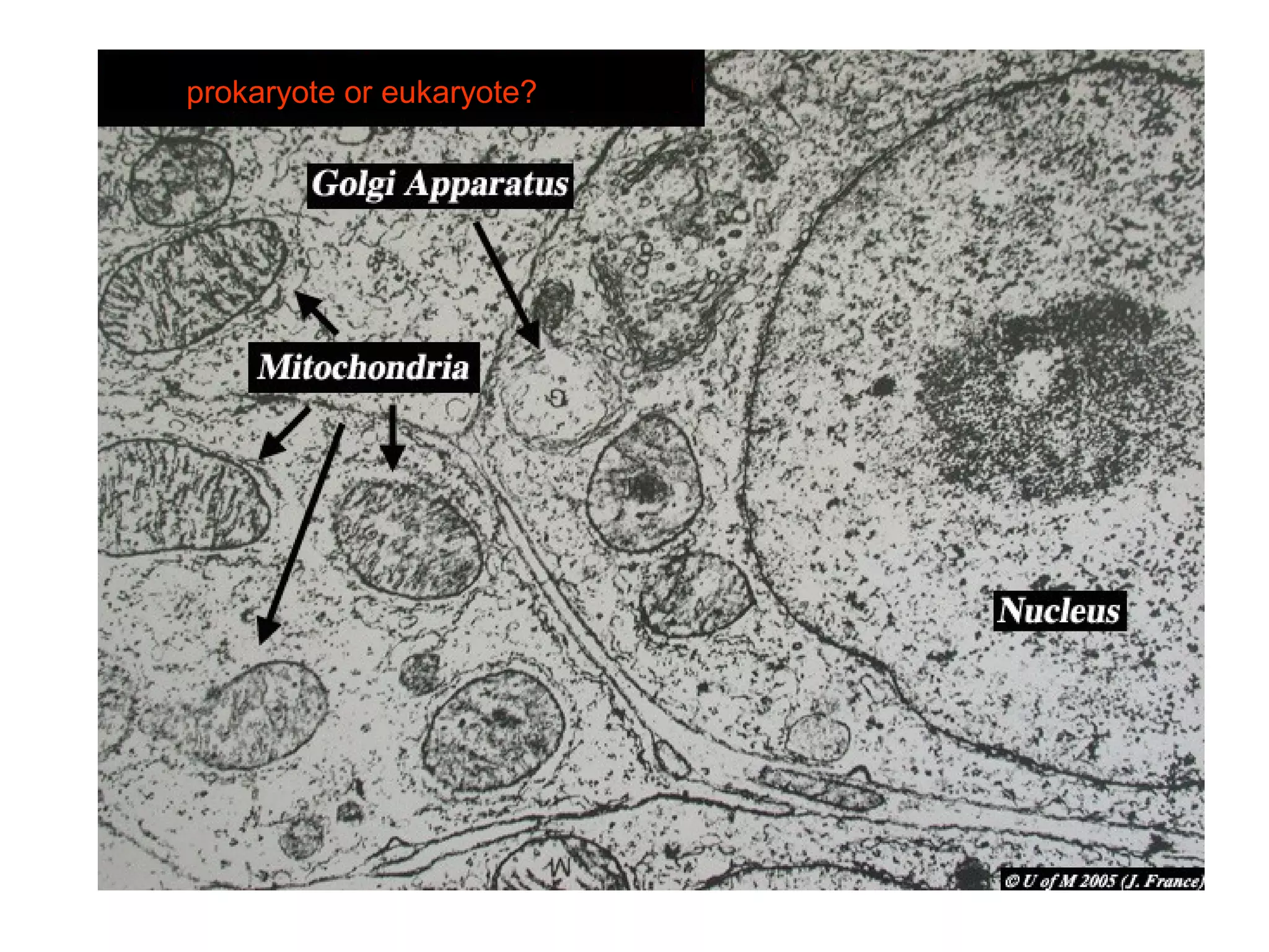
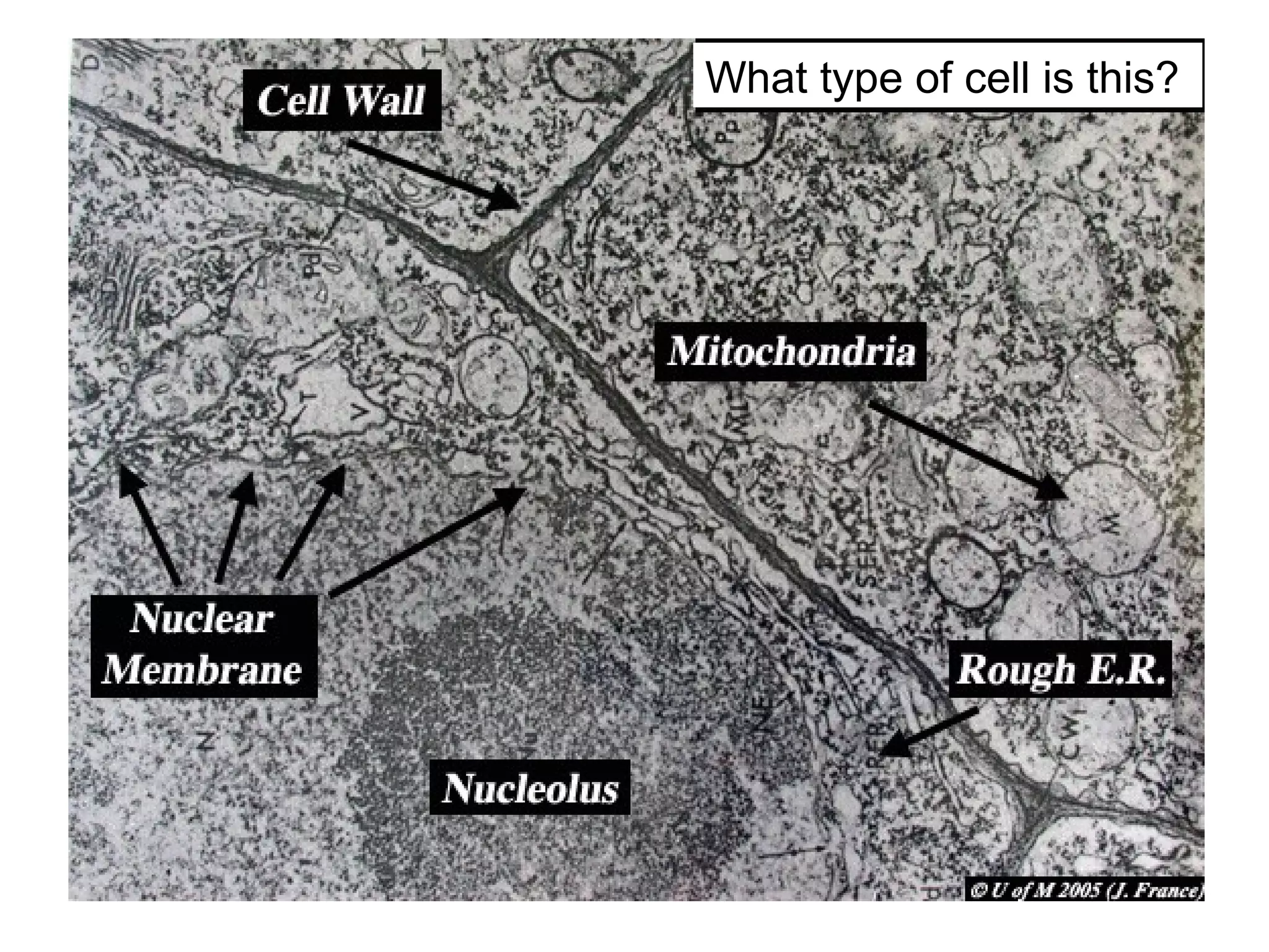
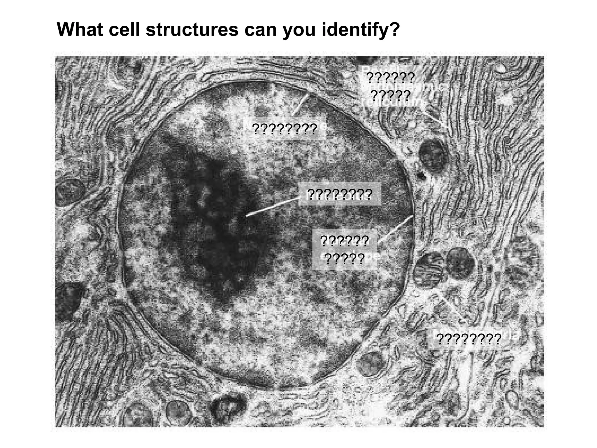
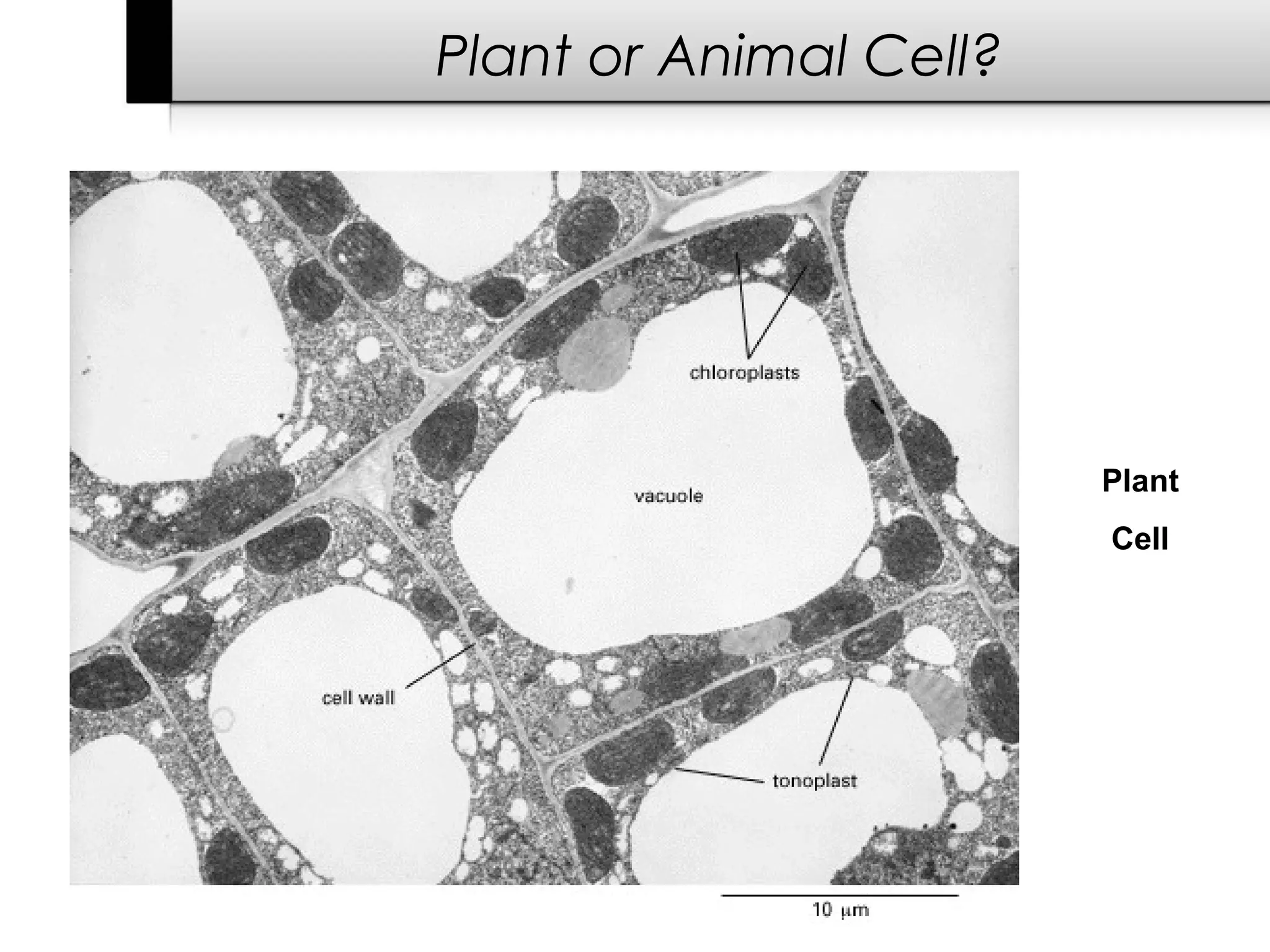
Cells: Structure
The Building Blocks of Life


      Heartlife 2012-2013
          Teacher: Julie Pen


          Science is Organized Knowledge
The Discovery of Cells


• Robert Hooke (mid-1600s)
  – Observed a sliver of cork with a hand-made microscope
  – Saw a “row of empty boxes”
  – Coined the term cell
Cell Theory


The Cell Theory states:
1. Cells are the basic units of life.
2. All living organisms are made up of one or more cells.
3. New cells can only come from already existing cells.
   – no spontaneous generation


• Francisco Redi's
  experiment:
Louis Pasteur


• Biogenesis: the theory that life can only arise
  from another living thing
Cell Size & Shape
Cell Size & Shape


             • All cells have:
                – a membrane
                – cytoplasm
                – DNA
Cell Types

• All living organisms are made up of cells.
   – There are two major cell types:
PROKARYOTIC Cells
   – All bacteria and archaebacteria
   – Lack a nucleus
   – Lack membrane-bound organelles

EUKARYOTIC Cells
   – Plants, animals, fungi and Protists
   – Have an enclosed nucleus
   – Have membrane-bound organelles
Prokaryotic Cell

Bacteria often have a rigid cell wall and capsule around
  the plasma membrane for extra protection.
Eukaryotic Cells

Plant Cell              Animal Cell
Cell Organelles


• Organelle = “little tool”

• Organelles found only
  inside eukaryotic cells

• Watery stuff between
  the organelles is cytosol

• Area inside a cell is the
  cytoplasm
    – includes the organelles
      but NOT the nucleus
Cell Membrane

Cell Membrane
• Boundary of the cell
   – a.k.a plasma membrane
• Holds cell contents together


                                 • Semi-permeable:
                                   acts like a filter or
                                   screen
                                    – some things pass
                                      while others cannot
Cell Membrane

• Made of a phospholipid bilayer


Semi-permeable:
A few molecules
move freely across
the membrane
(in and out of the cell)
   • water
   • carbon dioxide
   • ammonia
   • oxygen

Other molecules need the help of
transport proteins to get across
Nucleus


• Easiest cell structure to see
  under a microscope
• Contains chromatin
    – genetic material including
      DNA, RNA and proteins
    – form chromosomes
• “Control center of the cell”
Nucleus


• Surrounded by a double
  membrane

• Nuclear Pores
   – holes in membrane allow
     material to enter and leave
     the nucleus

• Nucleolus is an area of
  concentrated RNA
Endoplasmic Reticulum


• Abbreviated “ER”
• Connected to nuclear
  membrane
• “Highway of the cell”
• Smooth ER
   – makes lipids
       • hormones, steroids,
         some vitamins, etc.
• Rough ER
   – studded with ribosomes
   – makes proteins
Endoplasmic Reticulum
Golgi Apparatus


• Looks like a stack
  of pancakes
• Stores, sorts,
  modifies and
  packages proteins
• Molecules
  transported to
  and from the
  Golgi by means of
  vesicles
Lysosomes


• "Garbage disposal
  of the cell"
• Contain hydrolytic
  enzymes that break
  down food & wastes
• Destroy old cell
  parts as well as
  invaders
• Formed from the
  Golgi apparatus
Mitochondria


• “Powerhouse of the cell”
• Cellular respiration occurs
  here to convert and release
  energy for the cell to use
• Has its own DNA and
  ribosomes
• Folded parts called cristae
  increase surface area
Mitochondria
Vacuoles


• Storage containers for water, food, enzymes, wastes,
  pigments, etc.
Vacuoles


Animal cells have
multiple, smaller vacuoles


                             Plant cells have one
                             large central vacuole
Cell Wall


• Found in plant and
  bacterial cells
• Rigid, protective
  barrier composed of
  cellulose fibers
• Located outside the
  cell membrane
Chloroplast


• Found only in plant cells
• Contains the green
  pigment chlorophyll
• Site of food (glucose)
  production known as
  photosynthesis
• Contains own DNA and
  replicates independently
The green bubbles are the chloroplasts filled with Chlorophyll
Week 5 Lab

                       Review
          Intro to Microscopy
Laboratory Science as a Career
Cytoskeleton

• Microfilaments and
  microtubules provide
  a scaffolding on which
  organelles can anchor
  or move themselves
   – contracting muscles
     cells
   – dividing cells pinch in
     half
   – waving cilia and
     rotating flagella
Cilia & Flagella


• Hair or tail-like
  structures that provide
  motility (movement)
• Formed from
  microtubules that
  arrange themselves
  into centrioles
• Generally occur only in
  animal and bacteria
  cells
Peroxisomes


• Small sacks enclosed in a
  single membrane
• Contains enzymes that
  break down fatty acids &
  toxic peroxides (found in
  alcohol, formaldehyde, and
  other poisons)
• Found mostly in kidney and
  liver cells
• Formed from budding off
  the ER
Compound Microscope
Quick Review


•   Which organelle is the "control center" of the cell?
          nucleus (containing DNA, the blueprints)

•   Which organelle holds the cell together?
          plasma membrane (made of a phospholipid bilayer)

•   Which organelles are NOT found in animal cells?
          cell wall (and capsule), chloroplasts, large/central vacuole

•   Which organelle helps plant cells make food?
          chloroplasts (via photosynthesis, using chlorophyll)

•   What does E.R. stand for?
          endoplasmic reticulum
This cell has no clear nucleus, but it does have a CELL WALL.
  Is it prokaryotic or eukaryotic?
prokaryote or eukaryote?
What type of cell is this?
What cell structures can you identify?

                                     ??????
                                     ?????

                     ????????


                           ????????


                                ??????
                                ?????


                                              ????????
Plant or Animal Cell?


                        Plant
                        Cell
Careers in Laboratory Science

• Researcher
   – PhD or MD/PhD
   – Design experiments, publish
     conclusions
   – Apply for grants, hire staff
• Research Assistant
   – Manages experiments
   – Assists with data collection, data
     entry, and statistical analysis
• Medical Lab Tech
   – Run lab tests and reports data
       • research or medical treatment
       • Forensic science
       • genetic counseling
• Pharmaceutical Production & Sales
   – Requires business skills & knowledge of chemistry and human
     physiology

Cells: structure

  • 1.
    Cells: Structure The BuildingBlocks of Life Heartlife 2012-2013 Teacher: Julie Pen Science is Organized Knowledge
  • 2.
    The Discovery ofCells • Robert Hooke (mid-1600s) – Observed a sliver of cork with a hand-made microscope – Saw a “row of empty boxes” – Coined the term cell
  • 3.
    Cell Theory The CellTheory states: 1. Cells are the basic units of life. 2. All living organisms are made up of one or more cells. 3. New cells can only come from already existing cells. – no spontaneous generation • Francisco Redi's experiment:
  • 4.
    Louis Pasteur • Biogenesis:the theory that life can only arise from another living thing
  • 5.
  • 6.
    Cell Size &Shape • All cells have: – a membrane – cytoplasm – DNA
  • 7.
    Cell Types • Allliving organisms are made up of cells. – There are two major cell types: PROKARYOTIC Cells – All bacteria and archaebacteria – Lack a nucleus – Lack membrane-bound organelles EUKARYOTIC Cells – Plants, animals, fungi and Protists – Have an enclosed nucleus – Have membrane-bound organelles
  • 8.
    Prokaryotic Cell Bacteria oftenhave a rigid cell wall and capsule around the plasma membrane for extra protection.
  • 9.
  • 10.
    Cell Organelles • Organelle= “little tool” • Organelles found only inside eukaryotic cells • Watery stuff between the organelles is cytosol • Area inside a cell is the cytoplasm – includes the organelles but NOT the nucleus
  • 11.
    Cell Membrane Cell Membrane •Boundary of the cell – a.k.a plasma membrane • Holds cell contents together • Semi-permeable: acts like a filter or screen – some things pass while others cannot
  • 12.
    Cell Membrane • Madeof a phospholipid bilayer Semi-permeable: A few molecules move freely across the membrane (in and out of the cell) • water • carbon dioxide • ammonia • oxygen Other molecules need the help of transport proteins to get across
  • 13.
    Nucleus • Easiest cellstructure to see under a microscope • Contains chromatin – genetic material including DNA, RNA and proteins – form chromosomes • “Control center of the cell”
  • 14.
    Nucleus • Surrounded bya double membrane • Nuclear Pores – holes in membrane allow material to enter and leave the nucleus • Nucleolus is an area of concentrated RNA
  • 15.
    Endoplasmic Reticulum • Abbreviated“ER” • Connected to nuclear membrane • “Highway of the cell” • Smooth ER – makes lipids • hormones, steroids, some vitamins, etc. • Rough ER – studded with ribosomes – makes proteins
  • 16.
  • 17.
    Golgi Apparatus • Lookslike a stack of pancakes • Stores, sorts, modifies and packages proteins • Molecules transported to and from the Golgi by means of vesicles
  • 18.
    Lysosomes • "Garbage disposal of the cell" • Contain hydrolytic enzymes that break down food & wastes • Destroy old cell parts as well as invaders • Formed from the Golgi apparatus
  • 19.
    Mitochondria • “Powerhouse ofthe cell” • Cellular respiration occurs here to convert and release energy for the cell to use • Has its own DNA and ribosomes • Folded parts called cristae increase surface area
  • 20.
  • 21.
    Vacuoles • Storage containersfor water, food, enzymes, wastes, pigments, etc.
  • 22.
    Vacuoles Animal cells have multiple,smaller vacuoles Plant cells have one large central vacuole
  • 23.
    Cell Wall • Foundin plant and bacterial cells • Rigid, protective barrier composed of cellulose fibers • Located outside the cell membrane
  • 24.
    Chloroplast • Found onlyin plant cells • Contains the green pigment chlorophyll • Site of food (glucose) production known as photosynthesis • Contains own DNA and replicates independently
  • 25.
    The green bubblesare the chloroplasts filled with Chlorophyll
  • 27.
    Week 5 Lab Review Intro to Microscopy Laboratory Science as a Career
  • 28.
    Cytoskeleton • Microfilaments and microtubules provide a scaffolding on which organelles can anchor or move themselves – contracting muscles cells – dividing cells pinch in half – waving cilia and rotating flagella
  • 29.
    Cilia & Flagella •Hair or tail-like structures that provide motility (movement) • Formed from microtubules that arrange themselves into centrioles • Generally occur only in animal and bacteria cells
  • 31.
    Peroxisomes • Small sacksenclosed in a single membrane • Contains enzymes that break down fatty acids & toxic peroxides (found in alcohol, formaldehyde, and other poisons) • Found mostly in kidney and liver cells • Formed from budding off the ER
  • 32.
  • 33.
    Quick Review • Which organelle is the "control center" of the cell? nucleus (containing DNA, the blueprints) • Which organelle holds the cell together? plasma membrane (made of a phospholipid bilayer) • Which organelles are NOT found in animal cells? cell wall (and capsule), chloroplasts, large/central vacuole • Which organelle helps plant cells make food? chloroplasts (via photosynthesis, using chlorophyll) • What does E.R. stand for? endoplasmic reticulum
  • 34.
    This cell hasno clear nucleus, but it does have a CELL WALL. Is it prokaryotic or eukaryotic?
  • 35.
  • 36.
    What type ofcell is this?
  • 37.
    What cell structurescan you identify? ?????? ????? ???????? ???????? ?????? ????? ????????
  • 38.
    Plant or AnimalCell? Plant Cell
  • 39.
    Careers in LaboratoryScience • Researcher – PhD or MD/PhD – Design experiments, publish conclusions – Apply for grants, hire staff • Research Assistant – Manages experiments – Assists with data collection, data entry, and statistical analysis • Medical Lab Tech – Run lab tests and reports data • research or medical treatment • Forensic science • genetic counseling • Pharmaceutical Production & Sales – Requires business skills & knowledge of chemistry and human physiology

Editor's Notes

  • #3 Anton van Leeuwenhoek, a Dutch scientist in the mid-1600s, was the first to record looking at water under a microscope. He found that water was full of living things.
  • #4 Italian physician Redi disproved spontaneous generation. Others observed maggots crawling out of meat and assumed the dead cow meat gave rise to baby flies. By performing this experiment, Redi disproved that theory and sought another explanation: that flies lay eggs on the meat, which only provides the food maggots need to survive. The source of the baby fly larvae is, therefore, not the dead meat, but the living fly parents. Experiment 1: Meat was placed in an unsealed jar. Observation: Flies laid eggs on the meat and maggots emerged.   Experiment 2: Meat was placed in a sealed jar. Observation: Flies could not enter the sealed jar and therefore, maggots did not form on the raw meat.   Experiment 3: Meat was placed in a jar covered with gauze. Observation: The gauze prevented flies from entering the jar, and therefore, maggots did not form on the raw meat.   Conclusion: Maggots arise from the eggs laid by flies, not by spontaneous generation. This supported the theory of biogenesis, which states that "all life arises from other life."
  • #5 Some of Pasteur's flasks, still free of microorganisms, are at the Pasteur Institute in Paris.
  • #8 What benefits could there be to having distinct organelles separated by membranes? Chemical reactions which interfere with each other can occur at the same time.
  • #24 This is an actual microscopic image. Colors are added digitally to enhance the different parts.
  • #35 E. Coli
  • #36 Animal (liver) cell
  • #40 Personal experience - U of P, mice stress-immune response - Good Sam, sleep lab/EKG - OHSU, melatonin research