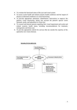
SlideShare a Scribd company logo
These existing classification doesn’t consider loss of function due to hemianopia, loss
of contrast sensitivity, photophobia, visual distortion, diplopia or visual mental difficulties
Introduction
Visual impairment is defined as a functional limitation of the eye(s) or visual system and can
manifest as reduced visual acuity or contrast sensitivity, visual field loss, photophobia,
diplopia, visual distortion, visual perceptual difficulties, or any combination of the above.
These functional limitations can result from congenital (e.g., prenatal or postnatal trauma,
genetic or developmental abnormalities), hereditary (e.g., retinitis pigmentosa or Stargardt's
macular degeneration), or acquired conditions (e.g., ocular infection or disease, trauma, age-
related changes, or systemic disease).
Medical definition: a functional limitation of the eye(s) or visual system manifesting as
reduced visual acuity or contrast sensitivity, visual field loss, photophobia, diplopia, visual
distortion, visual perceptual difficulties, or any combination of the above
Social definition: Vision inadequate for an individual’s needs. It can result in a visual
disability or a visual handicap. It limits the abilities of the individual and limits the
individual’s personal and socioeconomic independence
Legal definition: WHO defines “profound” visual impairment at 20/500 to 20/1000, CF at
less than 3m and VF 10 degrees or less.
Bangkok definition of Low Vision
“A person with low vision is one who has impairment of visual functioning even after
treatment and/or standard refractive correction, and has a visual acuity of less than 6/18 to
light perception, or a visual field less than 10 degrees from the point of fixation, but who uses
or is potentially able to use, vision for the planning and execution of the task.”
1
The World Health Organization (WHO) International Classification of Impairment,
Disabilities, and Handicaps (ICIDH) system is used to classify disorders (diseases),
impairments, disabilities, and handicaps. The definitions are as follows:
A disease is an illness or medical condition, irrespective of origin or source that represents or
could represent significant harm to humans.
An impairment is any loss or abnormality in an anatomical structure or a physiological or
psychological function.
A disability is any restriction or lack (resulting from an impairment) of ability to perform an
activity in the manner or within the range considered normal for a human being.
A handicap indicates a person’s disadvantaged position in society, resulting from impairment
and/or disabilities.
Categories of visual impairment
Fully sighted: normal or near normal vision
Visually impaired
Partially sighted: best corrected vision in the better eye 20/70 or worse but better than
PL and/or If maximum diameter of VF is ≤ 40 degrees.
Legally blind: best corrected vision in the better eye 20/700 or worse (USA), 20/400
(WHO and Many European countries) and/or if maximum diameter of visual field ≤20
degrees.
Functionally blind: best corrected vision with 20/4000 or worse, light projection or
light perception
Total blindness: No light perception
Low vision categories – ICD – 10
World Health Organization’s tenth revision of the International statistical classification of
diseases and related health problems (ICD –10) has included low vision therapy as category 1
and 3 of visual impairment.
This classification defines low vision as corresponding to
Visual acuity of less than 6/18 but equal to or better than 3/60, or
Corresponding visual field loss, to less than 20 degrees, in the better eye with best
possible correction
Blindness is defined as the best corrected visual acuity in the better eye less than 3/60 and/or
visual field less than 10 from the point of fixation.
2
Aspects of vision loss
Disorder: anatomic changes – inflammation, atrophy, scar
Impairment: functional changes – visual acuity, visual field contrast sensitivity
Disability: skills and abilities affected, reading, writing, Mobility
Handicap: socioeconomic consequences – extra effort, loss of independence
Ocular Disorders causing low vision in different age groups
Children Adult Elderly
Retinopathy of prematurity
Congenital Cataract
Nystagmus
Albinism
Microphthalmus
Lebers optic atrophy
Retinitis Pigmentosa
Heredo Macular
degeneration
Stargardts Disease
Pathological Myopia
Optic atrophy
Temporal Pallor
Retino Choroidal coloboma
Age related Macular
degeneration
Diabetic Retinopathy
Advanced Glaucoma
3
Low Vision Rehabilitation
The American Optometric Association defines vision rehabilitation as:
Vision rehabilitation is the process of treatment and education that helps individuals
who are visually disabled attain maximum function, a sense of wellbeing, a personally
satisfying level of independence, and optimum quality of life.
Function is maximized by evaluation, diagnosis and treatment including, but not limited to,
the prescription of optical, non-optical, electronic and/or other treatment
The rehabilitation process includes the development of an individual rehabilitation plan
specifying clinical therapy and/or instruction in compensatory approaches.
Objective of Rehabilitation
To develop the independent living skills of visually impaired people and help they regain
self-confidence for reintegrating into the community
• Analyse the visual elements of a task so that the task can be modified and the environment
adapted to the special equipment used
• Observe the visual environment and assess/observe the person under different
environmental conditions. Vary aspects of the environment also, e.g. distance from the task,
lighting, contrast, colour and time allowed
• Determine which sense is the most efficient for a particular task. For example, vision,
enhanced vision, auditory, tactual, or some combination of these senses. Confident living is
possible when you have the right skills.
The rehabilitation of a low vision patient is a joint responsibility involving the
ophthalmologist, optometrist and a rehabilitationist.
1. Ophthalmologist involves in the clinical evaluation of the patient, determines the
types of low vision aid most appropriate for the patient based on the diagnosis and
prognosis and future medical and surgical intervention for the patient.
2. Optometrist does the optometric low vision assessment, retinoscopy, best refractive
correction and involved in the dispensing of the low vision aids.
3. Rehabilitationist conducts functional vision assessment, giving psychological
counselling, Career guidance and independence in daily living activities, and teaches
orientation and mobility skills.
Goals of comprehensive optometric low vision care and rehabilitation
4
• To evaluate the functional status of the eyes and visual system
• To assess ocular health and related systemic health conditions and the impact of
disease or abnormal conditions on visual functioning
• To provide appropriate optometric rehabilitation intervention to improve the
patient's visual functioning, taking into account the patient's special vision
demands, needs, and adjustment to vision loss
• To counsel and educate patients regarding their visual impairment and ocular and
related systemic health status, including recommendations for treatment,
management, and future care
• To provide appropriate referral for services that are outside the expertise of the
optometric low vision clinician.
5
Central Visual Field Defects
Conditions Causing central visual field defects
 Optic neuritis
 Age related Macular degeneration
 Optic atrophy
 Macular hole/lesions
 Myopic maculopathy
 Occipital cortex lesions
 Stroke and trauma
 Central serous retinopathy
 Best’s and Vitelliform diseases
 Cone dystrophies
 Berlin’s edema
 Macular Coloboma
 Drug e.g. ethanol, lead, methanol, syphilis,
 Glaucoma
6
Distance vision
1. Achromatoposia
• Hereditary condition
• Cones are not developed properly so there is reduced central acuity in bright light
• Field can be slightly constricted particularly to colours
• Since the condition is non progressive, visual prognosis is good
2. Age Related Macular Degeneration
• Acquired retinal disorder caused by degenerative changes in RPE with subsequent
degeneration of overlying cones and rods
• ARMD results in progressive, irreversible loss of central vision fibrous scarring or
geographic atrophy of macular area
• Visual Acuity in dry ARMD ranges from 20/20 to 20/400 and in wet ARMD it is
worse than 20/400
• Visual Field may comprise of central or paracentral scotoma
3. Aniridia
• Congenital absence of iris tissue
• Might be associated with micro cornea with peripheral corneal pannus, congenital
polar cataract, lens dislocation and macular hypoplasia
• Photophobia is the major complaint
• VF defect is due to secondary glaucoma
4. Corneal Dystrophy
• Bilateral inherited entity and usually progressive
• E.g. lattice dystrophy, central crystalline dystrophy of Snyder
• Lacrimation, photophobia, monocular diplopia, foreign body sensation and pain
are the common symptoms
Near Vision
5. Macular Hole
• Appear as round red lesion in the fovea
• Caused by trauma, myopia, cystoids macular edema, inflammation or idiopathic
• Visual acuity ranges from 20/25 to 20/80 in stage 1 and 2 and 20/200 in stage 3
and 4
• Full thickness macular hole result in dense central scotoma
6. Multiple Sclerosis
• Demyelinating disease of CNS
• Several patterns of visual field loss occur in patients with optic neuritis secondary
to multiple sclerosis
• Central/caecoentral scotoma may be present although altitudinal defects occur
most commonly.
7. Optic atrophy
• 3 categories
• Heredodegenerative Optic Atrophy: insidious B/L loss of central vision
• Primary Optic Atrophy: results from no of condition that damage ganglion
cells, most common type
• Secondary Optic Atrophy: occurs subsequent to disc oedema
• Central/caeco central/ paracentral scotoma
8. Stargardt’s disease
• Condition is characterized by vermilion fundus caused by excessive lipofuscin
storage with macular atrophy or flecks at the level of RPE
• Relative central scotoma that may progress to absolute scotoma
9. Macular dystrophy
• Hereditary
• Malformation of light sensing cells of macula
• Painless, progressive, B/L reduced VA
• Central field defect
10. Diabetic retinopathy
• Vision loss occurs commonly from two major problems proliferative diabetic
retinopathy and macular oedema.
• Vision problems in diabetic retinopathy
 Loss of central sharp vision due to macular oedema. Vision loss caused by
macular oedema responds well to magnification.
 Profound loss in vision
 Colour vision impairment
 Reduced peripheral vision after laser causes difficulty in night vision
 Glaucoma
 Cataract
 Reduced contrast sensitivity and glare problem
Diabetics must be able to see to fill insulin syringes and take oral medications. They
need to see to test their blood sugar. Diabetics need to read labels on food containers to
control their intake of carbohydrates. Diabetics, additionally, may have neuropathies
affecting their feet.
Functional implications
• Difficulty seeing central field details for distance, intermediate and near tasks
• Complain with reading prints, recognizing faces and reading signs
• Blurred or distorted central vision; also grey or black area centrally if large dense
scotoma
• Lost individual letters or parts of letters when reading
Distance Vision
Near Vision
When scotoma is located right to the macula, reading becomes difficult as next word appears
If scotoma is to the left of macula, difficulty arises to read beginning of a line of print. So one should
use finger or marker to overcome the problem
• Complains of hearing when central vision impaired suddenly
• Colour vision can be affected
• No mobility problems
Tests to be performed
 Refraction
 Visuoscope
 Amsler grid
 prismatic scanning
 Visual acuity with slits
 visual acuity with isolated letters
 Vision with red/orange filters
 Colour vision
Functional test results
• visual acuity : 20/40 to 20/1000
• Amsler grid: distortion/scotoma/normal
• Glare: not necessary unless cataract, Corneal pathology
• Contrast: high and mid frequency depresses, retention of low frequency – predict
magnification
• Visual field: tangent screen, automated perimetry – extent/location
Rehabilitation of patients with Central Field defects
1. Refraction: Optimum distance and near correction, done as like low vision refraction.
o In patients with low-to-moderate vision loss, the near acuity often can be
improved by increasing the bifocal add alone. However, bifocal powers
greater than +5.00 D may impede mobility. Hence, separate glasses with a
more moderate add may be necessary. Also, demonstrating the reduced focal
distance (closer working distance) of high-add prescriptions helps to educate
patients so that they know exactly at what
distance the reading material should be held.
o Excess convergence demanded by add powers
greater than +4.00 D can be relieved by the use
of a base-in prism, 2 more than the dioptric
power of the add per eye. For example, +6.00 D
glasses would have 8 prism dioptres base-in for
both eyes. Standard prism half glasses are available in +4, +5, +6, +8, +10,
and +12 D powers. Optimal illumination with the light source directed at an
angle of 45° to the page minimizes glare.
o The advantage of high-add readers is that they leave the hands free and are
familiar to patients. The primary disadvantage of these readers is the need to
train patients to use a much closer reading distance.
2. Magnification: is the mainstay of treatment. Options are spectacles magnifier,
Hand/Stand magnifier, CCTV, Telescopes for distance magnification
Near aids
i. Microscopes
Microscopes typically are used when the
needed add power is greater than 12 D. Because of the
greatly reduced focal length, microscopes often are
prescribed monocularly.
In contrast, near telescopes allow the patient to read at a
more natural working distance. Near telescopes, like
surgical telescopes, have a narrow field of view, but they
can be manufactured to focus at any working distance.
However, good motor control is essential to maintain the
reading material within the field of view.
ii. Handheld magnifiers
To achieve full magnification, handheld magnifiers should be demonstrated
with the patient's distance correction in place. The demonstration should begin by
placing the magnifier on the page and, then, slowly raising it until the image is at the
maximum magnification. Although handheld magnifiers provide greater
magnification than high-add readers and allow for a greater reading distance than
microscopes, they require good motor control to be used effectively. Hence, handheld
magnifiers often are suggested for short-term reading tasks, such as checking prices in
a store or reading items on a menu.
iii. Stand magnifiers
Stand magnifiers are similar to handheld magnifiers, but they have a base that
is used to rest the magnifier on the reading page (see image below). Stand
magnifiers require less motor control than handheld magnifiers and allow for a
greater working distance than high-add reading glasses. Moreover, illuminated stand
magnifiers can provide the patient with direct illumination of the reading material.
Fixed-focus stand magnifiers are designed to be
viewed at a distance of 40 cm and require a reading add
to bring the image into focus. Patients often may wish to
use a greater add power than the standard +2.50 D to reduce the working distance
and to gain a wider field of view. They provide the clearest image when the patient
sights the object on axis.
An off-axis view of the object through the magnifiers often produces a
distorted image and results in reduced reading facility. If the patient desires to write
or sew under the magnifier and has only moderately impaired vision, then an
illuminated stand magnifier mounted on a flexible arm can be used. One such device
is the big eye magnifier with a +3.00 D or +6.00 D lens.
Stand magnifiers are especially useful for patients who have glaucoma or
retinitis pigmentosa with restricted visual field. For such patients, the reduced field
of view provided by the stand magnifier often results in better scanning skills while
reading. The 2 most common reasons for patients to reject these devices are postural
fatigue from having to bend over the magnifier and difficulty with manual
manipulation of the magnifier. Although practice makes the device more
manageable and an adjustable light source with clipboard can improve the posture,
patients may prefer the larger field of view provided by closed-circuit televisions
(CCTVs) when reading for an extended period of time.
iv. Close circuit television:
CCTVs provide a larger field of view than other
forms of magnification and greatly improve the reading
facility for patients with severely impaired vision.
Distance Aids
i. Telescopes
Telescopes are typically in clear focus for objects viewed from a distance of
20 feet or more. The doctor should prefocus the telescope on a distant object. The
telescope should be aligned by focusing a penlight beam through the telescope onto
the patient's pupil. The lowest possible magnification should be chosen to meet the
patient's needs, thereby keeping the field of view at a maximum. The patient should
wear the prescribed correction or have it placed within the lens of the telescope.
Improvements in telescopic systems include the ability to adjust for distance,
intermediate, and near vision. Furthermore, increased fields of view and decreased
weight allow patients to increase their reading duration and rate.
Hand-held telescopes are useful for short-
term spotting tasks. They are small and
inconspicuous and often are prescribed
monocularly.
Clip-on
telescopes are usually monocular. They are useful when
patients need their hands to be free. Patching the other eye is
helpful when learning how to see with a clip-on telescope.
Spectacle-mounted
telescopes are made by cutting out
a hole in the conventional lenses
and inserting a telescope. The telescopes may be mounted in
a central position or in a high position (bioptic position).
They are best made with the patient's prescription in the
telescope lens. Patients view through the conventional lens
most of the time and tip their heads down to look through the telescope only when
magnification is needed. In some states, they can be used to pass the acuity portion of
the driver’s license test. They are useful for long-term distance tasks, such as
watching sporting events or bird watching
3. Non optical devices: Felt tipped markers, large print reading materials, yellow filters
for outdoor use, Typoscope, reading table, etc.
Nonoptical aids can enhance greatly the patient's abilities to cope with reduced vision in daily
life. Adaptive equipment is offered by a wide variety of distributors, including the following:
• Large print material, such as the New York
Times, and the Reader's Digest, allow patients
with low-to-moderate vision loss to read with
greater facility.
• Dial markers offer tactile cues for patients to set
gauges on ovens, washers, dryers, and
thermostats.
• Self-threading needles allow patients to thread a needle without seeing the needle eye.
• Books on tape (talking books) allow patients to keep current on a plethora of topics
and to compensate for the lost pleasure of reading.
• Talking clocks and watches help patients to keep time verbally.
• Check signing and writing guides are stencils with openings cut out to allow the
patient to write the date, amount, or signature without writing above or below the line.
• Descriptive video service provides narrated descriptions of visual elements (e.g.
actions, costumes, gestures, scene changes) in program.
• Financial benefits: Patients who are certified as legally blind may be entitled to an
extra exemption on their state income tax form and/or a higher standard deduction on
their federal income tax form and may qualify for social security disability insurance
or supplemental security income benefits.
4. Lighting and glare control: increased illumination improves contrast and reduces the
amount of magnification needed. Tinted sunglasses, amber filters of varying density
with wide brimmed hats are recommended
5. Eccentric viewing : steady eye strategy (keep eye steady towards the best portion of
the field and moving the print across this region
Eccentric viewing training
Patients with macular disease may benefit from learning to view objects with their peripheral
vision. Instructions for eccentric viewing training are as follows:
• The boxed "E" is used to determine where the patient needs to look to see the E most
clearly. That area is marked on the box with an X. This is the eccentric viewing position.
• Next, the patient is instructed to practice looking away
and, then, back to the X.
• The patient should move the E left and right, while
maintaining fixation on the X. If the E becomes blurred or out
of focus, then the patient is instructed to place the E centrally
and refixate on the X. The exercise is repeated, while moving
the E up and down.
• Next, the patient has to repeat the exercise with an E
that does not have an X or lines to guide the patient to the
eccentric viewing position.
6. Prism therapy: to shift the image off a damaged macula
onto a useful adjacent area. An image that falls on non-functional macula can be
relocated a maximum of 10 degree to the healthier retinal area with prisms ( Base of
the prism should be placed corresponding to the useful area of retina) or base in the
direction the image is to be projected
Few note for magnifiers
People with dense, wide area of central visual field loss in an advanced stage may benefit
from stand magnifiers compared to high plus reading glass or hand magnifiers. In SM, patient
can use their peripheral vision to the best to read
High plus reading glasses are useful provided that field loss is not extensive
Prognosis of improving with optical device is bad if field loss is extensiv
Peripheral visual field defects
Conditions causing peripheral visual field defects
• Retinitis pigmentosa
• Degenerative myopia
• Rhegmatogenous retinal detachment
• Retinoschisis
• Retinal tumors
• Trauma
• Neurologic disease – CNS tumors, multiple sclerosis, neurotoxic drugs
• Vascular diseases – Diabetes mellitus,
• Glaucoma,
• Cerebrovascular accident, Aneurysms, etc
1. Glaucoma
• Is an optic neuropathy causing peripheral visual field loss progressing to central visual
field loss.
• Early glaucomatous visual field defects include paracentral scotomas, arcuate
scotomas, nasal steps and temporal wedges.
Tunnel vision in advanced glaucoma
2. Myopic degeneration
• Pathological myopia is an extremely high amount of nearsightedness that causes a
major alteration of the shape or globe of the eye, which may lead to profound vision
loss.
• Associated with other ocular and systemic diseases e.g.
Down’s syndrome
Ocular Albinism, Infantile Glaucoma
Distant vision
Near vision
Marfan’s Syndrome
Retinopathy of Prematurity
Ehler’s-Danlos Syndrome
Low birth weight, and maternal alcoholism
3. Retinitis pigmentosa
• Progressive loss of visual fields is a hallmark of RP. The visual field loss often begins
as a donut like ring in mid-periphery. As it progresses both centrally and peripherally,
the resultant tunnel vision begins to affect the patient’s activities, driving and
mobility.
Distant vision Near vision
• Night blindness, light and glare problem, diplopia (perceiving two separate worlds),
cataract, decreased visual acuity are the main problems.
• RP can be associated with different systemic symptoms/syndromes
Usher’s syndrome – RP with profound congenital hearing loss
Cockyane’s syndrome
Refsum’s disease
4. Traumatic Brain Injury/Stroke
Common form of visual field defect is a homonymous hemianopia.
• Visual Spatial Disorders and Visual Neglect: Patient may experience a variety of
visual spatial disorders. When certain portions of the brain are damaged, the
patient may fail to appreciate space to one side, which is usually to the left.
Unlike visual field loss, this problem is not a physical loss of sensation, but rather
a loss of attention to the area. A man with neglect may no longer shave one side
of his face. Patients with visual neglect have more difficulties than those with only
visual field loss. Unfortunately, both neglect and field loss may occur together.
Other visual-spatial disorders may occur as well. Patients may experience
difficulty navigating themselves even in familiar areas. Patients also misjudge the
straight-ahead position and can confuse right versus left.
• Vertigo, Dizziness and Impaired Eye Movements, Nystagmus
• Diplopia, dry eyes, visual hallucination, light sensitivity, vertigo
5. Anterior Ischemic Optic Neuropathy and Giant Cell Arteritis
• is a potentially visually devastating disease that occurs in the middle aged and the
elderly. Often referred to as a stroke of the optic nerve, and it usually begins suddenly
with little warning in one eye, but frequently progresses to the other eye over time.
• Vision loss often includes both the loss of visual field and visual acuity which can
vary from being nearly normal to severely impaired.
• Visual acuity after an AION may range from 20/20 to no light perception. Vision may
worsen slightly within the first few weeks. The arteritic form of AION (GCA) usually
causes a much worse or higher degree of vision loss than the non-arteritic form
(NAAION). Visual acuity is directly related to the location and amount of the optic
nerve affected by the stroke.
• There is loss of peripheral vision, usually first in the lower field of view. In severe
cases, tunnel vision may result. These field defects range in their severity and can be
either relative, where one can still perceive motion, or could be absolute, where no
motion or light can be seen in the area of the loss. Visual field defects may cause the
patient to be unsure in travel, causing one to trip or bump into objects and have
difficulty walking down stairs.
Importance of Peripheral Vision
1. Mobility and orientation
2. Motion perception
Orientation and mobility
Normal orientation ability and mobility require an intact peripheral visual field,
movement and contrast vision, and normal visual attention. The acoustic and vestibular
systems also contribute to spatial orientation. Patients with concentric constriction of the
visual field are impaired in their ability to orientate themselves. In practice, visual, tactile,
and acoustic aids are used. For patients with sufficient residual vision, small telescopes
(“monoculars”) allow street signs, bus numbers, etc., to be made out,
Left: With the gaze directed forward, the blind half-field is obscured. Right: With
compensatory eye movements to the hemianopic side, the blind half-field is scanned and the
whole field of view used for information gathering. This allows obstacles (in this example,
the suitcase) to be recognized in time. Saccade training is effective in training exploratory
saccades of this kind.
Functional Implications
• Unable to perceive people or object in their surroundings
• Difficulty ambulating in unfamiliar environments
• Bumping into peripheral objects
• Difficulty locating objects
• Two categories
Advanced B/L disease e.g. glaucoma
Sector/hemianopic field deficit – neurological disorders
• Reading problems
Have serious functional consequences
• It is more disabling than blurred vision over the entire field or central scotoma
• Patient must adopt the nonvisual techniques of travelling (long cane, dog guide,
sighted guide, etc.) in peripheral field loss
Objectives of rehabilitation in low vision patient with peripheral field defects
are:
1. Maximized visual acuity
2. Reduction of the effects of glare and photophobia both indoors and outdoors
3. Orientation and mobility management
4. Use of magnification
5. Field enhancement techniques
6. Referrals for additional rehabilitation services
I. Attaining maximum visual acuity
a. Visual acuity:
• Should be measured on charts with many increments of acuity and using
variable illumination
• In condition where pts. central visual field is cone shaped (it gets
proportionally larger the further away from eye), and therefore, if the
peripheral field is reduced, the pt. may have a problem seeing larger target at
near.
b.
Proper subjective and objective refraction
c. Binocularity which provide better visual acuity, greater depth of focus,
and larger field of view.
II. Glare and photophobia control
Glare and photophobia sensitivities are common problems with field restrictions
because of reduced contrast sensitivity, slower response to light and dark adaptation
and secondary media defects. In retinitis pigmentosa and glaucoma, the retinal
pigment epithelium tends to absorb less light and therefore photophobia and glare are
increased due to light scatter. Glare affects the middle and low spatial frequency
levels of a patient’s contrast sensitivity and therefore has an impact on contrast and
mobility.
The evaluation of a glare-or illumination-control lenses should be subjective and task
specific. Varying type of outdoor illumination must be considered when prescribing
any absorptive lens for outdoor glare or photophobia. Problem of ambient light can be
resolved using NoIR and Solar shields lenses or using side shields that can attached to
most frames. Slip-in wraparound, absorptive lenses can alleviate the ambient light and
may also be used in combination with spectacle tinted lenses for greater light
absorption
Glare testing indoors:
General room glare should be tested separately such as reading, watching television, or
using a computer or CCTV.
A tint for CCTV or computer screen can be provided in one of three ways:
a. The appropriate spectacle correction can be combined with the preferred tint.
b. A colored acetate filter can be placed over the entire screen.
b. Filters
Reduce glare, enhance contrast & reduce adaptation time
NOIR-reduce photophobia and glare by absorbing UV (200-
400nm) & near infrared (800-1400nm)
Younger protective lens: Filter UV and short wavelength blue
light
Corning photo chromatic filter: Protects eyes with progressive
retinal degeneration
/ CPF 550 nm filter- reduce scotopic transmittance and allow
photopic transmittance (Retinitis Pigmentosa)
CPF 450 – glare of fluorescent light, CPF 550XD-(aniridia, achromatopsia)
Glare cutter (390-410nm) - cuts 100%UVB, 99% UVA
c. Anti-reflective coatings 3.87 – 5.32% of light lost at each surface
Can be single layered or multiple layered. Higher the index – greater need of antireflective
coatings
Materials – metallic chlorides and fluorides, metals such as lithium sodium and alkaline earth
metals like magnesium.
/
d. Mirror coatings Reduce transmittance by absorption & reflection
/
e. Typo scopes Reduce unwanted glare from surface. Use of yellow acetate paper over the
reading material enhances contrast.
The evaluation for outdoor glare should take place under actual conditions by taking the
patient outdoors and judging his or her response to various tinted lenses.
Slip-in, wraparound, absorptive lenses can also be used.
The use of typo scope, ruler, or even a finger may provide refined tracking and thereby allow
the patient improved functioning. The reading material can be oriented to a position that
eliminates reading into a scotoma by having the material angled at a 45degree angle or even a
90 degree angle.
Glare cutting filter
CPF 450 (yellow color — for glaucoma patients
CPF 511 (yellow orange) — early cataracts
CPF 527 (orange) — for macular degeneration
Filters Properties
4 %Dark Plum this is a very dark, general purpose filter that
provides good contrast and glare reduction
20 % Medium Plum they let ‘in’ a lot more light, so they are good in
low light situations, and they can be
comfortably worn indoors
/2 % Dark Amber particularly helpful on very bright days, 100
percent UV protection, and 100 percent blue
light protection, and 100 percent infrared
protection
40% Light Amber Not only for protection from fluorescent lights,
as well as other indoor lighting sources,
comfortable when working on the computer.
58 % Light Grey Relieves indoor glare, especially under
fluorescent light.
32 % Medium Grey For macular degeneration.
13 % Standard Grey Good for postoperative cataract surgery,
glaucoma sufferers, diabetics or people who
have had corneal transplants.
65 % Yellow Heightens contrast in retinitis pigmentosa, and
macular degeneration.
49 % Orange For sufferers of retinitis pigmentosa.
44 % Red For achromatopsia or rod monochromacy
III. Illumination
Need for HIGHER levels of illumination are seen in patients
Who have?
a. Lost cone functions, like in macular degeneration or ARMD
b. Damaged nerve fibers layers like in glaucoma
c. Diabetic retinopathy
d. Patients on miotics
e. Degenerative myopia
f. Chorioretinitis
g. Retinitis pigmentosa
Eye diseases which require REDUCED illumination are:
a. Albinism
b. Aniridia
c. Corneal opacity
d Achromatopsia
IV. Use of Magnification
In the earlier stage of peripheral field loss, a patient’s central vision is generally no
impaired. When reading, therefore, nothing more than a low-powered reading lens may be
required. A cases of glaucoma or retinitis pigmentosa as the disease progresses, resulting in
increased constriction of the peripheral field along with reduced central vision, a low-
powered reading lenses may no longer be sufficient. If the patient now requires high powered
reading lens to read, he/she is told to hold the lens as closer at a much closer distance
The use of handheld and stand magnifiers should be investigated if reading lenses are not
accepted by the patient. When using magnifiers, the patient must be instructed to adjust
overhead illumination or angle the magnifier to avoid reflections from front surface of the
magnifier
The use of electric or projection-type magnification such as a CCTV, for writing, reading,
and other tasks is a major consideration for patients with peripheral field constrictions.
V. Field enhancement techniques
1. Fresnel Prisms
2. Semi reflective mirror
3. Field expanding channel lens
4. Reverse Telescope
5. Amorphic or new horizontal lens
6. Image Minifiers
7. Concave lenses
8. Convex mirror
1. Fresnel prisms
/
 Press on prism
 1mm plastic sheet of polyvinyl chloride (PVC)
 Applied in the back side of spectacle on carrier lens
Indications:
Overall constriction
Upper or lower field altitudinal defect
Homonymous hemianopia
Prism placement
 side of defect and base towards the defect
 E.g.: Bitemporal hemianopia prism placed base out in temporal part of the
spectacle
By glancing into the prism
 the patient can detect obstacles or targets in the non-seeing area with less eye
movement than would be required without the prism
Advantages
 Minimum weight /thickness /cost
 Wide range of power available (0.5 to 30 pd)
Disadvantages
 Decrease VA,
 Contrast sensitivity
 More distortion & chromatic aberration
 Temporary – fall off / discolored
Note: Use of prism is to create peripheral awareness not to sharpen the vision
Prism in retinitis pigmentosa:
a. Spectacle-mounted prisms effectively create visual field expansion and
noticeable spatial orientation benefits in patients with RP. This may provide
an adjunctive tool in low vision rehabilitation and should be considered in all
cases with RP with less than 10 degrees of visual field.
b. Prism glasses are “very helpful” for obstacle avoidance and reporting
significant benefits for obstacle avoidance in a variety of mobility situations
Prism in ARMD
a. Image Relocation with prisms in patients with ARMD results in a significant
and sizable improvement in BCVA. This effect is probably created by
facilitation of oculomotor functions resulting from direct reduction of fixation
instability.
b. Prismatic treatment in this population mainly improves contrast sensitivity and
visual comfort. It provides patients with better initiation and stabilization of
their eccentric viewing.
c. Prism spectacles are no more effective than conventional spectacles for people
with age-related macular degeneration
2. Semi-reflective mirror
 Semi reflective Plano mirror placed on the spectacle, in opposite side of field defect
 e.g.: on nasal side for a bitemporal hemianopia
 /Mirror reflect the image of an object on the non-seeing side to a more nasal
functional retinal area
Available in:
 Clip-on form or can be permanently affixed to the spectacle frame
 Plano-convex
 Semi-reflecting
 Fully reflecting
 Dichoric
 Mirror position; behind lens or, In front of lens
Disadvantages
 Bulky
 Poor cosmesis
 Confusion due to dual image
 Image reversal
3.Field expanding channel lens
 A pair of spectacle with each carrier lens having
 /Two 12 pd lateral prism and an inferior 8 pd lens
 Central non-prismatic channel (distance/near prescription)
 Apex towards the central non prismatic channel
Field loss Size of channel lens
needed
>20 degree 14mm
20 degree 12mm
15 degree 10mm
10 degree 8mm
6 degree 6mm
4. Reverse telescope
 Galilean or Keplerian telescope viewed through the objective lens causing
minification
 Minify the entire visual field in one or all meridians
 so that more information "fits" into the restricted area, but at the expense of
visual acuity
 Minification equal to power of telescope
 Eg: 3x telescope cause minification by 3x and reduce VA by three times
 Good visual acuity is required due to the minification effect of the telescope
 Only for static view not successful for mobility (due to peripheral distortion, motion
parallax)
 However, 1.3 X can be used on a constant basis
5. Amorphic or new horizontal lenses
 Use of +ve or – ve cylindrical lenses in combination, minification is limited only to
the horizontal meridian
 Resulting in horizontally compressed & vertically unchanged retinal image by a factor
of lens power
 Purpose: to minimize the VA degradation as minification is limited to one meridian
6. Image minifiers
 Principle:
 Reverse Galilean telescope that produces a 1/3rd
image size reduction without
producing excessive barrel distortion
 Features:
 Focusable, light weighted
 Handheld. Clip on, spectacle mounted system
7. Concave lenses
 Concave lens held at specific distance from the eye
 Act as a reverse telescope
 Minification is provided by
 Concave lens
 Accommodation or add power to see the image clearly
 Amount of Minification:
Concave lens power (D) / Add/Acco.)(A)
8. Convex mirror
 Enhance VF by minification
 Used as side mirrors of vehicle, hall ways, doorways
 Give wide field help patient with constricted field to be aware of the surroundings
VI. Orientation and mobility management
Orientation refers to a familiarization with and adaptation to a position
Or point in the environment.
Mobility refers to the safe travel from one point to another.
Peripheral field defects impair orientation:
a. Persons with a concentric field defect can be helped by tactile aids, such as a cane,
and with orientation and mobility training.
b. Persons with hemianopia can benefit from compensatory saccadic training
Orientation impairment
///
Vision aids
Long cane
Guide dog
Ultrasound
device
Acoustic aids
Obstacle detection
Electronic guidance
systems
Training
How to use aids
● learning possible
compensatory behaviors
– Orientation and mobility
training
– Saccade training
References:
1. Essentials of low vision: Richard L Brilliant
2. AOA Optometry clinical practical guide: Care of patient with visual impairment (Low
vision rehabilitation)
3. Silverstone, Barbara, ed. Lighthouse Handbook on Vision Impairment and Vision
Rehabilitation - Vol. 1 Vision Impairment/ Oxford: Oxford University Press, 2000
4. Lighthouse Handbook on Vision Impairment and Vision Rehabilitation - Vol 2,
Brilliant, Richard L
5. Goodrich GL. A trend analysis of the low-vision literature. BrJ Vis Impair2004;
22:105–6
6. Massof RW. A systems model for low vision rehabilitation. I. Basic concepts. Optom
Vis Sci1995; 72:725–36
7. International Society for Low Vision Research and Rehabilitation (ISLRR)
http://www.islrr.org
8. http://emedicine.medscape.com/article/1832033
~~~~

More Related Content

What's hot

Rgp lens
Rgp lensRgp lens
Causes of low vision in adult
Causes of low vision in adultCauses of low vision in adult
Causes of low vision in adult
Laxmi Eye Institute
 
Low vision in childhood
Low vision in childhoodLow vision in childhood
Low vision in childhoodbkoptom
 
Soft Contact Lens Fitting
Soft Contact Lens FittingSoft Contact Lens Fitting
Soft Contact Lens FittingVishakh Nair
 
low vision aids
low vision aidslow vision aids
low vision aidsAstha Jain
 
Sports vision
Sports visionSports vision
Sports vision
RAIN HEALTH CARE
 
Visiontherapy
VisiontherapyVisiontherapy
Visiontherapy
ShahanaSherin9
 
Test for stereopsis
Test for stereopsisTest for stereopsis
Test for stereopsis
Aliasger Fakhruddin
 
Visual Rehabilitation in low vision.pptx
Visual Rehabilitation in low vision.pptxVisual Rehabilitation in low vision.pptx
Visual Rehabilitation in low vision.pptx
Siddharth Duggal
 
Structural cnanges in the eyes by rb
Structural cnanges in the eyes by rbStructural cnanges in the eyes by rb
Structural cnanges in the eyes by rb
RajatBansal61
 
Insertion and removal of rgp contact lens.
Insertion and removal of rgp contact lens.Insertion and removal of rgp contact lens.
Insertion and removal of rgp contact lens.
Anandhan K
 
Rose K lens.pptx
Rose K lens.pptxRose K lens.pptx
Rose K lens.pptx
Bhuvaneswari Ganesan
 
Binocular refraction techniques, binocular balancing
Binocular refraction techniques, binocular balancing Binocular refraction techniques, binocular balancing
Binocular refraction techniques, binocular balancing Mohammad Arman Bin Aziz
 
Spherical RGP contact lens fitting and prescribing
Spherical RGP contact lens fitting and prescribingSpherical RGP contact lens fitting and prescribing
Spherical RGP contact lens fitting and prescribing
Pabita Dhungel
 
Special type of lenses
Special  type of lensesSpecial  type of lenses
Special type of lenses
OPTOM FASLU MUHAMMED
 
Basics of pediatric refraction by dr.adnan
 Basics of pediatric refraction by dr.adnan Basics of pediatric refraction by dr.adnan
Basics of pediatric refraction by dr.adnan
MahamudAdnan
 
Electronic Devices for Low Vision Patients
 Electronic Devices for Low Vision Patients Electronic Devices for Low Vision Patients
Electronic Devices for Low Vision Patients
Nilufa Akter
 
Diplopia charting
Diplopia chartingDiplopia charting
Diplopia charting
Mantu Akon
 

What's hot (20)

Rgp lens
Rgp lensRgp lens
Rgp lens
 
Causes of low vision in adult
Causes of low vision in adultCauses of low vision in adult
Causes of low vision in adult
 
Low vision in childhood
Low vision in childhoodLow vision in childhood
Low vision in childhood
 
Soft Contact Lens Fitting
Soft Contact Lens FittingSoft Contact Lens Fitting
Soft Contact Lens Fitting
 
low vision aids
low vision aidslow vision aids
low vision aids
 
Sports vision
Sports visionSports vision
Sports vision
 
Visiontherapy
VisiontherapyVisiontherapy
Visiontherapy
 
Maddox rod
Maddox rodMaddox rod
Maddox rod
 
Low vision chart
Low vision chart Low vision chart
Low vision chart
 
Test for stereopsis
Test for stereopsisTest for stereopsis
Test for stereopsis
 
Visual Rehabilitation in low vision.pptx
Visual Rehabilitation in low vision.pptxVisual Rehabilitation in low vision.pptx
Visual Rehabilitation in low vision.pptx
 
Structural cnanges in the eyes by rb
Structural cnanges in the eyes by rbStructural cnanges in the eyes by rb
Structural cnanges in the eyes by rb
 
Insertion and removal of rgp contact lens.
Insertion and removal of rgp contact lens.Insertion and removal of rgp contact lens.
Insertion and removal of rgp contact lens.
 
Rose K lens.pptx
Rose K lens.pptxRose K lens.pptx
Rose K lens.pptx
 
Binocular refraction techniques, binocular balancing
Binocular refraction techniques, binocular balancing Binocular refraction techniques, binocular balancing
Binocular refraction techniques, binocular balancing
 
Spherical RGP contact lens fitting and prescribing
Spherical RGP contact lens fitting and prescribingSpherical RGP contact lens fitting and prescribing
Spherical RGP contact lens fitting and prescribing
 
Special type of lenses
Special  type of lensesSpecial  type of lenses
Special type of lenses
 
Basics of pediatric refraction by dr.adnan
 Basics of pediatric refraction by dr.adnan Basics of pediatric refraction by dr.adnan
Basics of pediatric refraction by dr.adnan
 
Electronic Devices for Low Vision Patients
 Electronic Devices for Low Vision Patients Electronic Devices for Low Vision Patients
Electronic Devices for Low Vision Patients
 
Diplopia charting
Diplopia chartingDiplopia charting
Diplopia charting
 

Viewers also liked

The Low Vision Examination
The Low Vision ExaminationThe Low Vision Examination
The Low Vision ExaminationHossein Mirzaie
 
Low vision aid
Low vision aidLow vision aid
Low vision aid
SSSIHMS-PG
 
Calculation of magnification in low vision
Calculation of magnification in low visionCalculation of magnification in low vision
Calculation of magnification in low vision
Mohammad Arman Bin Aziz
 
Low Vision Aids
Low Vision AidsLow Vision Aids
Low Vision Aids
Ayinun Nahar
 
Presentation blindness
Presentation blindnessPresentation blindness
Presentation blindnesssmurfetteg2011
 
Survey of optometric low vision rehabilitation training
Survey of optometric low vision rehabilitation trainingSurvey of optometric low vision rehabilitation training
Survey of optometric low vision rehabilitation training
Juan Castaño
 
Low vision assessment
Low vision assessmentLow vision assessment
Low vision assessment
confusionexpert1
 
Low visual aids
Low visual aidsLow visual aids
Low visual aids
Doc Aaryan
 
Low vision aids dr. d p shah
Low vision aids   dr. d p shahLow vision aids   dr. d p shah
Low vision aids dr. d p shahlionsleaders
 
Low vision rehabilitation in patients with retinal dystrophy
Low vision rehabilitation in patients with retinal dystrophyLow vision rehabilitation in patients with retinal dystrophy
Low vision rehabilitation in patients with retinal dystrophy
Amrit Pokharel
 
National prevention of blindness program
National prevention of blindness programNational prevention of blindness program
National prevention of blindness programAbigail Abalos
 
NATIONAL PROGRAMME FOR CONTROL OF BLINDNESS
NATIONAL PROGRAMME FOR CONTROL OF BLINDNESSNATIONAL PROGRAMME FOR CONTROL OF BLINDNESS
NATIONAL PROGRAMME FOR CONTROL OF BLINDNESS
Sukruth Srinivas
 
understanding visual impairment
understanding visual impairmentunderstanding visual impairment
understanding visual impairment
Jomina Deri-Huerto
 
national blindness control programme
national blindness control programmenational blindness control programme
national blindness control programmevikramjit_singh
 

Viewers also liked (20)

The Low Vision Examination
The Low Vision ExaminationThe Low Vision Examination
The Low Vision Examination
 
Low vision aid
Low vision aidLow vision aid
Low vision aid
 
Blindness
BlindnessBlindness
Blindness
 
Blindness
BlindnessBlindness
Blindness
 
Magnification
MagnificationMagnification
Magnification
 
Calculation of magnification in low vision
Calculation of magnification in low visionCalculation of magnification in low vision
Calculation of magnification in low vision
 
Patel presentation
Patel presentationPatel presentation
Patel presentation
 
Low Vision Aids
Low Vision AidsLow Vision Aids
Low Vision Aids
 
Presentation blindness
Presentation blindnessPresentation blindness
Presentation blindness
 
Survey of optometric low vision rehabilitation training
Survey of optometric low vision rehabilitation trainingSurvey of optometric low vision rehabilitation training
Survey of optometric low vision rehabilitation training
 
Low vision assessment
Low vision assessmentLow vision assessment
Low vision assessment
 
Magnification
MagnificationMagnification
Magnification
 
Low visual aids
Low visual aidsLow visual aids
Low visual aids
 
Low vision aids dr. d p shah
Low vision aids   dr. d p shahLow vision aids   dr. d p shah
Low vision aids dr. d p shah
 
Low vision rehabilitation in patients with retinal dystrophy
Low vision rehabilitation in patients with retinal dystrophyLow vision rehabilitation in patients with retinal dystrophy
Low vision rehabilitation in patients with retinal dystrophy
 
National prevention of blindness program
National prevention of blindness programNational prevention of blindness program
National prevention of blindness program
 
NATIONAL PROGRAMME FOR CONTROL OF BLINDNESS
NATIONAL PROGRAMME FOR CONTROL OF BLINDNESSNATIONAL PROGRAMME FOR CONTROL OF BLINDNESS
NATIONAL PROGRAMME FOR CONTROL OF BLINDNESS
 
Tinted lenses
Tinted lensesTinted lenses
Tinted lenses
 
understanding visual impairment
understanding visual impairmentunderstanding visual impairment
understanding visual impairment
 
national blindness control programme
national blindness control programmenational blindness control programme
national blindness control programme
 

Similar to Low vision rehabilitation

Introduction to Low Vision
Introduction to Low VisionIntroduction to Low Vision
Introduction to Low Vision
PriyaMeenakshi
 
Comprehensive LOW VISION
Comprehensive LOW VISIONComprehensive LOW VISION
Comprehensive LOW VISION
Vivek Chaudhary
 
Lecture fakulti pendidikan 2011
Lecture fakulti pendidikan 2011Lecture fakulti pendidikan 2011
Lecture fakulti pendidikan 2011mfmy_fahmy
 
Low vision aids
Low vision aidsLow vision aids
Low vision aids
Krati Gupta
 
What every md should know about the eye
What every md should know about the eyeWhat every md should know about the eye
What every md should know about the eye
SDGWEP
 
13 blindness
13 blindness13 blindness
13 blindness
JAYDIP NINAMA
 
Enhancing Daily Living for Patients with Low Vision
Enhancing Daily Living for Patients with Low VisionEnhancing Daily Living for Patients with Low Vision
Enhancing Daily Living for Patients with Low Vision
Enhanced Vision
 
ED 7 REPORT VISUAL IMPAIRMENT INCLUDING BLINDNESS (INCLUSIVE EDUCATION)
ED 7 REPORT VISUAL IMPAIRMENT INCLUDING BLINDNESS (INCLUSIVE EDUCATION)ED 7 REPORT VISUAL IMPAIRMENT INCLUDING BLINDNESS (INCLUSIVE EDUCATION)
ED 7 REPORT VISUAL IMPAIRMENT INCLUDING BLINDNESS (INCLUSIVE EDUCATION)
lyracristel00
 
Vision in closed ocular trauma
Vision in closed ocular traumaVision in closed ocular trauma
Vision in closed ocular trauma
shemina aziz
 
Common Disorders in Low Vision
Common Disorders in Low VisionCommon Disorders in Low Vision
Common Disorders in Low Vision
RabindraAdhikary
 
Vision in older adults fall 2013 abridged
Vision in  older adults fall 2013 abridgedVision in  older adults fall 2013 abridged
Vision in older adults fall 2013 abridgedShepard Joy
 
Childhood Eye Problems.pptx
Childhood Eye Problems.pptxChildhood Eye Problems.pptx
Childhood Eye Problems.pptx
MohammadSalahUddin46
 
Biology Investigatory Project on Eye Diseases (class 12th)
Biology Investigatory Project on Eye Diseases (class 12th) Biology Investigatory Project on Eye Diseases (class 12th)
Biology Investigatory Project on Eye Diseases (class 12th)
MohitBhuraney
 
Promoting eye health for Elderly 1.ppt
Promoting eye health for Elderly 1.pptPromoting eye health for Elderly 1.ppt
Promoting eye health for Elderly 1.ppt
RitaOMOMOH
 
Blindness and its management
Blindness and its managementBlindness and its management
Blindness and its management
Archana Syiem
 
Aging and The Eye
Aging and The EyeAging and The Eye
Aging and The Eye
Rabia Ammer
 
Vision in older adults spring 2014 abridged
Vision in  older adults spring 2014 abridgedVision in  older adults spring 2014 abridged
Vision in older adults spring 2014 abridgedShepard Joy
 
Vision Deficits Following Traumatic Brain Injury
Vision Deficits Following Traumatic Brain InjuryVision Deficits Following Traumatic Brain Injury
Vision Deficits Following Traumatic Brain Injury
Brain Injury Alliance of New Jersey
 
Lazy Eye, Eye Turns and Other Functional Vision Disorders
Lazy Eye, Eye Turns and Other Functional Vision DisordersLazy Eye, Eye Turns and Other Functional Vision Disorders
Lazy Eye, Eye Turns and Other Functional Vision Disorders
Dominick Maino
 
visual impairements .
visual impairements .visual impairements .
visual impairements .
Mukesh Jangra
 

Similar to Low vision rehabilitation (20)

Introduction to Low Vision
Introduction to Low VisionIntroduction to Low Vision
Introduction to Low Vision
 
Comprehensive LOW VISION
Comprehensive LOW VISIONComprehensive LOW VISION
Comprehensive LOW VISION
 
Lecture fakulti pendidikan 2011
Lecture fakulti pendidikan 2011Lecture fakulti pendidikan 2011
Lecture fakulti pendidikan 2011
 
Low vision aids
Low vision aidsLow vision aids
Low vision aids
 
What every md should know about the eye
What every md should know about the eyeWhat every md should know about the eye
What every md should know about the eye
 
13 blindness
13 blindness13 blindness
13 blindness
 
Enhancing Daily Living for Patients with Low Vision
Enhancing Daily Living for Patients with Low VisionEnhancing Daily Living for Patients with Low Vision
Enhancing Daily Living for Patients with Low Vision
 
ED 7 REPORT VISUAL IMPAIRMENT INCLUDING BLINDNESS (INCLUSIVE EDUCATION)
ED 7 REPORT VISUAL IMPAIRMENT INCLUDING BLINDNESS (INCLUSIVE EDUCATION)ED 7 REPORT VISUAL IMPAIRMENT INCLUDING BLINDNESS (INCLUSIVE EDUCATION)
ED 7 REPORT VISUAL IMPAIRMENT INCLUDING BLINDNESS (INCLUSIVE EDUCATION)
 
Vision in closed ocular trauma
Vision in closed ocular traumaVision in closed ocular trauma
Vision in closed ocular trauma
 
Common Disorders in Low Vision
Common Disorders in Low VisionCommon Disorders in Low Vision
Common Disorders in Low Vision
 
Vision in older adults fall 2013 abridged
Vision in  older adults fall 2013 abridgedVision in  older adults fall 2013 abridged
Vision in older adults fall 2013 abridged
 
Childhood Eye Problems.pptx
Childhood Eye Problems.pptxChildhood Eye Problems.pptx
Childhood Eye Problems.pptx
 
Biology Investigatory Project on Eye Diseases (class 12th)
Biology Investigatory Project on Eye Diseases (class 12th) Biology Investigatory Project on Eye Diseases (class 12th)
Biology Investigatory Project on Eye Diseases (class 12th)
 
Promoting eye health for Elderly 1.ppt
Promoting eye health for Elderly 1.pptPromoting eye health for Elderly 1.ppt
Promoting eye health for Elderly 1.ppt
 
Blindness and its management
Blindness and its managementBlindness and its management
Blindness and its management
 
Aging and The Eye
Aging and The EyeAging and The Eye
Aging and The Eye
 
Vision in older adults spring 2014 abridged
Vision in  older adults spring 2014 abridgedVision in  older adults spring 2014 abridged
Vision in older adults spring 2014 abridged
 
Vision Deficits Following Traumatic Brain Injury
Vision Deficits Following Traumatic Brain InjuryVision Deficits Following Traumatic Brain Injury
Vision Deficits Following Traumatic Brain Injury
 
Lazy Eye, Eye Turns and Other Functional Vision Disorders
Lazy Eye, Eye Turns and Other Functional Vision DisordersLazy Eye, Eye Turns and Other Functional Vision Disorders
Lazy Eye, Eye Turns and Other Functional Vision Disorders
 
visual impairements .
visual impairements .visual impairements .
visual impairements .
 

More from Hira Dahal

Common binocular vision disorder neglected
Common binocular vision disorder neglectedCommon binocular vision disorder neglected
Common binocular vision disorder neglected
Hira Dahal
 
Gonioscopy presentation
Gonioscopy presentationGonioscopy presentation
Gonioscopy presentation
Hira Dahal
 
Glare testing and dark adaptation
Glare testing and dark adaptationGlare testing and dark adaptation
Glare testing and dark adaptation
Hira Dahal
 
Embryology and anatomy of human lens
Embryology and anatomy of human lensEmbryology and anatomy of human lens
Embryology and anatomy of human lens
Hira Dahal
 
Vitamins a
Vitamins aVitamins a
Vitamins a
Hira Dahal
 
Corneal transparency
Corneal transparencyCorneal transparency
Corneal transparency
Hira Dahal
 
Electrophysiology
ElectrophysiologyElectrophysiology
Electrophysiology
Hira Dahal
 
Anatomic and physiological ocular changes with age final
Anatomic and physiological ocular changes with age finalAnatomic and physiological ocular changes with age final
Anatomic and physiological ocular changes with age final
Hira Dahal
 
Visual field testing and interpretation
Visual field testing and interpretationVisual field testing and interpretation
Visual field testing and interpretation
Hira Dahal
 
Cyclorefraction
CyclorefractionCyclorefraction
Cyclorefraction
Hira Dahal
 
Colour vision examination
Colour vision examinationColour vision examination
Colour vision examination
Hira Dahal
 
Prisms in optometry practice
Prisms in optometry practicePrisms in optometry practice
Prisms in optometry practice
Hira Dahal
 
Therapeutic contact lens
Therapeutic contact lensTherapeutic contact lens
Therapeutic contact lens
Hira Dahal
 
Effects of radiation and glare in eye
Effects of radiation and glare in eyeEffects of radiation and glare in eye
Effects of radiation and glare in eye
Hira Dahal
 
Slit lamp biomicroscopy
Slit lamp biomicroscopySlit lamp biomicroscopy
Slit lamp biomicroscopy
Hira Dahal
 
General eye overview
General eye overviewGeneral eye overview
General eye overview
Hira Dahal
 
Frame slection
Frame slectionFrame slection
Frame slection
Hira Dahal
 
Circadian cycle
Circadian cycleCircadian cycle
Circadian cycle
Hira Dahal
 
RGP Complications
RGP ComplicationsRGP Complications
RGP Complications
Hira Dahal
 

More from Hira Dahal (20)

Common binocular vision disorder neglected
Common binocular vision disorder neglectedCommon binocular vision disorder neglected
Common binocular vision disorder neglected
 
Gonioscopy presentation
Gonioscopy presentationGonioscopy presentation
Gonioscopy presentation
 
Glare testing and dark adaptation
Glare testing and dark adaptationGlare testing and dark adaptation
Glare testing and dark adaptation
 
Embryology and anatomy of human lens
Embryology and anatomy of human lensEmbryology and anatomy of human lens
Embryology and anatomy of human lens
 
Vitamins a
Vitamins aVitamins a
Vitamins a
 
Corneal transparency
Corneal transparencyCorneal transparency
Corneal transparency
 
Electrophysiology
ElectrophysiologyElectrophysiology
Electrophysiology
 
Anatomic and physiological ocular changes with age final
Anatomic and physiological ocular changes with age finalAnatomic and physiological ocular changes with age final
Anatomic and physiological ocular changes with age final
 
Visual field testing and interpretation
Visual field testing and interpretationVisual field testing and interpretation
Visual field testing and interpretation
 
Cyclorefraction
CyclorefractionCyclorefraction
Cyclorefraction
 
Colour vision examination
Colour vision examinationColour vision examination
Colour vision examination
 
Prisms in optometry practice
Prisms in optometry practicePrisms in optometry practice
Prisms in optometry practice
 
Therapeutic contact lens
Therapeutic contact lensTherapeutic contact lens
Therapeutic contact lens
 
Effects of radiation and glare in eye
Effects of radiation and glare in eyeEffects of radiation and glare in eye
Effects of radiation and glare in eye
 
Slit lamp biomicroscopy
Slit lamp biomicroscopySlit lamp biomicroscopy
Slit lamp biomicroscopy
 
General eye overview
General eye overviewGeneral eye overview
General eye overview
 
Frame slection
Frame slectionFrame slection
Frame slection
 
Circadian cycle
Circadian cycleCircadian cycle
Circadian cycle
 
RGP Complications
RGP ComplicationsRGP Complications
RGP Complications
 
Retinoscopy
RetinoscopyRetinoscopy
Retinoscopy
 

Recently uploaded

Physiology of Special Chemical Sensation of Taste
Physiology of Special Chemical Sensation of TastePhysiology of Special Chemical Sensation of Taste
Physiology of Special Chemical Sensation of Taste
MedicoseAcademics
 
The hemodynamic and autonomic determinants of elevated blood pressure in obes...
The hemodynamic and autonomic determinants of elevated blood pressure in obes...The hemodynamic and autonomic determinants of elevated blood pressure in obes...
The hemodynamic and autonomic determinants of elevated blood pressure in obes...
Catherine Liao
 
Charaka Samhita Sutra sthana Chapter 15 Upakalpaniyaadhyaya
Charaka Samhita Sutra sthana Chapter 15 UpakalpaniyaadhyayaCharaka Samhita Sutra sthana Chapter 15 Upakalpaniyaadhyaya
Charaka Samhita Sutra sthana Chapter 15 Upakalpaniyaadhyaya
Dr KHALID B.M
 
TEST BANK for Operations Management, 14th Edition by William J. Stevenson, Ve...
TEST BANK for Operations Management, 14th Edition by William J. Stevenson, Ve...TEST BANK for Operations Management, 14th Edition by William J. Stevenson, Ve...
TEST BANK for Operations Management, 14th Edition by William J. Stevenson, Ve...
kevinkariuki227
 
ARTIFICIAL INTELLIGENCE IN HEALTHCARE.pdf
ARTIFICIAL INTELLIGENCE IN  HEALTHCARE.pdfARTIFICIAL INTELLIGENCE IN  HEALTHCARE.pdf
ARTIFICIAL INTELLIGENCE IN HEALTHCARE.pdf
Anujkumaranit
 
THOA 2.ppt Human Organ Transplantation Act
THOA 2.ppt Human Organ Transplantation ActTHOA 2.ppt Human Organ Transplantation Act
THOA 2.ppt Human Organ Transplantation Act
DrSathishMS1
 
ANATOMY AND PHYSIOLOGY OF URINARY SYSTEM.pptx
ANATOMY AND PHYSIOLOGY OF URINARY SYSTEM.pptxANATOMY AND PHYSIOLOGY OF URINARY SYSTEM.pptx
ANATOMY AND PHYSIOLOGY OF URINARY SYSTEM.pptx
Swetaba Besh
 
Ozempic: Preoperative Management of Patients on GLP-1 Receptor Agonists
Ozempic: Preoperative Management of Patients on GLP-1 Receptor Agonists  Ozempic: Preoperative Management of Patients on GLP-1 Receptor Agonists
Ozempic: Preoperative Management of Patients on GLP-1 Receptor Agonists
Saeid Safari
 
The Normal Electrocardiogram - Part I of II
The Normal Electrocardiogram - Part I of IIThe Normal Electrocardiogram - Part I of II
The Normal Electrocardiogram - Part I of II
MedicoseAcademics
 
Hemodialysis: Chapter 3, Dialysis Water Unit - Dr.Gawad
Hemodialysis: Chapter 3, Dialysis Water Unit - Dr.GawadHemodialysis: Chapter 3, Dialysis Water Unit - Dr.Gawad
Hemodialysis: Chapter 3, Dialysis Water Unit - Dr.Gawad
NephroTube - Dr.Gawad
 
Surgical Site Infections, pathophysiology, and prevention.pptx
Surgical Site Infections, pathophysiology, and prevention.pptxSurgical Site Infections, pathophysiology, and prevention.pptx
Surgical Site Infections, pathophysiology, and prevention.pptx
jval Landero
 
ARTHROLOGY PPT NCISM SYLLABUS AYURVEDA STUDENTS
ARTHROLOGY PPT NCISM SYLLABUS AYURVEDA STUDENTSARTHROLOGY PPT NCISM SYLLABUS AYURVEDA STUDENTS
ARTHROLOGY PPT NCISM SYLLABUS AYURVEDA STUDENTS
Dr. Vinay Pareek
 
Surat @ℂall @Girls ꧁❤8527049040❤꧂@ℂall @Girls Service Vip Top Model Safe
Surat @ℂall @Girls ꧁❤8527049040❤꧂@ℂall @Girls Service Vip Top Model SafeSurat @ℂall @Girls ꧁❤8527049040❤꧂@ℂall @Girls Service Vip Top Model Safe
Surat @ℂall @Girls ꧁❤8527049040❤꧂@ℂall @Girls Service Vip Top Model Safe
Savita Shen $i11
 
POST OPERATIVE OLIGURIA and its management
POST OPERATIVE OLIGURIA and its managementPOST OPERATIVE OLIGURIA and its management
POST OPERATIVE OLIGURIA and its management
touseefaziz1
 
Charaka Samhita Sutra Sthana 9 Chapter khuddakachatuspadadhyaya
Charaka Samhita Sutra Sthana 9 Chapter khuddakachatuspadadhyayaCharaka Samhita Sutra Sthana 9 Chapter khuddakachatuspadadhyaya
Charaka Samhita Sutra Sthana 9 Chapter khuddakachatuspadadhyaya
Dr KHALID B.M
 
Physiology of Chemical Sensation of smell.pdf
Physiology of Chemical Sensation of smell.pdfPhysiology of Chemical Sensation of smell.pdf
Physiology of Chemical Sensation of smell.pdf
MedicoseAcademics
 
MANAGEMENT OF ATRIOVENTRICULAR CONDUCTION BLOCK.pdf
MANAGEMENT OF ATRIOVENTRICULAR CONDUCTION BLOCK.pdfMANAGEMENT OF ATRIOVENTRICULAR CONDUCTION BLOCK.pdf
MANAGEMENT OF ATRIOVENTRICULAR CONDUCTION BLOCK.pdf
Jim Jacob Roy
 
Report Back from SGO 2024: What’s the Latest in Cervical Cancer?
Report Back from SGO 2024: What’s the Latest in Cervical Cancer?Report Back from SGO 2024: What’s the Latest in Cervical Cancer?
Report Back from SGO 2024: What’s the Latest in Cervical Cancer?
bkling
 
basicmodesofventilation2022-220313203758.pdf
basicmodesofventilation2022-220313203758.pdfbasicmodesofventilation2022-220313203758.pdf
basicmodesofventilation2022-220313203758.pdf
aljamhori teaching hospital
 
The POPPY STUDY (Preconception to post-partum cardiovascular function in prim...
The POPPY STUDY (Preconception to post-partum cardiovascular function in prim...The POPPY STUDY (Preconception to post-partum cardiovascular function in prim...
The POPPY STUDY (Preconception to post-partum cardiovascular function in prim...
Catherine Liao
 

Recently uploaded (20)

Physiology of Special Chemical Sensation of Taste
Physiology of Special Chemical Sensation of TastePhysiology of Special Chemical Sensation of Taste
Physiology of Special Chemical Sensation of Taste
 
The hemodynamic and autonomic determinants of elevated blood pressure in obes...
The hemodynamic and autonomic determinants of elevated blood pressure in obes...The hemodynamic and autonomic determinants of elevated blood pressure in obes...
The hemodynamic and autonomic determinants of elevated blood pressure in obes...
 
Charaka Samhita Sutra sthana Chapter 15 Upakalpaniyaadhyaya
Charaka Samhita Sutra sthana Chapter 15 UpakalpaniyaadhyayaCharaka Samhita Sutra sthana Chapter 15 Upakalpaniyaadhyaya
Charaka Samhita Sutra sthana Chapter 15 Upakalpaniyaadhyaya
 
TEST BANK for Operations Management, 14th Edition by William J. Stevenson, Ve...
TEST BANK for Operations Management, 14th Edition by William J. Stevenson, Ve...TEST BANK for Operations Management, 14th Edition by William J. Stevenson, Ve...
TEST BANK for Operations Management, 14th Edition by William J. Stevenson, Ve...
 
ARTIFICIAL INTELLIGENCE IN HEALTHCARE.pdf
ARTIFICIAL INTELLIGENCE IN  HEALTHCARE.pdfARTIFICIAL INTELLIGENCE IN  HEALTHCARE.pdf
ARTIFICIAL INTELLIGENCE IN HEALTHCARE.pdf
 
THOA 2.ppt Human Organ Transplantation Act
THOA 2.ppt Human Organ Transplantation ActTHOA 2.ppt Human Organ Transplantation Act
THOA 2.ppt Human Organ Transplantation Act
 
ANATOMY AND PHYSIOLOGY OF URINARY SYSTEM.pptx
ANATOMY AND PHYSIOLOGY OF URINARY SYSTEM.pptxANATOMY AND PHYSIOLOGY OF URINARY SYSTEM.pptx
ANATOMY AND PHYSIOLOGY OF URINARY SYSTEM.pptx
 
Ozempic: Preoperative Management of Patients on GLP-1 Receptor Agonists
Ozempic: Preoperative Management of Patients on GLP-1 Receptor Agonists  Ozempic: Preoperative Management of Patients on GLP-1 Receptor Agonists
Ozempic: Preoperative Management of Patients on GLP-1 Receptor Agonists
 
The Normal Electrocardiogram - Part I of II
The Normal Electrocardiogram - Part I of IIThe Normal Electrocardiogram - Part I of II
The Normal Electrocardiogram - Part I of II
 
Hemodialysis: Chapter 3, Dialysis Water Unit - Dr.Gawad
Hemodialysis: Chapter 3, Dialysis Water Unit - Dr.GawadHemodialysis: Chapter 3, Dialysis Water Unit - Dr.Gawad
Hemodialysis: Chapter 3, Dialysis Water Unit - Dr.Gawad
 
Surgical Site Infections, pathophysiology, and prevention.pptx
Surgical Site Infections, pathophysiology, and prevention.pptxSurgical Site Infections, pathophysiology, and prevention.pptx
Surgical Site Infections, pathophysiology, and prevention.pptx
 
ARTHROLOGY PPT NCISM SYLLABUS AYURVEDA STUDENTS
ARTHROLOGY PPT NCISM SYLLABUS AYURVEDA STUDENTSARTHROLOGY PPT NCISM SYLLABUS AYURVEDA STUDENTS
ARTHROLOGY PPT NCISM SYLLABUS AYURVEDA STUDENTS
 
Surat @ℂall @Girls ꧁❤8527049040❤꧂@ℂall @Girls Service Vip Top Model Safe
Surat @ℂall @Girls ꧁❤8527049040❤꧂@ℂall @Girls Service Vip Top Model SafeSurat @ℂall @Girls ꧁❤8527049040❤꧂@ℂall @Girls Service Vip Top Model Safe
Surat @ℂall @Girls ꧁❤8527049040❤꧂@ℂall @Girls Service Vip Top Model Safe
 
POST OPERATIVE OLIGURIA and its management
POST OPERATIVE OLIGURIA and its managementPOST OPERATIVE OLIGURIA and its management
POST OPERATIVE OLIGURIA and its management
 
Charaka Samhita Sutra Sthana 9 Chapter khuddakachatuspadadhyaya
Charaka Samhita Sutra Sthana 9 Chapter khuddakachatuspadadhyayaCharaka Samhita Sutra Sthana 9 Chapter khuddakachatuspadadhyaya
Charaka Samhita Sutra Sthana 9 Chapter khuddakachatuspadadhyaya
 
Physiology of Chemical Sensation of smell.pdf
Physiology of Chemical Sensation of smell.pdfPhysiology of Chemical Sensation of smell.pdf
Physiology of Chemical Sensation of smell.pdf
 
MANAGEMENT OF ATRIOVENTRICULAR CONDUCTION BLOCK.pdf
MANAGEMENT OF ATRIOVENTRICULAR CONDUCTION BLOCK.pdfMANAGEMENT OF ATRIOVENTRICULAR CONDUCTION BLOCK.pdf
MANAGEMENT OF ATRIOVENTRICULAR CONDUCTION BLOCK.pdf
 
Report Back from SGO 2024: What’s the Latest in Cervical Cancer?
Report Back from SGO 2024: What’s the Latest in Cervical Cancer?Report Back from SGO 2024: What’s the Latest in Cervical Cancer?
Report Back from SGO 2024: What’s the Latest in Cervical Cancer?
 
basicmodesofventilation2022-220313203758.pdf
basicmodesofventilation2022-220313203758.pdfbasicmodesofventilation2022-220313203758.pdf
basicmodesofventilation2022-220313203758.pdf
 
The POPPY STUDY (Preconception to post-partum cardiovascular function in prim...
The POPPY STUDY (Preconception to post-partum cardiovascular function in prim...The POPPY STUDY (Preconception to post-partum cardiovascular function in prim...
The POPPY STUDY (Preconception to post-partum cardiovascular function in prim...
 

Low vision rehabilitation

  • 1. These existing classification doesn’t consider loss of function due to hemianopia, loss of contrast sensitivity, photophobia, visual distortion, diplopia or visual mental difficulties Introduction Visual impairment is defined as a functional limitation of the eye(s) or visual system and can manifest as reduced visual acuity or contrast sensitivity, visual field loss, photophobia, diplopia, visual distortion, visual perceptual difficulties, or any combination of the above. These functional limitations can result from congenital (e.g., prenatal or postnatal trauma, genetic or developmental abnormalities), hereditary (e.g., retinitis pigmentosa or Stargardt's macular degeneration), or acquired conditions (e.g., ocular infection or disease, trauma, age- related changes, or systemic disease). Medical definition: a functional limitation of the eye(s) or visual system manifesting as reduced visual acuity or contrast sensitivity, visual field loss, photophobia, diplopia, visual distortion, visual perceptual difficulties, or any combination of the above Social definition: Vision inadequate for an individual’s needs. It can result in a visual disability or a visual handicap. It limits the abilities of the individual and limits the individual’s personal and socioeconomic independence Legal definition: WHO defines “profound” visual impairment at 20/500 to 20/1000, CF at less than 3m and VF 10 degrees or less. Bangkok definition of Low Vision “A person with low vision is one who has impairment of visual functioning even after treatment and/or standard refractive correction, and has a visual acuity of less than 6/18 to light perception, or a visual field less than 10 degrees from the point of fixation, but who uses or is potentially able to use, vision for the planning and execution of the task.” 1
  • 2. The World Health Organization (WHO) International Classification of Impairment, Disabilities, and Handicaps (ICIDH) system is used to classify disorders (diseases), impairments, disabilities, and handicaps. The definitions are as follows: A disease is an illness or medical condition, irrespective of origin or source that represents or could represent significant harm to humans. An impairment is any loss or abnormality in an anatomical structure or a physiological or psychological function. A disability is any restriction or lack (resulting from an impairment) of ability to perform an activity in the manner or within the range considered normal for a human being. A handicap indicates a person’s disadvantaged position in society, resulting from impairment and/or disabilities. Categories of visual impairment Fully sighted: normal or near normal vision Visually impaired Partially sighted: best corrected vision in the better eye 20/70 or worse but better than PL and/or If maximum diameter of VF is ≤ 40 degrees. Legally blind: best corrected vision in the better eye 20/700 or worse (USA), 20/400 (WHO and Many European countries) and/or if maximum diameter of visual field ≤20 degrees. Functionally blind: best corrected vision with 20/4000 or worse, light projection or light perception Total blindness: No light perception Low vision categories – ICD – 10 World Health Organization’s tenth revision of the International statistical classification of diseases and related health problems (ICD –10) has included low vision therapy as category 1 and 3 of visual impairment. This classification defines low vision as corresponding to Visual acuity of less than 6/18 but equal to or better than 3/60, or Corresponding visual field loss, to less than 20 degrees, in the better eye with best possible correction Blindness is defined as the best corrected visual acuity in the better eye less than 3/60 and/or visual field less than 10 from the point of fixation. 2
  • 3. Aspects of vision loss Disorder: anatomic changes – inflammation, atrophy, scar Impairment: functional changes – visual acuity, visual field contrast sensitivity Disability: skills and abilities affected, reading, writing, Mobility Handicap: socioeconomic consequences – extra effort, loss of independence Ocular Disorders causing low vision in different age groups Children Adult Elderly Retinopathy of prematurity Congenital Cataract Nystagmus Albinism Microphthalmus Lebers optic atrophy Retinitis Pigmentosa Heredo Macular degeneration Stargardts Disease Pathological Myopia Optic atrophy Temporal Pallor Retino Choroidal coloboma Age related Macular degeneration Diabetic Retinopathy Advanced Glaucoma 3
  • 4. Low Vision Rehabilitation The American Optometric Association defines vision rehabilitation as: Vision rehabilitation is the process of treatment and education that helps individuals who are visually disabled attain maximum function, a sense of wellbeing, a personally satisfying level of independence, and optimum quality of life. Function is maximized by evaluation, diagnosis and treatment including, but not limited to, the prescription of optical, non-optical, electronic and/or other treatment The rehabilitation process includes the development of an individual rehabilitation plan specifying clinical therapy and/or instruction in compensatory approaches. Objective of Rehabilitation To develop the independent living skills of visually impaired people and help they regain self-confidence for reintegrating into the community • Analyse the visual elements of a task so that the task can be modified and the environment adapted to the special equipment used • Observe the visual environment and assess/observe the person under different environmental conditions. Vary aspects of the environment also, e.g. distance from the task, lighting, contrast, colour and time allowed • Determine which sense is the most efficient for a particular task. For example, vision, enhanced vision, auditory, tactual, or some combination of these senses. Confident living is possible when you have the right skills. The rehabilitation of a low vision patient is a joint responsibility involving the ophthalmologist, optometrist and a rehabilitationist. 1. Ophthalmologist involves in the clinical evaluation of the patient, determines the types of low vision aid most appropriate for the patient based on the diagnosis and prognosis and future medical and surgical intervention for the patient. 2. Optometrist does the optometric low vision assessment, retinoscopy, best refractive correction and involved in the dispensing of the low vision aids. 3. Rehabilitationist conducts functional vision assessment, giving psychological counselling, Career guidance and independence in daily living activities, and teaches orientation and mobility skills. Goals of comprehensive optometric low vision care and rehabilitation 4
  • 5. • To evaluate the functional status of the eyes and visual system • To assess ocular health and related systemic health conditions and the impact of disease or abnormal conditions on visual functioning • To provide appropriate optometric rehabilitation intervention to improve the patient's visual functioning, taking into account the patient's special vision demands, needs, and adjustment to vision loss • To counsel and educate patients regarding their visual impairment and ocular and related systemic health status, including recommendations for treatment, management, and future care • To provide appropriate referral for services that are outside the expertise of the optometric low vision clinician. 5
  • 6. Central Visual Field Defects Conditions Causing central visual field defects  Optic neuritis  Age related Macular degeneration  Optic atrophy  Macular hole/lesions  Myopic maculopathy  Occipital cortex lesions  Stroke and trauma  Central serous retinopathy  Best’s and Vitelliform diseases  Cone dystrophies  Berlin’s edema  Macular Coloboma  Drug e.g. ethanol, lead, methanol, syphilis,  Glaucoma 6
  • 7. Distance vision 1. Achromatoposia • Hereditary condition • Cones are not developed properly so there is reduced central acuity in bright light • Field can be slightly constricted particularly to colours • Since the condition is non progressive, visual prognosis is good 2. Age Related Macular Degeneration • Acquired retinal disorder caused by degenerative changes in RPE with subsequent degeneration of overlying cones and rods • ARMD results in progressive, irreversible loss of central vision fibrous scarring or geographic atrophy of macular area • Visual Acuity in dry ARMD ranges from 20/20 to 20/400 and in wet ARMD it is worse than 20/400 • Visual Field may comprise of central or paracentral scotoma 3. Aniridia • Congenital absence of iris tissue • Might be associated with micro cornea with peripheral corneal pannus, congenital polar cataract, lens dislocation and macular hypoplasia • Photophobia is the major complaint • VF defect is due to secondary glaucoma 4. Corneal Dystrophy • Bilateral inherited entity and usually progressive • E.g. lattice dystrophy, central crystalline dystrophy of Snyder • Lacrimation, photophobia, monocular diplopia, foreign body sensation and pain are the common symptoms Near Vision
  • 8. 5. Macular Hole • Appear as round red lesion in the fovea • Caused by trauma, myopia, cystoids macular edema, inflammation or idiopathic • Visual acuity ranges from 20/25 to 20/80 in stage 1 and 2 and 20/200 in stage 3 and 4 • Full thickness macular hole result in dense central scotoma 6. Multiple Sclerosis • Demyelinating disease of CNS • Several patterns of visual field loss occur in patients with optic neuritis secondary to multiple sclerosis • Central/caecoentral scotoma may be present although altitudinal defects occur most commonly. 7. Optic atrophy • 3 categories • Heredodegenerative Optic Atrophy: insidious B/L loss of central vision • Primary Optic Atrophy: results from no of condition that damage ganglion cells, most common type • Secondary Optic Atrophy: occurs subsequent to disc oedema • Central/caeco central/ paracentral scotoma 8. Stargardt’s disease • Condition is characterized by vermilion fundus caused by excessive lipofuscin storage with macular atrophy or flecks at the level of RPE • Relative central scotoma that may progress to absolute scotoma 9. Macular dystrophy • Hereditary • Malformation of light sensing cells of macula • Painless, progressive, B/L reduced VA • Central field defect 10. Diabetic retinopathy
  • 9. • Vision loss occurs commonly from two major problems proliferative diabetic retinopathy and macular oedema. • Vision problems in diabetic retinopathy  Loss of central sharp vision due to macular oedema. Vision loss caused by macular oedema responds well to magnification.  Profound loss in vision  Colour vision impairment  Reduced peripheral vision after laser causes difficulty in night vision  Glaucoma  Cataract  Reduced contrast sensitivity and glare problem Diabetics must be able to see to fill insulin syringes and take oral medications. They need to see to test their blood sugar. Diabetics need to read labels on food containers to control their intake of carbohydrates. Diabetics, additionally, may have neuropathies affecting their feet. Functional implications • Difficulty seeing central field details for distance, intermediate and near tasks • Complain with reading prints, recognizing faces and reading signs • Blurred or distorted central vision; also grey or black area centrally if large dense scotoma • Lost individual letters or parts of letters when reading Distance Vision Near Vision
  • 10. When scotoma is located right to the macula, reading becomes difficult as next word appears If scotoma is to the left of macula, difficulty arises to read beginning of a line of print. So one should use finger or marker to overcome the problem • Complains of hearing when central vision impaired suddenly • Colour vision can be affected • No mobility problems Tests to be performed  Refraction  Visuoscope  Amsler grid  prismatic scanning  Visual acuity with slits  visual acuity with isolated letters  Vision with red/orange filters  Colour vision Functional test results • visual acuity : 20/40 to 20/1000 • Amsler grid: distortion/scotoma/normal • Glare: not necessary unless cataract, Corneal pathology • Contrast: high and mid frequency depresses, retention of low frequency – predict magnification • Visual field: tangent screen, automated perimetry – extent/location
  • 11. Rehabilitation of patients with Central Field defects 1. Refraction: Optimum distance and near correction, done as like low vision refraction. o In patients with low-to-moderate vision loss, the near acuity often can be improved by increasing the bifocal add alone. However, bifocal powers greater than +5.00 D may impede mobility. Hence, separate glasses with a more moderate add may be necessary. Also, demonstrating the reduced focal distance (closer working distance) of high-add prescriptions helps to educate patients so that they know exactly at what distance the reading material should be held. o Excess convergence demanded by add powers greater than +4.00 D can be relieved by the use of a base-in prism, 2 more than the dioptric power of the add per eye. For example, +6.00 D glasses would have 8 prism dioptres base-in for both eyes. Standard prism half glasses are available in +4, +5, +6, +8, +10, and +12 D powers. Optimal illumination with the light source directed at an angle of 45° to the page minimizes glare. o The advantage of high-add readers is that they leave the hands free and are familiar to patients. The primary disadvantage of these readers is the need to train patients to use a much closer reading distance.
  • 12. 2. Magnification: is the mainstay of treatment. Options are spectacles magnifier, Hand/Stand magnifier, CCTV, Telescopes for distance magnification Near aids i. Microscopes Microscopes typically are used when the needed add power is greater than 12 D. Because of the greatly reduced focal length, microscopes often are prescribed monocularly. In contrast, near telescopes allow the patient to read at a more natural working distance. Near telescopes, like surgical telescopes, have a narrow field of view, but they can be manufactured to focus at any working distance. However, good motor control is essential to maintain the reading material within the field of view. ii. Handheld magnifiers To achieve full magnification, handheld magnifiers should be demonstrated with the patient's distance correction in place. The demonstration should begin by placing the magnifier on the page and, then, slowly raising it until the image is at the maximum magnification. Although handheld magnifiers provide greater magnification than high-add readers and allow for a greater reading distance than microscopes, they require good motor control to be used effectively. Hence, handheld magnifiers often are suggested for short-term reading tasks, such as checking prices in a store or reading items on a menu. iii. Stand magnifiers Stand magnifiers are similar to handheld magnifiers, but they have a base that is used to rest the magnifier on the reading page (see image below). Stand magnifiers require less motor control than handheld magnifiers and allow for a greater working distance than high-add reading glasses. Moreover, illuminated stand magnifiers can provide the patient with direct illumination of the reading material. Fixed-focus stand magnifiers are designed to be viewed at a distance of 40 cm and require a reading add to bring the image into focus. Patients often may wish to
  • 13. use a greater add power than the standard +2.50 D to reduce the working distance and to gain a wider field of view. They provide the clearest image when the patient sights the object on axis. An off-axis view of the object through the magnifiers often produces a distorted image and results in reduced reading facility. If the patient desires to write or sew under the magnifier and has only moderately impaired vision, then an illuminated stand magnifier mounted on a flexible arm can be used. One such device is the big eye magnifier with a +3.00 D or +6.00 D lens. Stand magnifiers are especially useful for patients who have glaucoma or retinitis pigmentosa with restricted visual field. For such patients, the reduced field of view provided by the stand magnifier often results in better scanning skills while reading. The 2 most common reasons for patients to reject these devices are postural fatigue from having to bend over the magnifier and difficulty with manual manipulation of the magnifier. Although practice makes the device more manageable and an adjustable light source with clipboard can improve the posture, patients may prefer the larger field of view provided by closed-circuit televisions (CCTVs) when reading for an extended period of time. iv. Close circuit television: CCTVs provide a larger field of view than other forms of magnification and greatly improve the reading facility for patients with severely impaired vision. Distance Aids i. Telescopes Telescopes are typically in clear focus for objects viewed from a distance of 20 feet or more. The doctor should prefocus the telescope on a distant object. The telescope should be aligned by focusing a penlight beam through the telescope onto the patient's pupil. The lowest possible magnification should be chosen to meet the patient's needs, thereby keeping the field of view at a maximum. The patient should wear the prescribed correction or have it placed within the lens of the telescope. Improvements in telescopic systems include the ability to adjust for distance, intermediate, and near vision. Furthermore, increased fields of view and decreased weight allow patients to increase their reading duration and rate.
  • 14. Hand-held telescopes are useful for short- term spotting tasks. They are small and inconspicuous and often are prescribed monocularly. Clip-on telescopes are usually monocular. They are useful when patients need their hands to be free. Patching the other eye is helpful when learning how to see with a clip-on telescope. Spectacle-mounted telescopes are made by cutting out a hole in the conventional lenses and inserting a telescope. The telescopes may be mounted in a central position or in a high position (bioptic position). They are best made with the patient's prescription in the telescope lens. Patients view through the conventional lens most of the time and tip their heads down to look through the telescope only when magnification is needed. In some states, they can be used to pass the acuity portion of the driver’s license test. They are useful for long-term distance tasks, such as watching sporting events or bird watching 3. Non optical devices: Felt tipped markers, large print reading materials, yellow filters for outdoor use, Typoscope, reading table, etc. Nonoptical aids can enhance greatly the patient's abilities to cope with reduced vision in daily life. Adaptive equipment is offered by a wide variety of distributors, including the following: • Large print material, such as the New York Times, and the Reader's Digest, allow patients with low-to-moderate vision loss to read with greater facility. • Dial markers offer tactile cues for patients to set gauges on ovens, washers, dryers, and thermostats. • Self-threading needles allow patients to thread a needle without seeing the needle eye. • Books on tape (talking books) allow patients to keep current on a plethora of topics and to compensate for the lost pleasure of reading. • Talking clocks and watches help patients to keep time verbally. • Check signing and writing guides are stencils with openings cut out to allow the patient to write the date, amount, or signature without writing above or below the line. • Descriptive video service provides narrated descriptions of visual elements (e.g. actions, costumes, gestures, scene changes) in program.
  • 15. • Financial benefits: Patients who are certified as legally blind may be entitled to an extra exemption on their state income tax form and/or a higher standard deduction on their federal income tax form and may qualify for social security disability insurance or supplemental security income benefits. 4. Lighting and glare control: increased illumination improves contrast and reduces the amount of magnification needed. Tinted sunglasses, amber filters of varying density with wide brimmed hats are recommended 5. Eccentric viewing : steady eye strategy (keep eye steady towards the best portion of the field and moving the print across this region Eccentric viewing training Patients with macular disease may benefit from learning to view objects with their peripheral vision. Instructions for eccentric viewing training are as follows: • The boxed "E" is used to determine where the patient needs to look to see the E most clearly. That area is marked on the box with an X. This is the eccentric viewing position. • Next, the patient is instructed to practice looking away and, then, back to the X. • The patient should move the E left and right, while maintaining fixation on the X. If the E becomes blurred or out of focus, then the patient is instructed to place the E centrally and refixate on the X. The exercise is repeated, while moving the E up and down. • Next, the patient has to repeat the exercise with an E that does not have an X or lines to guide the patient to the eccentric viewing position. 6. Prism therapy: to shift the image off a damaged macula onto a useful adjacent area. An image that falls on non-functional macula can be relocated a maximum of 10 degree to the healthier retinal area with prisms ( Base of the prism should be placed corresponding to the useful area of retina) or base in the direction the image is to be projected Few note for magnifiers People with dense, wide area of central visual field loss in an advanced stage may benefit from stand magnifiers compared to high plus reading glass or hand magnifiers. In SM, patient can use their peripheral vision to the best to read High plus reading glasses are useful provided that field loss is not extensive Prognosis of improving with optical device is bad if field loss is extensiv
  • 16. Peripheral visual field defects Conditions causing peripheral visual field defects • Retinitis pigmentosa • Degenerative myopia • Rhegmatogenous retinal detachment • Retinoschisis • Retinal tumors
  • 17. • Trauma • Neurologic disease – CNS tumors, multiple sclerosis, neurotoxic drugs • Vascular diseases – Diabetes mellitus, • Glaucoma, • Cerebrovascular accident, Aneurysms, etc 1. Glaucoma • Is an optic neuropathy causing peripheral visual field loss progressing to central visual field loss. • Early glaucomatous visual field defects include paracentral scotomas, arcuate scotomas, nasal steps and temporal wedges. Tunnel vision in advanced glaucoma 2. Myopic degeneration • Pathological myopia is an extremely high amount of nearsightedness that causes a major alteration of the shape or globe of the eye, which may lead to profound vision loss. • Associated with other ocular and systemic diseases e.g. Down’s syndrome Ocular Albinism, Infantile Glaucoma Distant vision Near vision
  • 18. Marfan’s Syndrome Retinopathy of Prematurity Ehler’s-Danlos Syndrome Low birth weight, and maternal alcoholism 3. Retinitis pigmentosa • Progressive loss of visual fields is a hallmark of RP. The visual field loss often begins as a donut like ring in mid-periphery. As it progresses both centrally and peripherally, the resultant tunnel vision begins to affect the patient’s activities, driving and mobility. Distant vision Near vision • Night blindness, light and glare problem, diplopia (perceiving two separate worlds), cataract, decreased visual acuity are the main problems. • RP can be associated with different systemic symptoms/syndromes Usher’s syndrome – RP with profound congenital hearing loss Cockyane’s syndrome Refsum’s disease 4. Traumatic Brain Injury/Stroke Common form of visual field defect is a homonymous hemianopia. • Visual Spatial Disorders and Visual Neglect: Patient may experience a variety of visual spatial disorders. When certain portions of the brain are damaged, the patient may fail to appreciate space to one side, which is usually to the left. Unlike visual field loss, this problem is not a physical loss of sensation, but rather a loss of attention to the area. A man with neglect may no longer shave one side of his face. Patients with visual neglect have more difficulties than those with only
  • 19. visual field loss. Unfortunately, both neglect and field loss may occur together. Other visual-spatial disorders may occur as well. Patients may experience difficulty navigating themselves even in familiar areas. Patients also misjudge the straight-ahead position and can confuse right versus left. • Vertigo, Dizziness and Impaired Eye Movements, Nystagmus • Diplopia, dry eyes, visual hallucination, light sensitivity, vertigo 5. Anterior Ischemic Optic Neuropathy and Giant Cell Arteritis • is a potentially visually devastating disease that occurs in the middle aged and the elderly. Often referred to as a stroke of the optic nerve, and it usually begins suddenly with little warning in one eye, but frequently progresses to the other eye over time. • Vision loss often includes both the loss of visual field and visual acuity which can vary from being nearly normal to severely impaired. • Visual acuity after an AION may range from 20/20 to no light perception. Vision may worsen slightly within the first few weeks. The arteritic form of AION (GCA) usually causes a much worse or higher degree of vision loss than the non-arteritic form (NAAION). Visual acuity is directly related to the location and amount of the optic nerve affected by the stroke. • There is loss of peripheral vision, usually first in the lower field of view. In severe cases, tunnel vision may result. These field defects range in their severity and can be either relative, where one can still perceive motion, or could be absolute, where no motion or light can be seen in the area of the loss. Visual field defects may cause the patient to be unsure in travel, causing one to trip or bump into objects and have difficulty walking down stairs. Importance of Peripheral Vision 1. Mobility and orientation 2. Motion perception Orientation and mobility Normal orientation ability and mobility require an intact peripheral visual field, movement and contrast vision, and normal visual attention. The acoustic and vestibular systems also contribute to spatial orientation. Patients with concentric constriction of the visual field are impaired in their ability to orientate themselves. In practice, visual, tactile, and acoustic aids are used. For patients with sufficient residual vision, small telescopes (“monoculars”) allow street signs, bus numbers, etc., to be made out,
  • 20. Left: With the gaze directed forward, the blind half-field is obscured. Right: With compensatory eye movements to the hemianopic side, the blind half-field is scanned and the whole field of view used for information gathering. This allows obstacles (in this example, the suitcase) to be recognized in time. Saccade training is effective in training exploratory saccades of this kind. Functional Implications • Unable to perceive people or object in their surroundings • Difficulty ambulating in unfamiliar environments • Bumping into peripheral objects • Difficulty locating objects • Two categories Advanced B/L disease e.g. glaucoma Sector/hemianopic field deficit – neurological disorders • Reading problems Have serious functional consequences • It is more disabling than blurred vision over the entire field or central scotoma • Patient must adopt the nonvisual techniques of travelling (long cane, dog guide, sighted guide, etc.) in peripheral field loss
  • 21. Objectives of rehabilitation in low vision patient with peripheral field defects are: 1. Maximized visual acuity 2. Reduction of the effects of glare and photophobia both indoors and outdoors 3. Orientation and mobility management 4. Use of magnification 5. Field enhancement techniques 6. Referrals for additional rehabilitation services I. Attaining maximum visual acuity a. Visual acuity: • Should be measured on charts with many increments of acuity and using variable illumination • In condition where pts. central visual field is cone shaped (it gets proportionally larger the further away from eye), and therefore, if the peripheral field is reduced, the pt. may have a problem seeing larger target at near. b. Proper subjective and objective refraction c. Binocularity which provide better visual acuity, greater depth of focus, and larger field of view. II. Glare and photophobia control
  • 22. Glare and photophobia sensitivities are common problems with field restrictions because of reduced contrast sensitivity, slower response to light and dark adaptation and secondary media defects. In retinitis pigmentosa and glaucoma, the retinal pigment epithelium tends to absorb less light and therefore photophobia and glare are increased due to light scatter. Glare affects the middle and low spatial frequency levels of a patient’s contrast sensitivity and therefore has an impact on contrast and mobility. The evaluation of a glare-or illumination-control lenses should be subjective and task specific. Varying type of outdoor illumination must be considered when prescribing any absorptive lens for outdoor glare or photophobia. Problem of ambient light can be resolved using NoIR and Solar shields lenses or using side shields that can attached to most frames. Slip-in wraparound, absorptive lenses can alleviate the ambient light and may also be used in combination with spectacle tinted lenses for greater light absorption Glare testing indoors: General room glare should be tested separately such as reading, watching television, or using a computer or CCTV. A tint for CCTV or computer screen can be provided in one of three ways: a. The appropriate spectacle correction can be combined with the preferred tint. b. A colored acetate filter can be placed over the entire screen. b. Filters Reduce glare, enhance contrast & reduce adaptation time NOIR-reduce photophobia and glare by absorbing UV (200- 400nm) & near infrared (800-1400nm) Younger protective lens: Filter UV and short wavelength blue light Corning photo chromatic filter: Protects eyes with progressive retinal degeneration / CPF 550 nm filter- reduce scotopic transmittance and allow photopic transmittance (Retinitis Pigmentosa) CPF 450 – glare of fluorescent light, CPF 550XD-(aniridia, achromatopsia) Glare cutter (390-410nm) - cuts 100%UVB, 99% UVA c. Anti-reflective coatings 3.87 – 5.32% of light lost at each surface Can be single layered or multiple layered. Higher the index – greater need of antireflective coatings
  • 23. Materials – metallic chlorides and fluorides, metals such as lithium sodium and alkaline earth metals like magnesium. / d. Mirror coatings Reduce transmittance by absorption & reflection / e. Typo scopes Reduce unwanted glare from surface. Use of yellow acetate paper over the reading material enhances contrast. The evaluation for outdoor glare should take place under actual conditions by taking the patient outdoors and judging his or her response to various tinted lenses. Slip-in, wraparound, absorptive lenses can also be used. The use of typo scope, ruler, or even a finger may provide refined tracking and thereby allow the patient improved functioning. The reading material can be oriented to a position that eliminates reading into a scotoma by having the material angled at a 45degree angle or even a 90 degree angle. Glare cutting filter CPF 450 (yellow color — for glaucoma patients CPF 511 (yellow orange) — early cataracts CPF 527 (orange) — for macular degeneration Filters Properties 4 %Dark Plum this is a very dark, general purpose filter that provides good contrast and glare reduction 20 % Medium Plum they let ‘in’ a lot more light, so they are good in low light situations, and they can be comfortably worn indoors /2 % Dark Amber particularly helpful on very bright days, 100 percent UV protection, and 100 percent blue light protection, and 100 percent infrared protection 40% Light Amber Not only for protection from fluorescent lights, as well as other indoor lighting sources, comfortable when working on the computer. 58 % Light Grey Relieves indoor glare, especially under fluorescent light. 32 % Medium Grey For macular degeneration.
  • 24. 13 % Standard Grey Good for postoperative cataract surgery, glaucoma sufferers, diabetics or people who have had corneal transplants. 65 % Yellow Heightens contrast in retinitis pigmentosa, and macular degeneration. 49 % Orange For sufferers of retinitis pigmentosa. 44 % Red For achromatopsia or rod monochromacy III. Illumination Need for HIGHER levels of illumination are seen in patients Who have? a. Lost cone functions, like in macular degeneration or ARMD b. Damaged nerve fibers layers like in glaucoma c. Diabetic retinopathy d. Patients on miotics e. Degenerative myopia f. Chorioretinitis g. Retinitis pigmentosa Eye diseases which require REDUCED illumination are: a. Albinism b. Aniridia c. Corneal opacity d Achromatopsia IV. Use of Magnification In the earlier stage of peripheral field loss, a patient’s central vision is generally no impaired. When reading, therefore, nothing more than a low-powered reading lens may be required. A cases of glaucoma or retinitis pigmentosa as the disease progresses, resulting in increased constriction of the peripheral field along with reduced central vision, a low- powered reading lenses may no longer be sufficient. If the patient now requires high powered reading lens to read, he/she is told to hold the lens as closer at a much closer distance The use of handheld and stand magnifiers should be investigated if reading lenses are not accepted by the patient. When using magnifiers, the patient must be instructed to adjust overhead illumination or angle the magnifier to avoid reflections from front surface of the magnifier The use of electric or projection-type magnification such as a CCTV, for writing, reading, and other tasks is a major consideration for patients with peripheral field constrictions. V. Field enhancement techniques 1. Fresnel Prisms 2. Semi reflective mirror
  • 25. 3. Field expanding channel lens 4. Reverse Telescope 5. Amorphic or new horizontal lens 6. Image Minifiers 7. Concave lenses 8. Convex mirror 1. Fresnel prisms /  Press on prism  1mm plastic sheet of polyvinyl chloride (PVC)  Applied in the back side of spectacle on carrier lens Indications: Overall constriction Upper or lower field altitudinal defect Homonymous hemianopia Prism placement  side of defect and base towards the defect  E.g.: Bitemporal hemianopia prism placed base out in temporal part of the spectacle By glancing into the prism  the patient can detect obstacles or targets in the non-seeing area with less eye movement than would be required without the prism Advantages  Minimum weight /thickness /cost  Wide range of power available (0.5 to 30 pd) Disadvantages  Decrease VA,
  • 26.  Contrast sensitivity  More distortion & chromatic aberration  Temporary – fall off / discolored Note: Use of prism is to create peripheral awareness not to sharpen the vision Prism in retinitis pigmentosa: a. Spectacle-mounted prisms effectively create visual field expansion and noticeable spatial orientation benefits in patients with RP. This may provide an adjunctive tool in low vision rehabilitation and should be considered in all cases with RP with less than 10 degrees of visual field. b. Prism glasses are “very helpful” for obstacle avoidance and reporting significant benefits for obstacle avoidance in a variety of mobility situations Prism in ARMD a. Image Relocation with prisms in patients with ARMD results in a significant and sizable improvement in BCVA. This effect is probably created by facilitation of oculomotor functions resulting from direct reduction of fixation instability. b. Prismatic treatment in this population mainly improves contrast sensitivity and visual comfort. It provides patients with better initiation and stabilization of their eccentric viewing. c. Prism spectacles are no more effective than conventional spectacles for people with age-related macular degeneration 2. Semi-reflective mirror  Semi reflective Plano mirror placed on the spectacle, in opposite side of field defect  e.g.: on nasal side for a bitemporal hemianopia  /Mirror reflect the image of an object on the non-seeing side to a more nasal functional retinal area Available in:  Clip-on form or can be permanently affixed to the spectacle frame  Plano-convex
  • 27.  Semi-reflecting  Fully reflecting  Dichoric  Mirror position; behind lens or, In front of lens Disadvantages  Bulky  Poor cosmesis  Confusion due to dual image  Image reversal 3.Field expanding channel lens  A pair of spectacle with each carrier lens having  /Two 12 pd lateral prism and an inferior 8 pd lens  Central non-prismatic channel (distance/near prescription)  Apex towards the central non prismatic channel Field loss Size of channel lens needed >20 degree 14mm 20 degree 12mm 15 degree 10mm 10 degree 8mm 6 degree 6mm
  • 28. 4. Reverse telescope  Galilean or Keplerian telescope viewed through the objective lens causing minification  Minify the entire visual field in one or all meridians  so that more information "fits" into the restricted area, but at the expense of visual acuity  Minification equal to power of telescope  Eg: 3x telescope cause minification by 3x and reduce VA by three times  Good visual acuity is required due to the minification effect of the telescope  Only for static view not successful for mobility (due to peripheral distortion, motion parallax)  However, 1.3 X can be used on a constant basis 5. Amorphic or new horizontal lenses  Use of +ve or – ve cylindrical lenses in combination, minification is limited only to the horizontal meridian  Resulting in horizontally compressed & vertically unchanged retinal image by a factor of lens power  Purpose: to minimize the VA degradation as minification is limited to one meridian 6. Image minifiers  Principle:  Reverse Galilean telescope that produces a 1/3rd image size reduction without producing excessive barrel distortion  Features:  Focusable, light weighted  Handheld. Clip on, spectacle mounted system
  • 29. 7. Concave lenses  Concave lens held at specific distance from the eye  Act as a reverse telescope  Minification is provided by  Concave lens  Accommodation or add power to see the image clearly  Amount of Minification: Concave lens power (D) / Add/Acco.)(A) 8. Convex mirror  Enhance VF by minification  Used as side mirrors of vehicle, hall ways, doorways  Give wide field help patient with constricted field to be aware of the surroundings VI. Orientation and mobility management Orientation refers to a familiarization with and adaptation to a position Or point in the environment. Mobility refers to the safe travel from one point to another. Peripheral field defects impair orientation: a. Persons with a concentric field defect can be helped by tactile aids, such as a cane, and with orientation and mobility training. b. Persons with hemianopia can benefit from compensatory saccadic training
  • 30. Orientation impairment /// Vision aids Long cane Guide dog Ultrasound device Acoustic aids Obstacle detection Electronic guidance systems Training How to use aids ● learning possible compensatory behaviors – Orientation and mobility training – Saccade training
  • 31. References: 1. Essentials of low vision: Richard L Brilliant 2. AOA Optometry clinical practical guide: Care of patient with visual impairment (Low vision rehabilitation) 3. Silverstone, Barbara, ed. Lighthouse Handbook on Vision Impairment and Vision Rehabilitation - Vol. 1 Vision Impairment/ Oxford: Oxford University Press, 2000 4. Lighthouse Handbook on Vision Impairment and Vision Rehabilitation - Vol 2, Brilliant, Richard L 5. Goodrich GL. A trend analysis of the low-vision literature. BrJ Vis Impair2004; 22:105–6 6. Massof RW. A systems model for low vision rehabilitation. I. Basic concepts. Optom Vis Sci1995; 72:725–36 7. International Society for Low Vision Research and Rehabilitation (ISLRR) http://www.islrr.org 8. http://emedicine.medscape.com/article/1832033 ~~~~