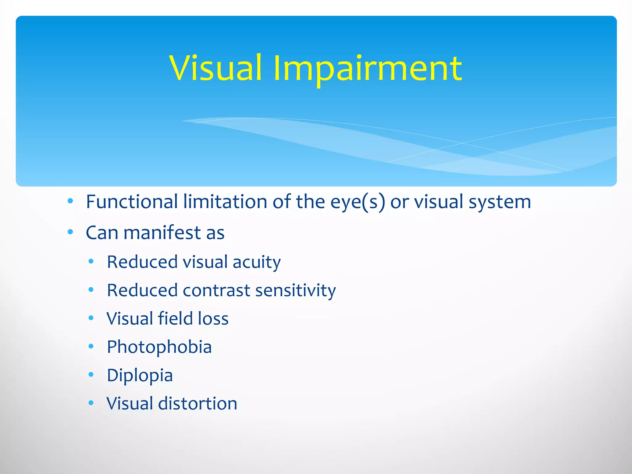
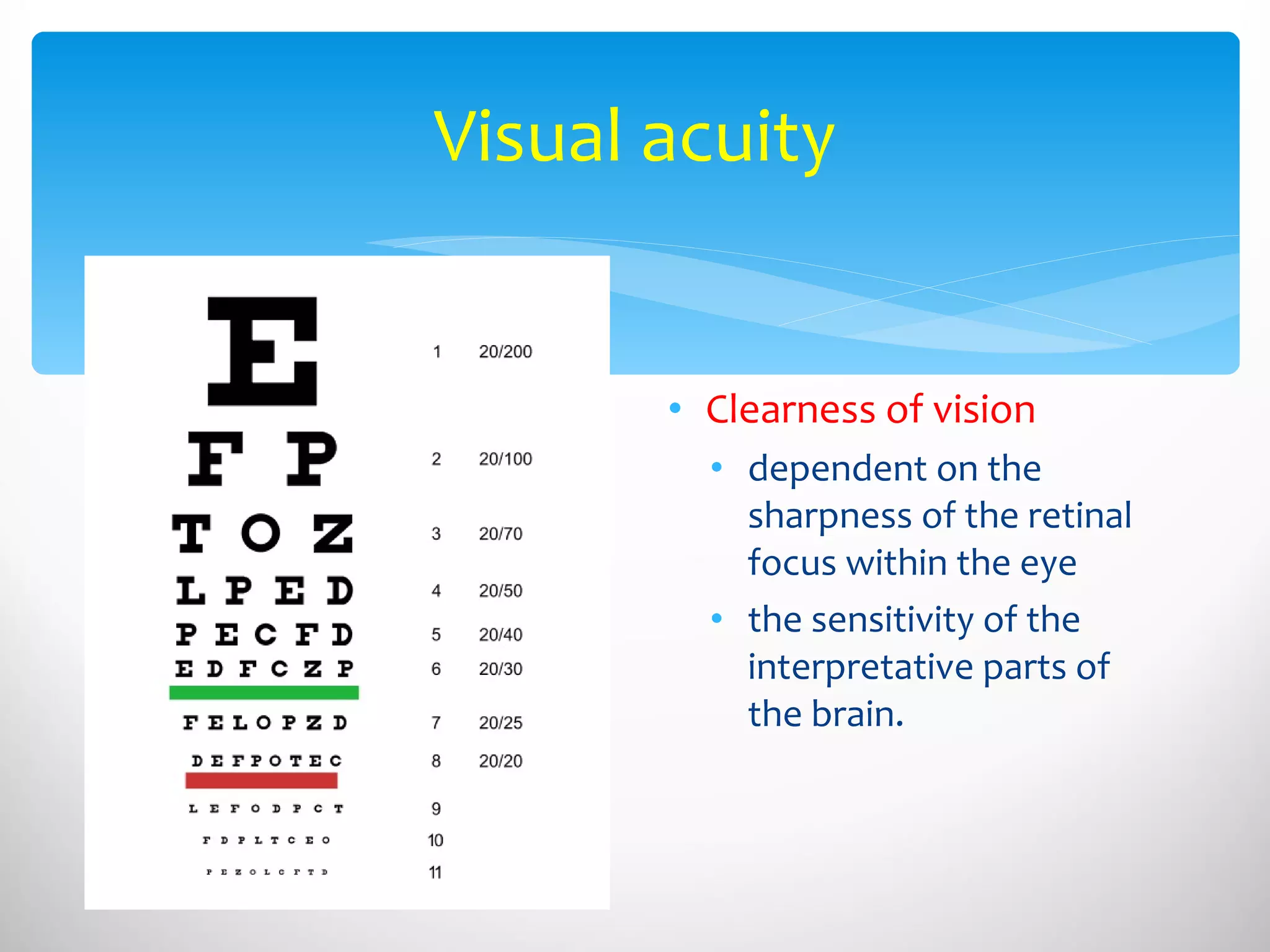
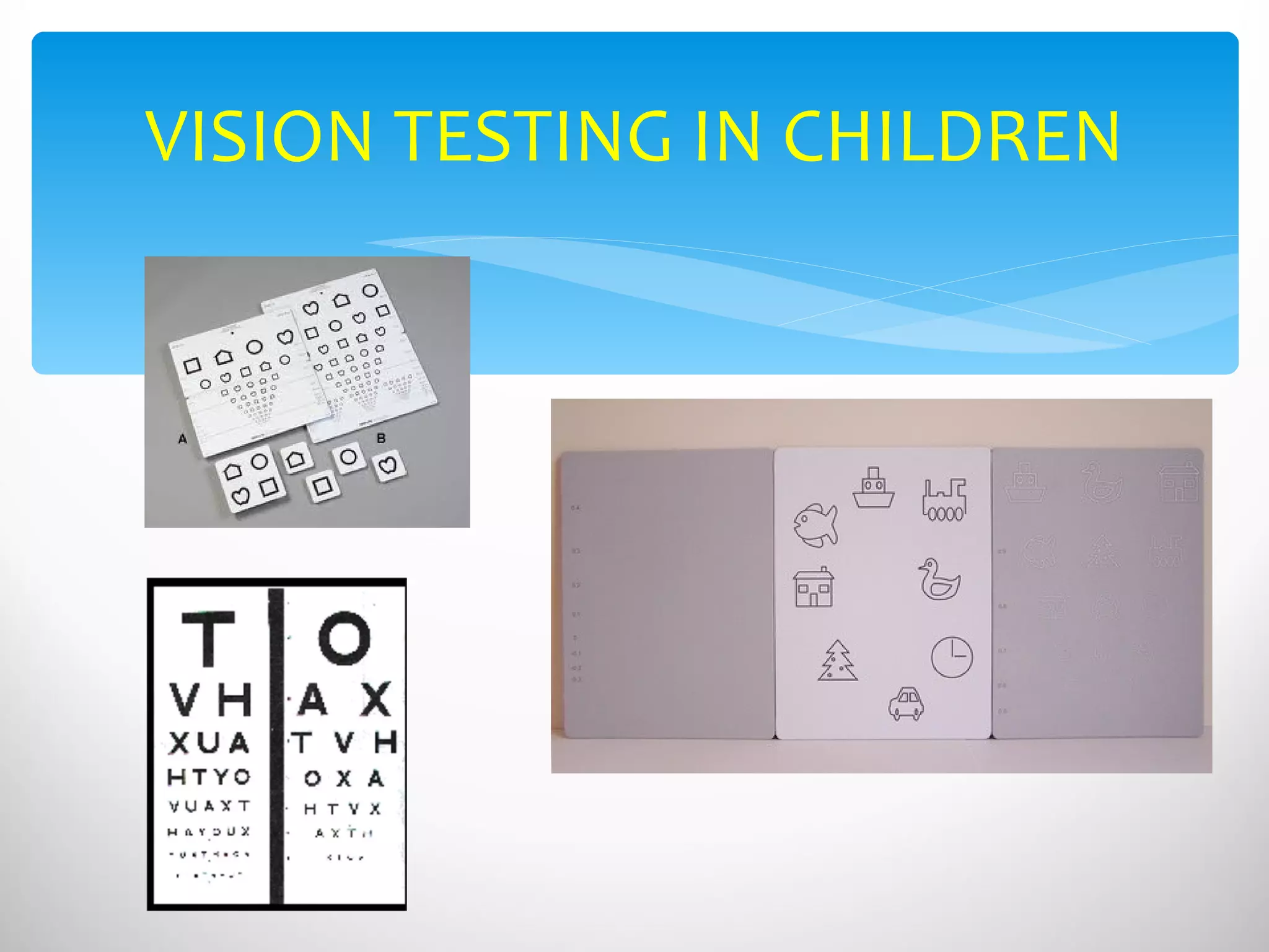
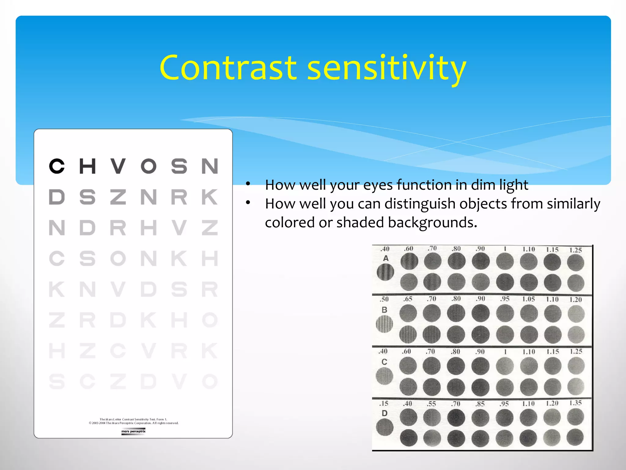
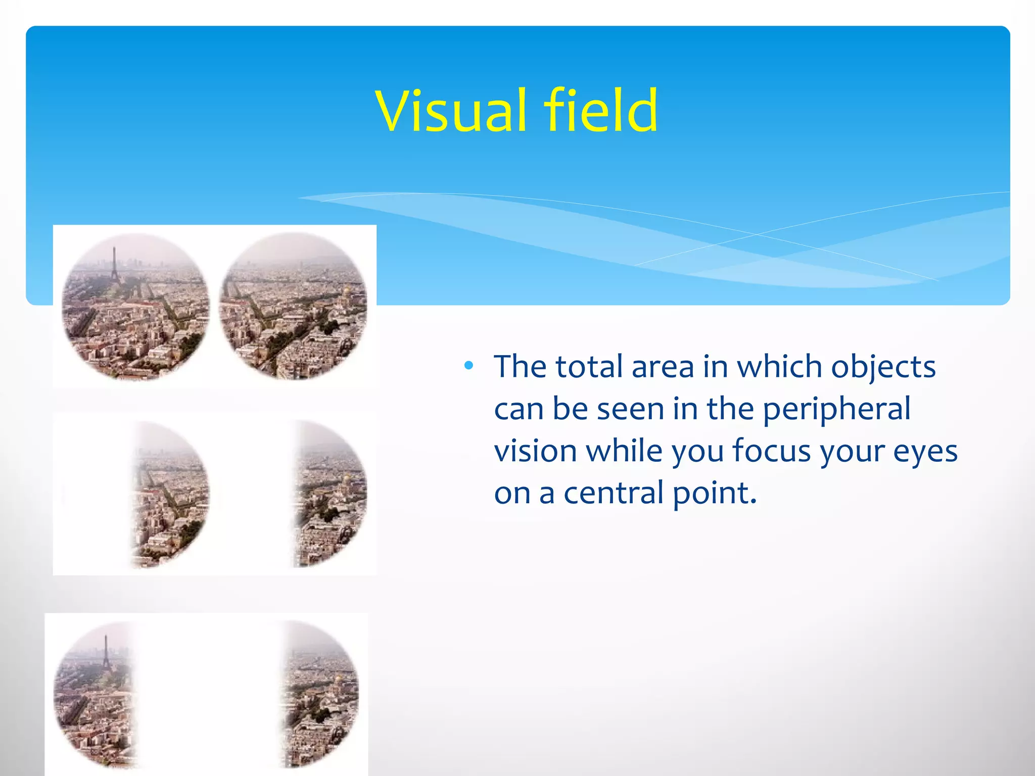
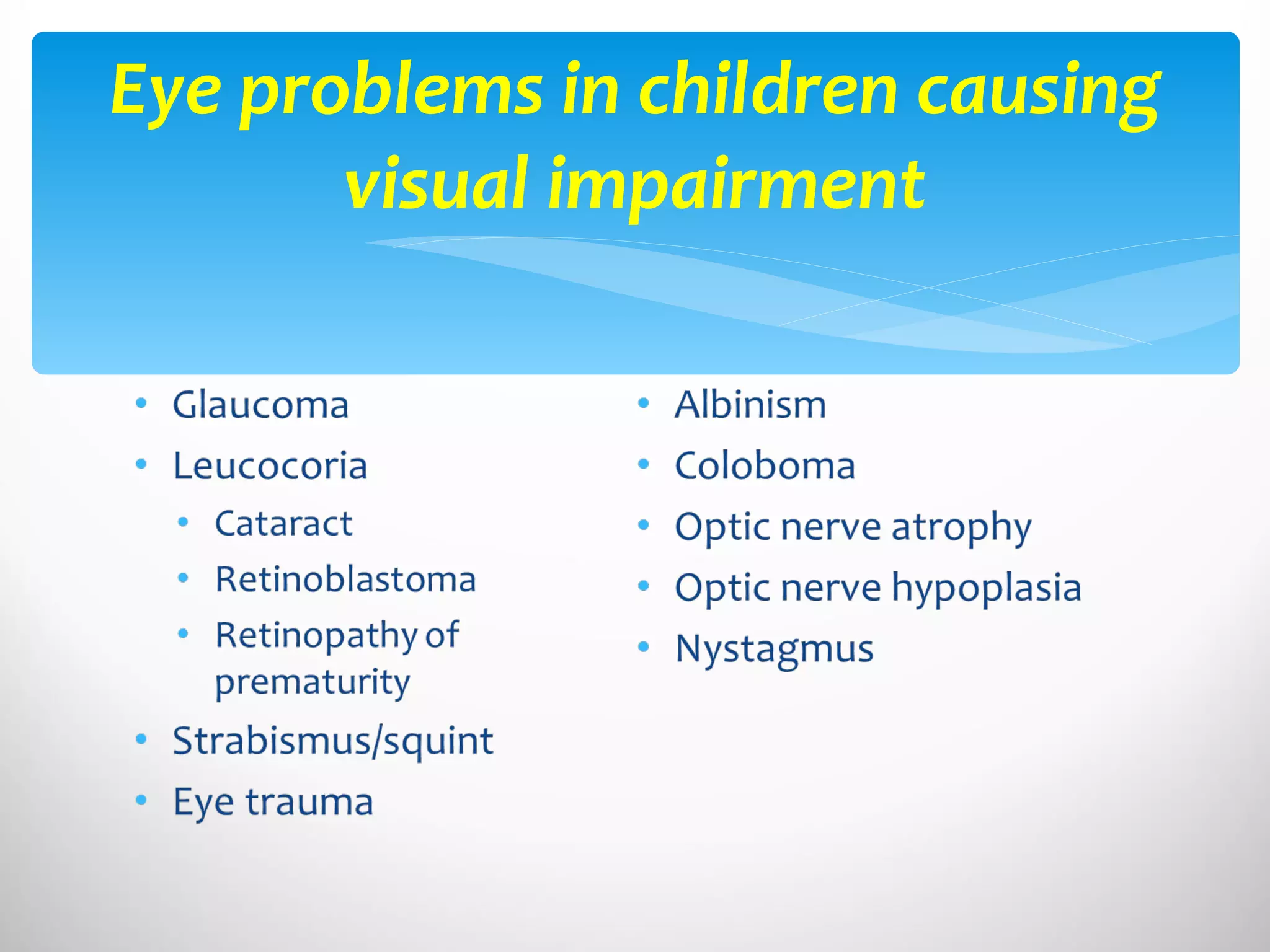
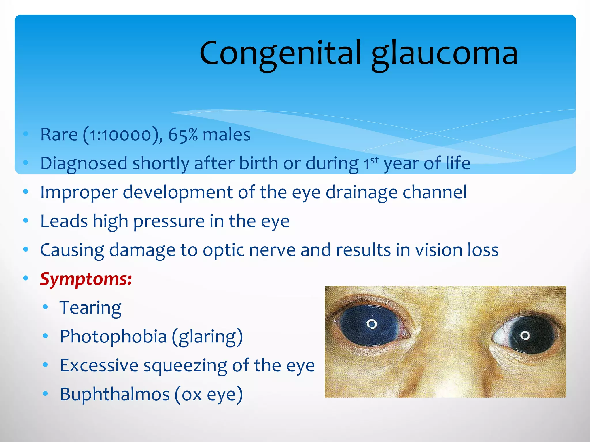
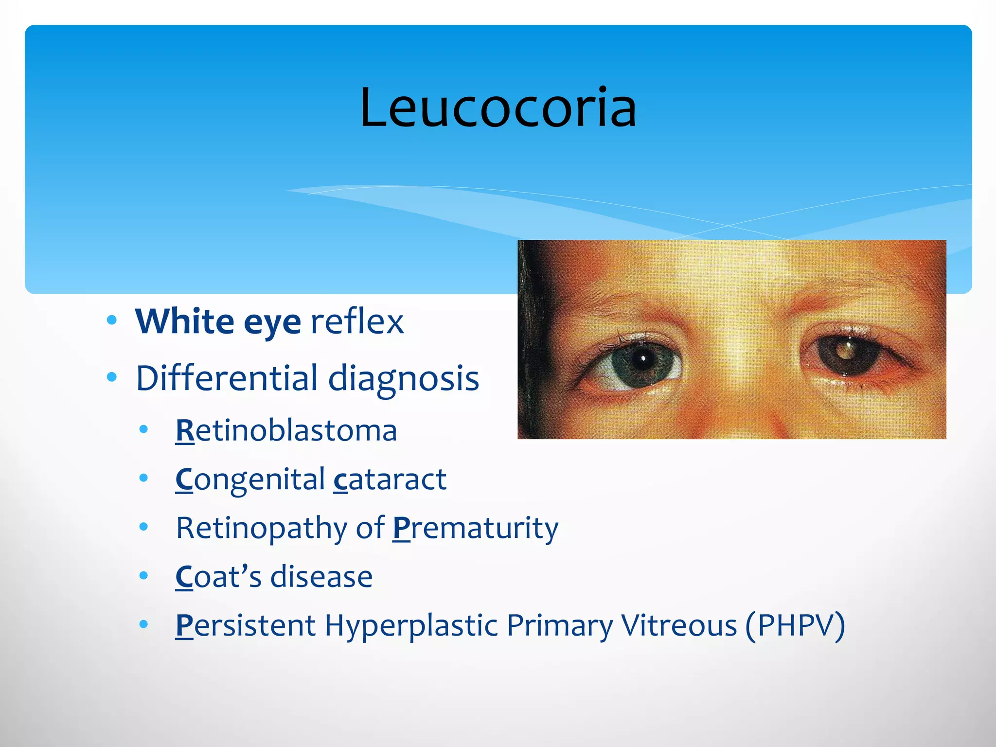
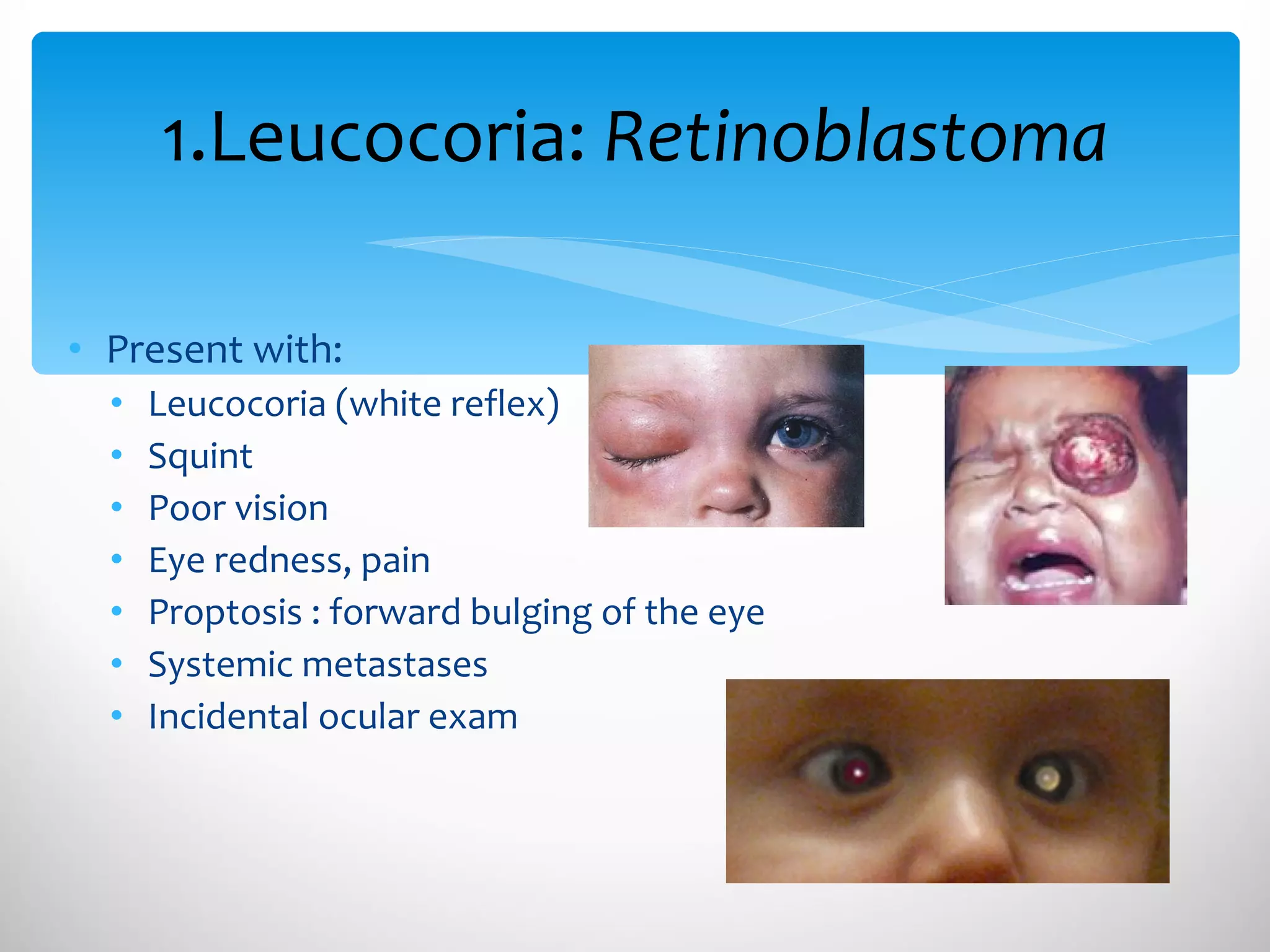
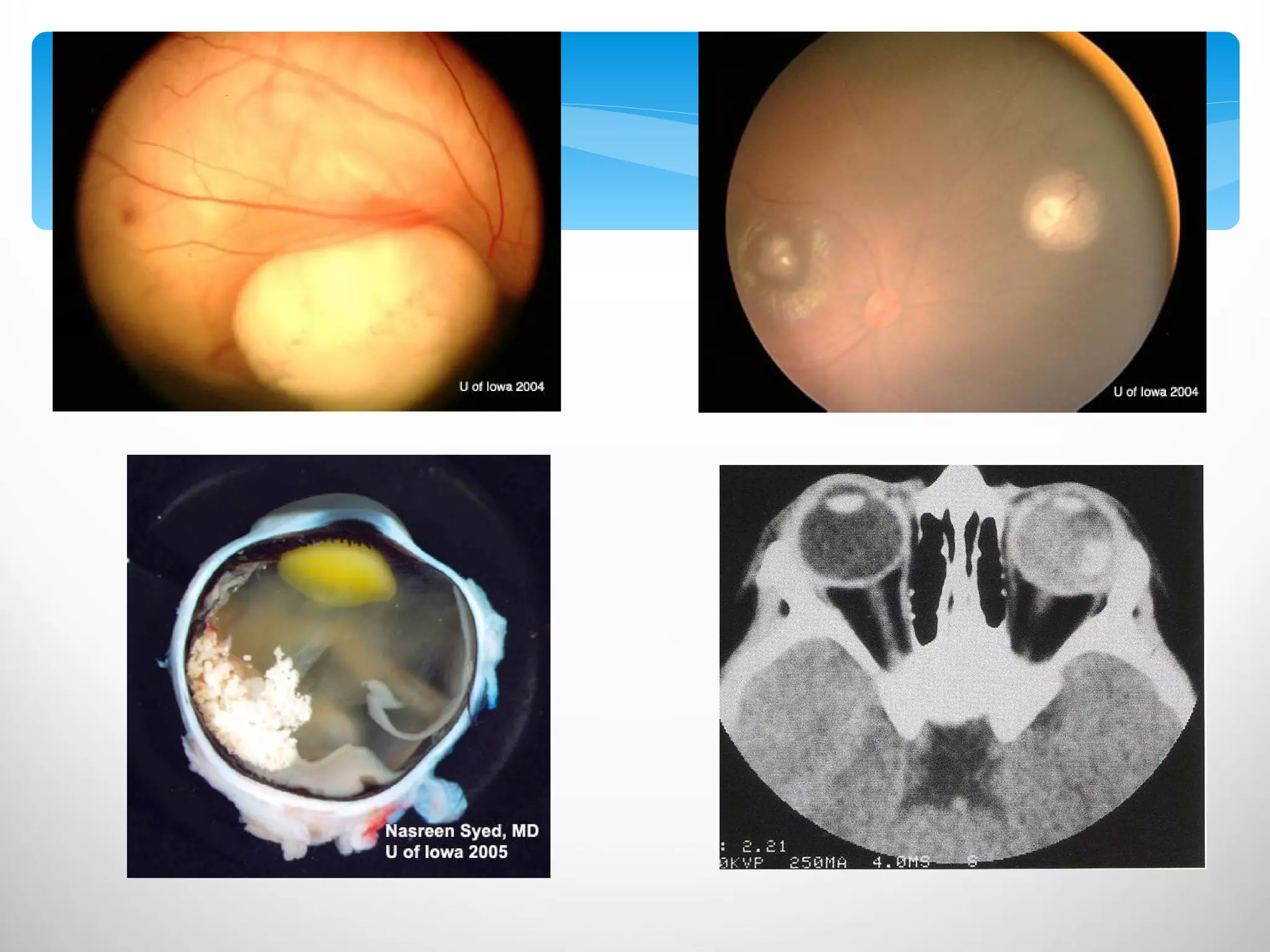
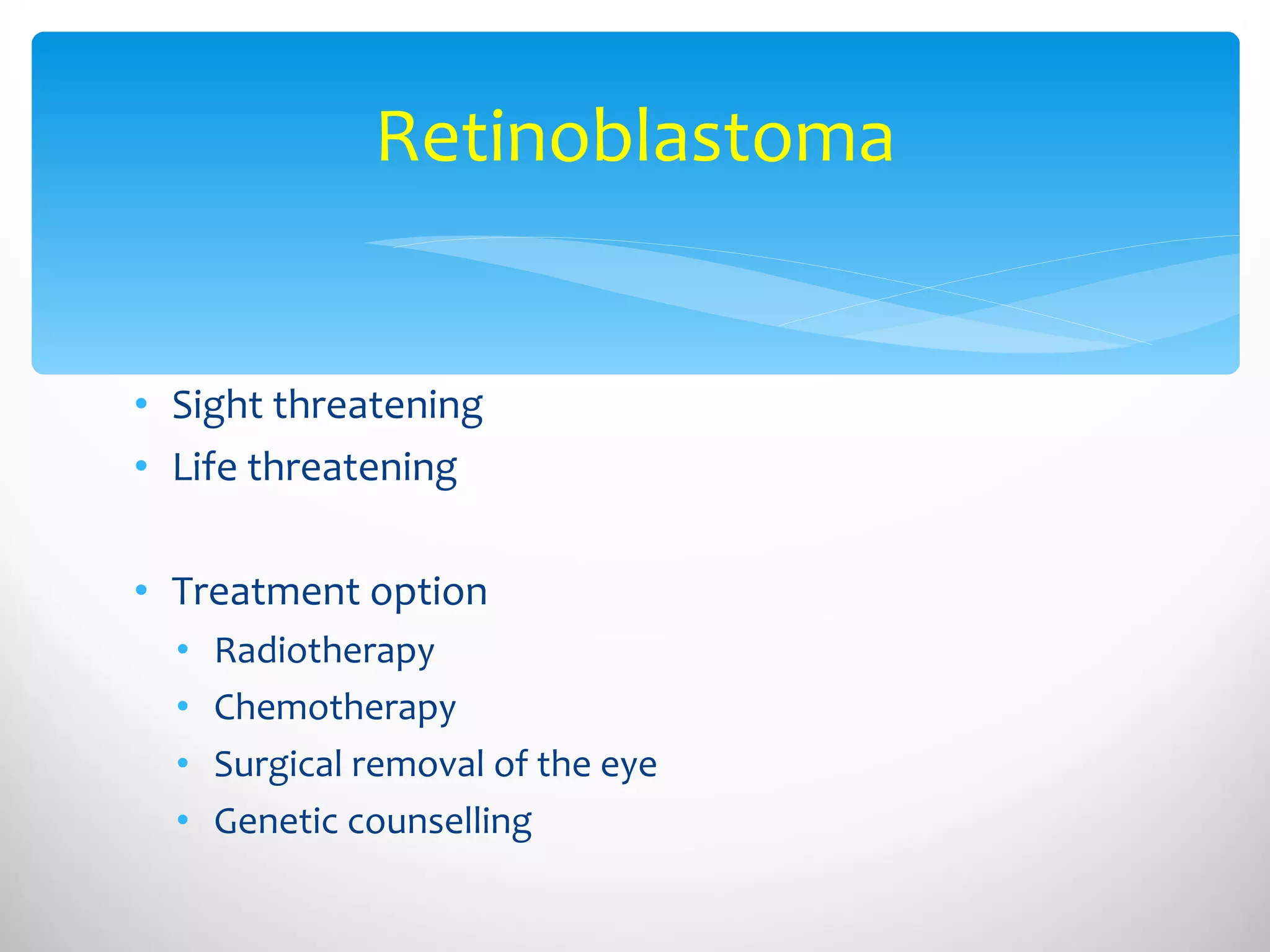
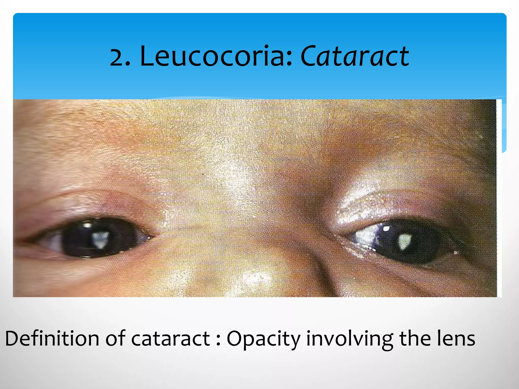
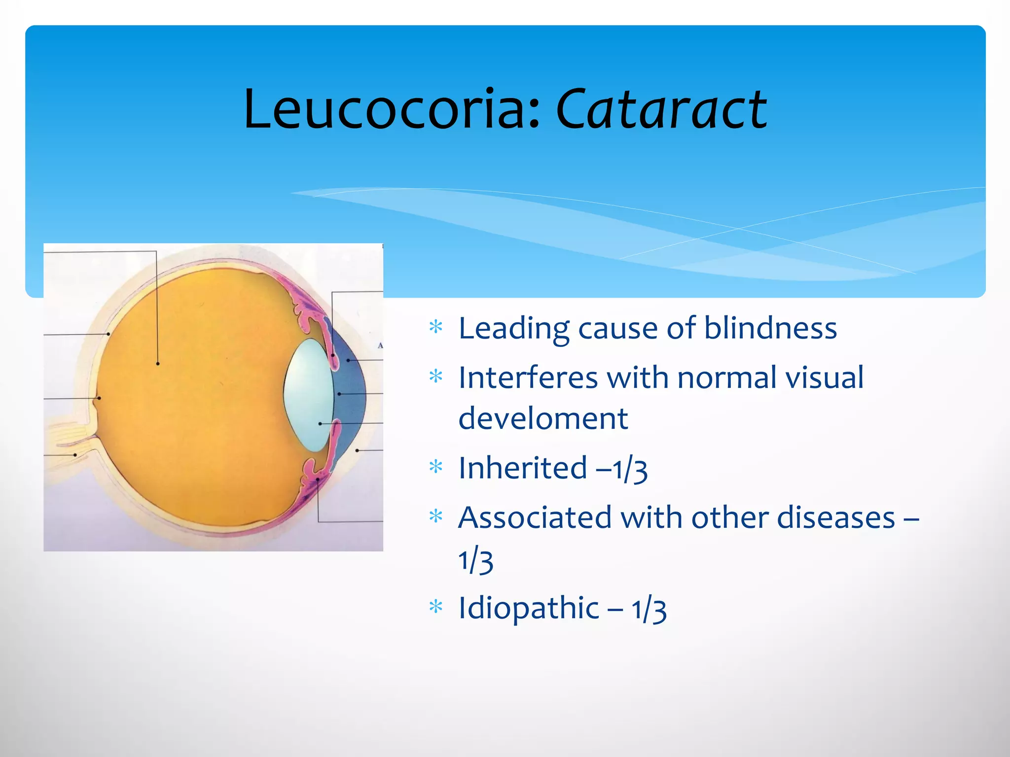
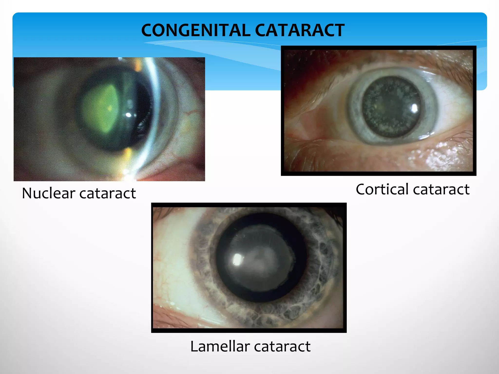
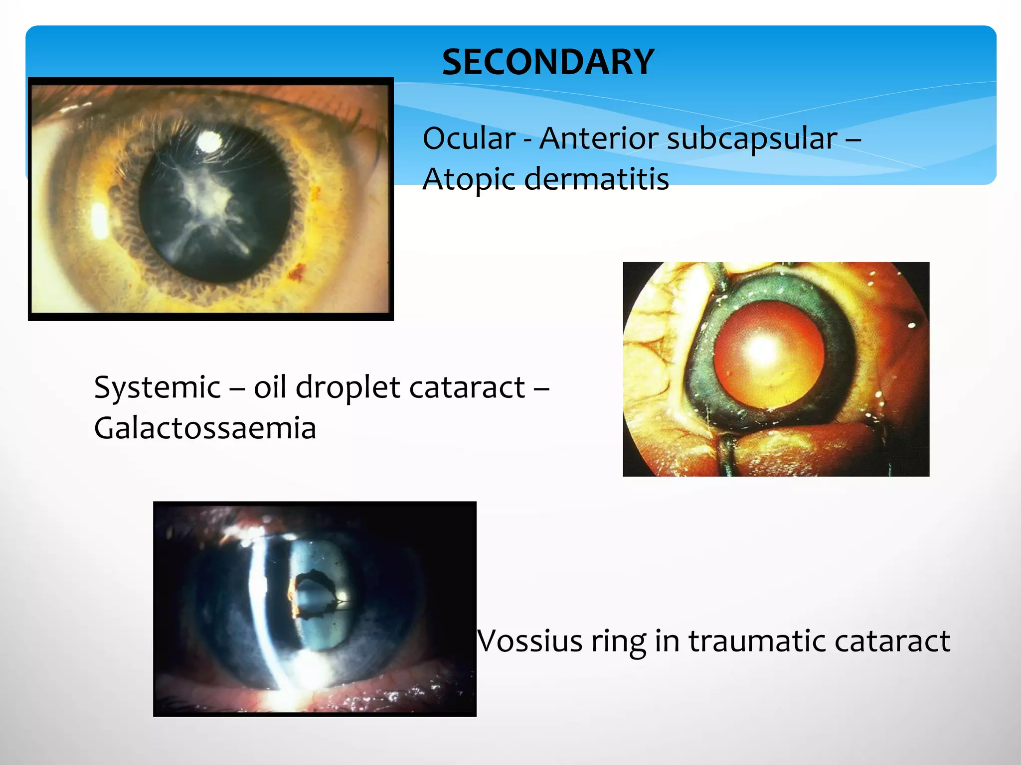
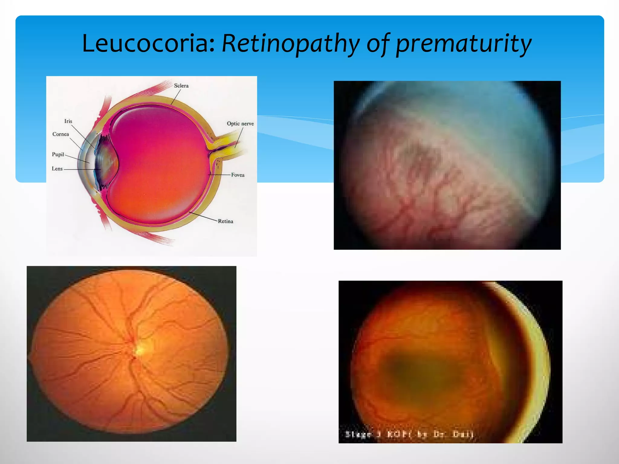
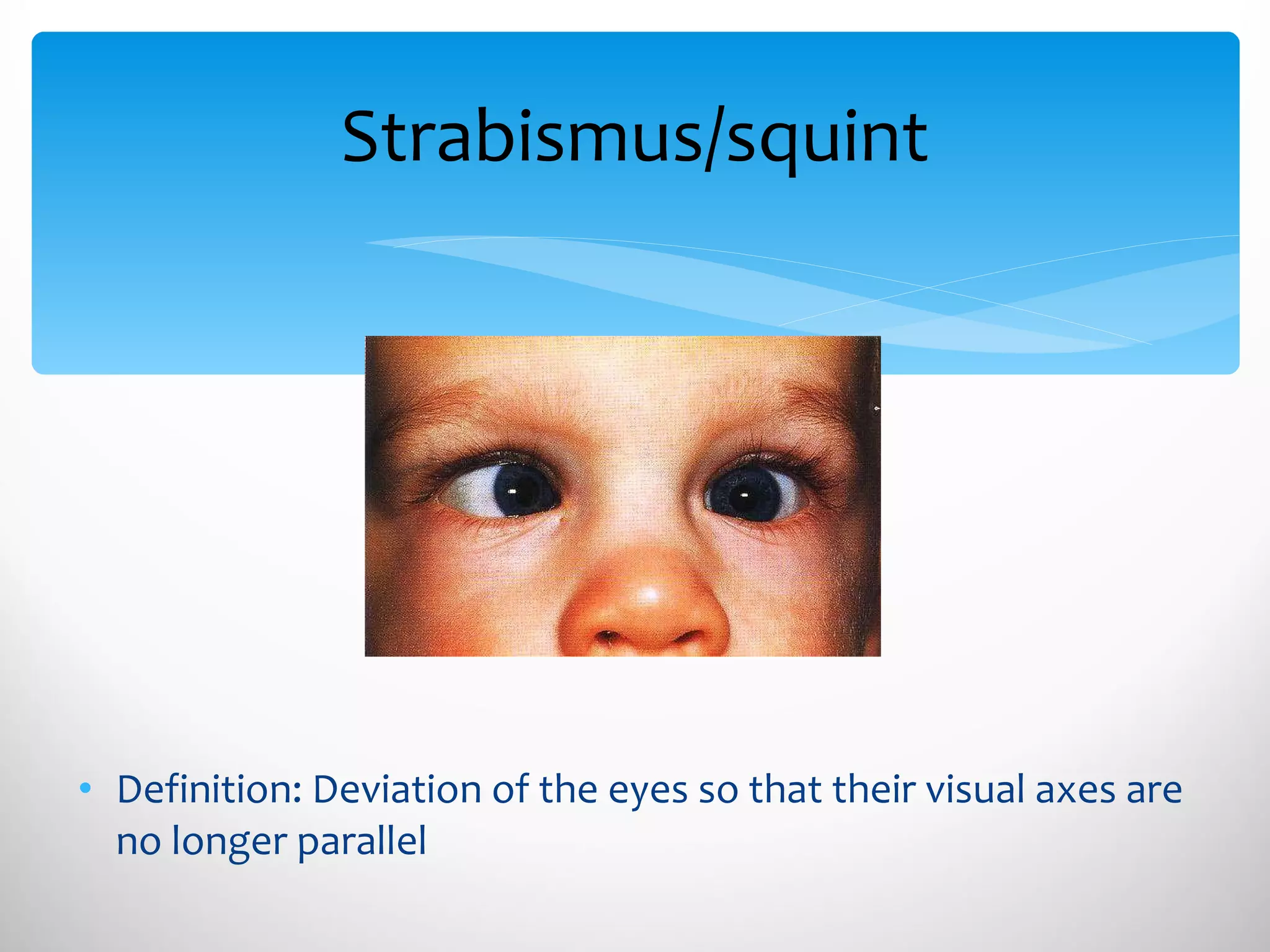
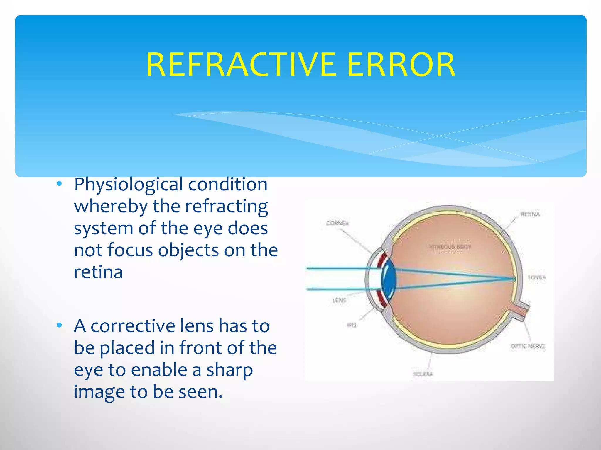
Penaksiran dan Diagnosis Kanak-kanak Bermasalah Penglihatan Dr Safinaz Mohd Khialdin Pensyarah & Pakar Oftalmologi Jabatan Oftalmologi PPUKM
 
Visual impairment or low vision  reduction in vision that cannot be corrected with standard glasses or contact lenses  reduces a person's ability to function at certain or all tasks Visual Impairment
Functional limitation of the eye(s) or visual system Can manifest as Reduced visual acuity  Reduced contrast sensitivity Visual field loss Photophobia Diplopia Visual distortion Visual Impairment
Clearness of vision dependent on the sharpness of the retinal focus within the eye  the sensitivity of the interpretative parts of the brain. Visual acuity
VISION TESTING IN CHILDREN
Contrast sensitivity How well your eyes function in dim light  How well you can distinguish objects from similarly colored or shaded backgrounds.
The total area in which objects can be seen in the peripheral vision while you focus your eyes on a central point. Visual field
PHOTOPHOBIA – glaring DIPLOPIA – double vision VISUAL DISTORTION
Definition: Visual impairment :  visual acuity of less than  6/18,  but equal or better than  3/60 in the better eye Visual field less than 20 degree from fixation  Blindness:  visual acuity  is less than 3/60  in the better eye Visual field less than 10 degree from fixation  Visual Impairment
6/60 6/18
HISTORY TAKING – SYMPTOMS EXAMINATIONS – SIGNS INVESTIGATIONS – CONFIRM DIAGNOSIS EVALUATION & DIAGNOSIS OF VISUAL IMPAIRMENT
Ocular History Diagnosis and onset of symptoms Past, current, or planned surgeries or treatments Stability of vision Family history of eye disease Previous history of eye disease or vision problems Current or previous use of spectacles, contact lenses, or low vision aids EVALUATION & DIAGNOSIS: HISTORY
Visual Functioning Ability to read print and specific reading needs (schooling) Intermediate visual ability and needs (e.g., use of computer, reading music) Distance visual ability and needs Photophobia, glare sensitivity, and lighting requirements EVALUATION & DIAGNOSIS: HISTORY
Medical History General health review Current medications Hearing impairment or other disability Developmental delay EVALUATION & DIAGNOSIS: HISTORY
EXAMINATION Visual acuity Visual field Refraction Eye movement Detailed examination of the eye structures EVALUATION & DIAGNOSIS: EXAMINATION
To confirm eye problem Ultrasound CT scan To assess visual impairment Contrast sensitivity testing Colour vision testing Visual evoked potential EVALUATION & DIAGNOSIS: INVESTIGATIONS
Eye problems in children Importance  Early detection & treatment of peadiatric ocular disease is critical Delay in diagnosis results in irreversible vision loss Assessment should begin at birth
Eye pathology :  Structural impairment or damage to one or more parts of the eye Refractive errors : or an inability of the eye to sharply focus images on the back of retina Cortical visual impairments : damage to the part of the brain that interprets visual information CAUSES OF VISUAL IMPAIRMENT IN CHILDREN
Eye problems in children causing visual impairment
Anatomy of the eye
Congenital glaucoma Rare (1:10000), 65% males Diagnosed shortly after birth or during 1 st  year of life Improper development of the eye drainage channel Leads high pressure in the eye Causing damage to optic nerve and results in vision loss Symptoms: Tearing  Photophobia (glaring) Excessive squeezing of the eye Buphthalmos (ox eye)
Leucocoria White   eye  reflex Differential diagnosis R etinoblastoma  C ongenital  c ataract Retinopathy of  P rematurity C oat’s disease P ersistent Hyperplastic Primary Vitreous (PHPV)
1.Leucocoria:  Retinoblastoma Most common intraocular malignancy of childhood Rare (1 in 14000-20000) 3% of all childhood cancers Bilateral in 30-35% Age at diagnosis : 18 months (90% less than 3 years old) May be inherited
1.Leucocoria:  Retinoblastoma Present with: Leucocoria (white reflex) Squint  Poor vision Eye redness, pain Proptosis : forward bulging of the eye Systemic metastases Incidental ocular exam
 
Retinoblastoma Sight threatening Life threatening Treatment option Radiotherapy Chemotherapy Surgical removal of the eye Genetic counselling
2. Leucocoria:  Cataract Definition of cataract : Opacity involving the lens
Leucocoria:  Cataract Leading cause of blindness Interferes with normal visual develoment Inherited –1/3 Associated with other diseases – 1/3 Idiopathic – 1/3
Nuclear cataract Lamellar cataract Cortical cataract CONGENITAL CATARACT
SECONDARY  Ocular - Anterior subcapsular –  Atopic dermatitis Systemic – oil droplet cataract –  Galactossaemia Vossius ring in traumatic cataract
Presentations of childhood cataract Lack of visual interest Strabismus/squint Nystagmus : abnormal movement of the eye Developmental delay Associated with systemic/ ocular abnormalities
Action Prompt detection & dedicated screening Referral to Ophthalmologist (urgent) Why? Monocular/ binocular cataract in infants results in significant visual deprivation Early visual rehabilitation can prevent amblyopia (lazy eye)
Eye disease that affects prematurely born babies Caused by disorganized growth of retinal blood vessels May be mild and resolves spontaneously Severe cases can lead to scarring and retinal detachment. 3. Leucocoria:  Retinopathy of prematurity
Leucocoria:  Retinopathy of prematurity
Risk factor for ROP Pre-term babies – screening Low birth weight Exposure to oxygen Screening of premature babies has to continue till school-going age Risk of myopia, even with no ROP Leucocoria:  Retinopathy of prematurity
Strabismus/squint Definition: Deviation of the eyes so that their visual axes are no longer parallel
Causes of squint Muscle imbalance Refractive errors Ocular abnormalities – cataract, macular scar, optic disc pathology Special syndromes – Duanes, Brown Associated disease – hydrocephalus, cerebral palsy, meningitis
Other presenting features Symptoms None Noted by parents Poor vision Funny eye movements Signs  Poor vision Abnormal head posture
Management for squint Refer to ophthalmologist (urgent) Treatment of refractive errors Treatment of amblyopia (lazy eye) Ocular exercise Surgery – mucsle imbalance Why?  Amblyopia (Lazy eye) Loss of binocular single vision Cosmetic blemish
REFRACTIVE ERROR Physiological condition whereby the refracting system of the eye does not focus objects on the retina  A corrective lens has to be placed in front of the eye to enable a sharp image to be seen.
Myopia Hyperopia Astigmatism Refractive errors
Refractive errors Various presentations: Squeezing eyes Squint Headache – ocular/ frontal/ diffuse Tearing Complains of tired eyes Child is slow at school Has problems with attentiveness Strong family history of myopia or other refractive error Importance: Untreated or undertreated will cause AMBLYOPIA
Visual acuity screening in schools Performed in Standard 1 Referral to Ophthalmologist/Optometrist Refractive errors
Amblyopia ‘ Lazy eye’ Definition:  Unilateral/ bilateral decrease of  best corrected  visual acuity  Is  potentially reversible  during the  critical period  (before 7-9 years old)
Critical period: Why is visual development important? Sensory function continues to develop after birth Requires proper visual stimulation Normal vision – receptive cells, clear media and normal retina Normal alignment  Normal brain development
How does amblyopia develop? Brain receives stimuli from both eyes Child’s maturing brain will select the better image and ignore the blurry image Vision not properly developed in the bad eye (AMBLYOPE)
Causes of amblyopia Ammetropia (bilateral high refractive error) Anisometropia (large/ asymmetrical refractive error difference between eyes) Strabismic (squint) Occlusional / form deprivation (media problem)
Treatment Can be treated or reversed if detected earlier (critical period) Treat underlying condition – cataract, squint Prescribe spectacles correction – refractive error Force to use the amblyopic eye Patching the good eye Putting atropine eye drops to blur image in the good eye
Visual pathway
As a result of the damage to the brain. Cerebral palsy Seizure disorder Hydrocephalus, Microcephaly Infants and children with cortical vision impairment Delayed in reaching developmental milestones  Sensory motor & activities and social development. Intellectual disability Cortical visual impairments
The process of treatment and education that helps individuals who are visually disable attain maximum function, a sense of well being,  a personally satisfying level of independence,  optimum quality of life. Visual Rehabilitation
OPTICAL DEVICES NON OPTICAL DEVICES HOLISTIC APPROACH Clinicians – ophthalmologist, peadiatrician, optometrist Teachers, Education ministry Occupational therapist Counsellors : psychologist, psychiatrist, social worker Vocational Rehabilitation centers Visual Rehabilitation
OPTICAL DEVICES Reduced Visual Acuity Magnification for near Magnification for distance Central visual field defect Peripheral visual field defect Reduced contrast sensitivity Glare sensitivity Visual Rehabilitation
HAND HELD MAGNIFIERS MAGNIFICATION FOR NEAR STAND MAGNIFIERS SPECTACLE-MOUNTED MAGNIFIER CLOSE-CIRCUIT TELEVISION SYSTEM
TELESCOPE MAGNIFICATION FOR FAR
TREATMENT OF CENTRAL VISUAL FIELD LOSS MODIFIED TEXT Training for  eccentric viewing- avoid fixating using the fovea Using large print material Using prism relocation
TREATMENT OF PERIPHERAL VISUAL FIELD LOSS MIRROR PRISMS Acts like a side mirror of a car Able to view objects at the non-seeing area of the eye when glancing into the mirror or prism
TREATMENT  FOR REDUCED CONTRAST SENSITIVITY AND GLARE TINTED LENSES COLOUR CONTRAST TYPOSCOPE : reduce reflected glare from printed pages
NON-OPTICAL METHODS  Move  CLOSER : use an angled reading desk Use  COLOUR  to show objects more clearly Use  CONTRAST : eat white rice off a coloured plate Pay attention to  LIGHTING : sit near a window in class Make objects  LARGER:   write with larger letters Use a  LINE-GUIDE  such as a ruler when reading and writing. Visual Rehabilitation
Prognosis for success depends on: Ocular condition Vision loss – nature & extent Patient – physical & mental abilities, attitude, expectation, motivation Clinician – attitude & motivation Visual rehabilitation
THANK YOU

Lecture fakulti pendidikan 2011

  • 1.
    Penaksiran dan DiagnosisKanak-kanak Bermasalah Penglihatan Dr Safinaz Mohd Khialdin Pensyarah & Pakar Oftalmologi Jabatan Oftalmologi PPUKM
  • 2.
  • 3.
    Visual impairment orlow vision reduction in vision that cannot be corrected with standard glasses or contact lenses reduces a person's ability to function at certain or all tasks Visual Impairment
  • 4.
    Functional limitation ofthe eye(s) or visual system Can manifest as Reduced visual acuity Reduced contrast sensitivity Visual field loss Photophobia Diplopia Visual distortion Visual Impairment
  • 5.
    Clearness of visiondependent on the sharpness of the retinal focus within the eye the sensitivity of the interpretative parts of the brain. Visual acuity
  • 6.
  • 7.
    Contrast sensitivity Howwell your eyes function in dim light How well you can distinguish objects from similarly colored or shaded backgrounds.
  • 8.
    The total areain which objects can be seen in the peripheral vision while you focus your eyes on a central point. Visual field
  • 9.
    PHOTOPHOBIA – glaringDIPLOPIA – double vision VISUAL DISTORTION
  • 10.
    Definition: Visual impairment: visual acuity of less than 6/18, but equal or better than 3/60 in the better eye Visual field less than 20 degree from fixation Blindness: visual acuity is less than 3/60 in the better eye Visual field less than 10 degree from fixation Visual Impairment
  • 11.
  • 12.
    HISTORY TAKING –SYMPTOMS EXAMINATIONS – SIGNS INVESTIGATIONS – CONFIRM DIAGNOSIS EVALUATION & DIAGNOSIS OF VISUAL IMPAIRMENT
  • 13.
    Ocular History Diagnosisand onset of symptoms Past, current, or planned surgeries or treatments Stability of vision Family history of eye disease Previous history of eye disease or vision problems Current or previous use of spectacles, contact lenses, or low vision aids EVALUATION & DIAGNOSIS: HISTORY
  • 14.
    Visual Functioning Abilityto read print and specific reading needs (schooling) Intermediate visual ability and needs (e.g., use of computer, reading music) Distance visual ability and needs Photophobia, glare sensitivity, and lighting requirements EVALUATION & DIAGNOSIS: HISTORY
  • 15.
    Medical History Generalhealth review Current medications Hearing impairment or other disability Developmental delay EVALUATION & DIAGNOSIS: HISTORY
  • 16.
    EXAMINATION Visual acuityVisual field Refraction Eye movement Detailed examination of the eye structures EVALUATION & DIAGNOSIS: EXAMINATION
  • 17.
    To confirm eyeproblem Ultrasound CT scan To assess visual impairment Contrast sensitivity testing Colour vision testing Visual evoked potential EVALUATION & DIAGNOSIS: INVESTIGATIONS
  • 18.
    Eye problems inchildren Importance Early detection & treatment of peadiatric ocular disease is critical Delay in diagnosis results in irreversible vision loss Assessment should begin at birth
  • 19.
    Eye pathology : Structural impairment or damage to one or more parts of the eye Refractive errors : or an inability of the eye to sharply focus images on the back of retina Cortical visual impairments : damage to the part of the brain that interprets visual information CAUSES OF VISUAL IMPAIRMENT IN CHILDREN
  • 20.
    Eye problems inchildren causing visual impairment
  • 21.
  • 22.
    Congenital glaucoma Rare(1:10000), 65% males Diagnosed shortly after birth or during 1 st year of life Improper development of the eye drainage channel Leads high pressure in the eye Causing damage to optic nerve and results in vision loss Symptoms: Tearing Photophobia (glaring) Excessive squeezing of the eye Buphthalmos (ox eye)
  • 23.
    Leucocoria White eye reflex Differential diagnosis R etinoblastoma C ongenital c ataract Retinopathy of P rematurity C oat’s disease P ersistent Hyperplastic Primary Vitreous (PHPV)
  • 24.
    1.Leucocoria: RetinoblastomaMost common intraocular malignancy of childhood Rare (1 in 14000-20000) 3% of all childhood cancers Bilateral in 30-35% Age at diagnosis : 18 months (90% less than 3 years old) May be inherited
  • 25.
    1.Leucocoria: RetinoblastomaPresent with: Leucocoria (white reflex) Squint Poor vision Eye redness, pain Proptosis : forward bulging of the eye Systemic metastases Incidental ocular exam
  • 26.
  • 27.
    Retinoblastoma Sight threateningLife threatening Treatment option Radiotherapy Chemotherapy Surgical removal of the eye Genetic counselling
  • 28.
    2. Leucocoria: Cataract Definition of cataract : Opacity involving the lens
  • 29.
    Leucocoria: CataractLeading cause of blindness Interferes with normal visual develoment Inherited –1/3 Associated with other diseases – 1/3 Idiopathic – 1/3
  • 30.
    Nuclear cataract Lamellarcataract Cortical cataract CONGENITAL CATARACT
  • 31.
    SECONDARY Ocular- Anterior subcapsular – Atopic dermatitis Systemic – oil droplet cataract – Galactossaemia Vossius ring in traumatic cataract
  • 32.
    Presentations of childhoodcataract Lack of visual interest Strabismus/squint Nystagmus : abnormal movement of the eye Developmental delay Associated with systemic/ ocular abnormalities
  • 33.
    Action Prompt detection& dedicated screening Referral to Ophthalmologist (urgent) Why? Monocular/ binocular cataract in infants results in significant visual deprivation Early visual rehabilitation can prevent amblyopia (lazy eye)
  • 34.
    Eye disease thataffects prematurely born babies Caused by disorganized growth of retinal blood vessels May be mild and resolves spontaneously Severe cases can lead to scarring and retinal detachment. 3. Leucocoria: Retinopathy of prematurity
  • 35.
  • 36.
    Risk factor forROP Pre-term babies – screening Low birth weight Exposure to oxygen Screening of premature babies has to continue till school-going age Risk of myopia, even with no ROP Leucocoria: Retinopathy of prematurity
  • 37.
    Strabismus/squint Definition: Deviationof the eyes so that their visual axes are no longer parallel
  • 38.
    Causes of squintMuscle imbalance Refractive errors Ocular abnormalities – cataract, macular scar, optic disc pathology Special syndromes – Duanes, Brown Associated disease – hydrocephalus, cerebral palsy, meningitis
  • 39.
    Other presenting featuresSymptoms None Noted by parents Poor vision Funny eye movements Signs Poor vision Abnormal head posture
  • 40.
    Management for squintRefer to ophthalmologist (urgent) Treatment of refractive errors Treatment of amblyopia (lazy eye) Ocular exercise Surgery – mucsle imbalance Why? Amblyopia (Lazy eye) Loss of binocular single vision Cosmetic blemish
  • 41.
    REFRACTIVE ERROR Physiologicalcondition whereby the refracting system of the eye does not focus objects on the retina A corrective lens has to be placed in front of the eye to enable a sharp image to be seen.
  • 42.
  • 43.
    Refractive errors Variouspresentations: Squeezing eyes Squint Headache – ocular/ frontal/ diffuse Tearing Complains of tired eyes Child is slow at school Has problems with attentiveness Strong family history of myopia or other refractive error Importance: Untreated or undertreated will cause AMBLYOPIA
  • 44.
    Visual acuity screeningin schools Performed in Standard 1 Referral to Ophthalmologist/Optometrist Refractive errors
  • 45.
    Amblyopia ‘ Lazyeye’ Definition: Unilateral/ bilateral decrease of best corrected visual acuity Is potentially reversible during the critical period (before 7-9 years old)
  • 46.
    Critical period: Whyis visual development important? Sensory function continues to develop after birth Requires proper visual stimulation Normal vision – receptive cells, clear media and normal retina Normal alignment Normal brain development
  • 47.
    How does amblyopiadevelop? Brain receives stimuli from both eyes Child’s maturing brain will select the better image and ignore the blurry image Vision not properly developed in the bad eye (AMBLYOPE)
  • 48.
    Causes of amblyopiaAmmetropia (bilateral high refractive error) Anisometropia (large/ asymmetrical refractive error difference between eyes) Strabismic (squint) Occlusional / form deprivation (media problem)
  • 49.
    Treatment Can betreated or reversed if detected earlier (critical period) Treat underlying condition – cataract, squint Prescribe spectacles correction – refractive error Force to use the amblyopic eye Patching the good eye Putting atropine eye drops to blur image in the good eye
  • 50.
  • 51.
    As a resultof the damage to the brain. Cerebral palsy Seizure disorder Hydrocephalus, Microcephaly Infants and children with cortical vision impairment Delayed in reaching developmental milestones Sensory motor & activities and social development. Intellectual disability Cortical visual impairments
  • 52.
    The process oftreatment and education that helps individuals who are visually disable attain maximum function, a sense of well being, a personally satisfying level of independence, optimum quality of life. Visual Rehabilitation
  • 53.
    OPTICAL DEVICES NONOPTICAL DEVICES HOLISTIC APPROACH Clinicians – ophthalmologist, peadiatrician, optometrist Teachers, Education ministry Occupational therapist Counsellors : psychologist, psychiatrist, social worker Vocational Rehabilitation centers Visual Rehabilitation
  • 54.
    OPTICAL DEVICES ReducedVisual Acuity Magnification for near Magnification for distance Central visual field defect Peripheral visual field defect Reduced contrast sensitivity Glare sensitivity Visual Rehabilitation
  • 55.
    HAND HELD MAGNIFIERSMAGNIFICATION FOR NEAR STAND MAGNIFIERS SPECTACLE-MOUNTED MAGNIFIER CLOSE-CIRCUIT TELEVISION SYSTEM
  • 56.
  • 57.
    TREATMENT OF CENTRALVISUAL FIELD LOSS MODIFIED TEXT Training for eccentric viewing- avoid fixating using the fovea Using large print material Using prism relocation
  • 58.
    TREATMENT OF PERIPHERALVISUAL FIELD LOSS MIRROR PRISMS Acts like a side mirror of a car Able to view objects at the non-seeing area of the eye when glancing into the mirror or prism
  • 59.
    TREATMENT FORREDUCED CONTRAST SENSITIVITY AND GLARE TINTED LENSES COLOUR CONTRAST TYPOSCOPE : reduce reflected glare from printed pages
  • 60.
    NON-OPTICAL METHODS Move CLOSER : use an angled reading desk Use COLOUR to show objects more clearly Use CONTRAST : eat white rice off a coloured plate Pay attention to LIGHTING : sit near a window in class Make objects LARGER: write with larger letters Use a LINE-GUIDE such as a ruler when reading and writing. Visual Rehabilitation
  • 61.
    Prognosis for successdepends on: Ocular condition Vision loss – nature & extent Patient – physical & mental abilities, attitude, expectation, motivation Clinician – attitude & motivation Visual rehabilitation
  • 62.

Editor's Notes

  • #9 60 Nasal /100 degrees temporal / 60 degrees above / 75 below the horizontal meridian
  • #11 Resnikoff et al. (2004),
  • #20 (Stiles & Knox, 1996):
  • #28 Affected pt – 40% children affected, 10 % carriers, 50 % normal
  • #52 (Hughes 1995).