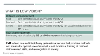
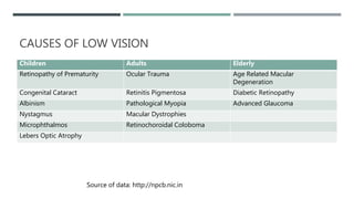
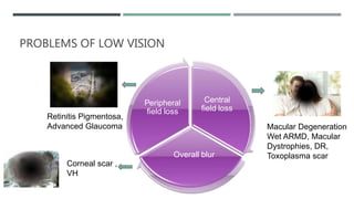
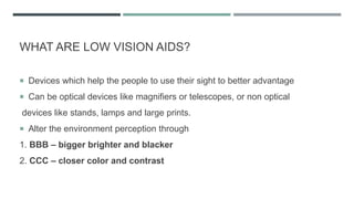
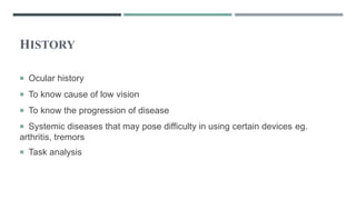
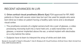
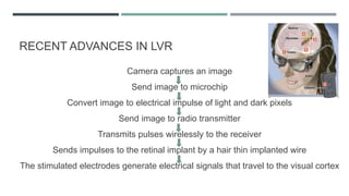
The document discusses low vision rehabilitation, including definitions of low vision, epidemiology of visual impairment, approaches to assessing patients with low vision, types of low vision aids, and recent advances in the field. It provides details on the history and assessments involved in low vision rehabilitation, as well as optical and non-optical devices that can help patients with low vision maximize their remaining vision.
This document provides information on low vision assessment, including its purpose and steps. The purpose is to evaluate a person's residual vision and determine how to enhance their visual function based on their needs. The main steps are reviewing medical records, observation, interview, assessing visual acuity, visual fields, contrast sensitivity, and refraction. The assessment helps identify appropriate aids like magnification, filters, or training to help low vision patients perform daily activities.
This document discusses contact lens fitting following various refractive surgeries. It begins with an introduction to refractive surgeries like radial keratotomy, PRK, LASIK, LASEK, SMILE, and others. It then discusses considerations and techniques for fitting contact lenses after different surgeries, focusing on fitting rigid gas permeable lenses, mini-scleral lenses, and hybrid lenses following procedures like radial keratotomy that can result in irregular astigmatism. The document provides guidance on lens parameters and fitting criteria to achieve a stable, comfortable fit while maintaining corneal health after refractive surgery.
This document discusses several key considerations for providing eye care to elderly patients. It notes that the elderly population is one of the fastest growing internet users and will require more frequent eye exams. It highlights that aging brings natural changes to vision that should be addressed sensitively. Examinations and dispensing processes should be thorough and explain recommendations in detail while showing personal attention. Multiple pairs of eyewear are often needed to meet the varied visual needs of elderly patients for tasks like reading, computers, and driving. Lens material, coatings, and frame fit considerations are especially important for comfort.
This document discusses low vision aids and their use for people with visual impairments. It defines low vision according to the WHO and describes common causes of visual dysfunction like macular degeneration and glaucoma. The goals of low vision rehabilitation are to maintain and improve visual function through clinical assessment and optometric intervention. Low vision aids can be optical devices like magnifying glasses, telescopes, or non-optical devices that alter lighting, contrast and size of objects. Common optical devices discussed include magnifying spectacles, hand magnifiers, stand magnifiers, and telescopes.
The synoptophore is an ophthalmic instrument used to diagnose and treat imbalances of the eye muscles. It consists of two cylindrical tubes with mirrored bends that allow pictures to be presented simultaneously to both eyes, compensating for any angle of squint. It is used to investigate binocular function in patients with a manifest squint, detect suppression and abnormal retinal correspondence, and measure horizontal, vertical and torsional misalignments. The synoptophore can test for three grades of binocular vision - simultaneous perception, fusion, and stereopsis - and detect whether a patient has normal or abnormal retinal correspondence based on differences between subjective and objective angles of squint.
Vision therapy is an individualized treatment program designed to correct visual and perceptual deficiencies and improve vision skills through activities like eye tracking and coordination exercises. It can help with conditions like amblyopia, strabismus, and convergence insufficiency by strengthening the visual system and improving connections in the brain. Research studies have found vision therapy to be an effective treatment for improving visual acuity and stereopsis in patients with refractive amblyopia and for establishing sensory fusion in patients with strabismus.
This document discusses the fitting of toric contact lenses. It begins with an introduction and discusses preliminary testing, fitting steps, and different toric lens designs. Stabilization techniques for toric lenses like prism ballast, truncation, and reverse prism are explained. The conclusion emphasizes measuring axis mislocation and compensating for lens rotation when determining the final prescription.
This document discusses low vision aids and their use for people with low vision. It defines low vision as visual acuity between 6/18 and 3/60 in the better eye after correction, or a field of vision between 20 to 30 degrees. Common causes of low vision include macular degeneration, glaucoma, and diabetic retinopathy. Optical low vision aids like magnifying spectacles, hand magnifiers, and telescopes use magnification to improve vision. Non-optical aids include increased lighting, contrast enhancement, and electronic magnifiers. Proper evaluation and prescribing of low vision aids depends on the patient's needs, vision status, and motivation. The goal is to prescribe simple, portable devices to help low vision
The document summarizes a case study of a 20-year-old male patient with left eye vision loss since childhood due to corneal scarring who was fitted for a prosthetic soft contact lens. Details are provided on the patient's history and examination, differential diagnosis, types and fitting criteria of prosthetic contact lenses, fitting of a medium brown type D prosthetic lens, and fitting assessment showing good coverage, centration, and movement. The plan is for the patient to be fitted with a single purecon prosthetic soft contact lens.
The document discusses the major causes of low vision in adults, including age-related macular degeneration, cataracts, diabetic retinopathy, multiple sclerosis, myopic degeneration, retinal detachment, and glaucoma. For each condition, it describes how visual acuity and visual fields are typically affected and recommends approaches to low vision management such as magnification, filters, prisms, lighting aids, and mobility training. Overall, the document provides an overview of the leading causes and treatment considerations for low vision in the adult population.
VISUAL AIDS FOR CHILDREN IN LOW VISION AND CONTACT LENSITM UNIVERSITY
This document discusses visual aids for children with low vision. It begins by defining low vision according to WHO classifications. Common causes of low vision in children are then outlined, including albinism, retinopathy, and macular degeneration. Both optical and non-optical low vision aids are described. Optical aids discussed include magnifying spectacles, hand magnifiers, stand magnifiers, telescopes, tinted lenses, and filters. Non-optical aids include reading lamps, writing guides, and bold line paper. The principles of low vision aids and factors to consider when prescribing them are also summarized. Contact lenses that can help children with low vision conditions such as aphakia and corneal problems are briefly mentioned.
This document discusses low vision in childhood, including various pathologies that can cause low vision such as Leber's amaurosis, optic atrophy, and retinitis pigmentosa. It outlines the visual prognosis and visual field defects associated with each condition. The document also discusses the use of low vision aids in children, noting that children are more accepting of aids and that aids should be introduced early. Various types of aids are described, from magnifiers to closed-circuit television. The document concludes with references.
Therapeutic contact lenses are used for therapeutic, diagnostic and cosmetic purposes to treat various ocular surface diseases and conditions. They provide mechanical protection and support to the cornea, maintain corneal epithelial hydration, and can be used to deliver medications to the eye. The type of therapeutic contact lens chosen depends on the specific condition being treated and should aim to maximize oxygen to the cornea unless the eye has no vision. Common complications include ocular redness, minor corneal edema and lens deposits which require regular follow up visits.
- Aphakia is the absence of the crystalline lens from the eye. It can be congenital or caused by surgery or trauma.
- In aphakia, the eye becomes highly hyperopic, the anterior focal point moves forward, and the retinal image is magnified. This decreases visual acuity and field of view.
- Aphakia is treated with spectacles, contact lenses, or intraocular lenses. Spectacles cause issues like increased image size, ring scotomas, and reduced field of view. Contact lenses and IOLs provide better image quality but have risks of complications.
The document provides an introduction to low vision, including definitions and classifications. It discusses common causes of low vision such as macular degeneration, glaucoma, diabetic retinopathy, and cataracts. Functional effects of low vision include loss of central and peripheral vision, problems with glare and contrast sensitivity. Goals of low vision management are to increase functionality, make the most of remaining vision, and provide support services.
The document summarizes the Amsler grid, a diagnostic tool used since 1945 to screen for and monitor macular diseases. It consists of a grid with a central dot that patients look at to detect any distortions, gaps, or blurred areas in their central vision. Various versions are available, including ones with different colors, patterns of lines, or dot sizes to test specific parts of the visual field and detect different types of visual abnormalities that could indicate conditions like macular degeneration or glaucoma. The procedure involves having patients view the grid with each eye separately at 16 inches and report any anomalies in the lines of the grid.
Keratometry is used to measure the curvature of the cornea by analyzing the reflection of light off its surface. It works by projecting illuminated circles called mires onto the cornea and measuring the size of the reflected image to calculate the radius of curvature. The main uses of keratometry include measuring corneal astigmatism, estimating contact lens power, and detecting irregularities like keratoconus. Modern instruments automate the process but traditional keratometers require aligning the mires and adjusting knobs until the doubled images come into close alignment. Factors like blinking, eye movements, and irregular corneas can impact the accuracy of measurements.
This document discusses rigid gas permeable (RGP) contact lenses. It notes that RGP lenses are made of oxygen permeable materials and are better than soft lenses for vision, durability, correcting astigmatism, eye health, and ease of care. RGP lenses are recommended for conditions like keratoconus or high refractive errors. The fitting process involves screening patients, measuring the eye, trial fittings, and dynamic and static assessments. Proper care and maintenance of RGP lenses is also discussed.
This document provides an overview of corneal topography. It begins by defining corneal topography as the study of the shape of the corneal surface. It then describes several techniques for evaluating corneal topography including keratometry, keratoscopy using Placido discs and photokeratoscopy, rasterstereography, and interferometry. Computerized topography systems that provide detailed maps of the corneal surface are also discussed. The document outlines clinical applications of corneal topography and variations in topographic patterns seen in normal and diseased corneas.
The cover test is used to qualitatively measure strabismus. It involves covering each eye separately while having the patient fixate on a target. This allows the examiner to observe any movement in the uncovered eye, indicating the presence or absence of a manifest deviation. There are three main types of cover tests: direct cover test to detect manifest squint, cover-uncover test to detect heterophoria, and alternate cover test to differentiate between unilateral and alternating squint and determine if the deviation is concomitant or paralytic. The results of the cover test help diagnose the type of strabismus present.
Aniseikonia refers to an unequal apparent size of images seen by the two eyes. It can result from differences in refractive errors between the eyes (refractive aniseikonia) or differences in the distribution of retinal elements (basic aniseikonia). Symptoms include headaches, asthenopia, and difficulties with mobility or fusion. Aniseikonia is usually caused by anisometropia above 1.50-2.00 diopters and analyzing ocular components can help determine if it is due to refractive or axial differences.
This document discusses soft toric contact lenses for correcting astigmatism. It defines astigmatism and describes various types. It explains that toric lenses contain a cylindrical component to correct astigmatism unlike standard soft lenses. The document outlines several designs and methods for stabilizing toric soft contact lenses, including prism ballast, dynamic stabilization, and reverse prism designs. It provides steps for fitting toric lenses including diagnosis, trial lenses, and assessing lens rotation to finalize the axis. Examples of toric lens prescriptions and assessments of fit are also summarized.
This document discusses pediatric refraction and various techniques used for refracting children. Pediatric refraction is different from adult refraction due to active accommodation in children. Cycloplegic refraction is preferable to paralyze accommodation. Different techniques are used based on the age of the child, including near retinoscopy, dynamic retinoscopy, and book retinoscopy. Cycloplegics help obtain an accurate refraction by paralyzing accommodation.
To know Humphrey visual field analyser
To know about various types of perimetry
To identify field defect
To recognize that field defect is due to glaucoma or neurological lesion
To know that field defect is progressive or not
Interpretation of HVFA
This document discusses the importance of sports vision and the role of optometrists in evaluating various visual skills needed for athletes. It outlines how optometrists can test visual acuity, depth perception, eye tracking, hand-eye coordination and more. The document also describes various vision training techniques and devices that can help improve these skills. Maintaining good ocular health and using protective eyewear can help prevent sports-related eye injuries.
This document provides an overview of retinoscopy, including:
- A brief history and the development of different types of retinoscopes.
- The optical principles behind how retinoscopy works to assess refractive error.
- Different techniques for performing retinoscopy, including static, dynamic, and near retinoscopy.
- Factors that can impact the results or make retinoscopy more difficult in some patients.
- Uses of retinoscopy beyond assessing refractive error, such as detecting other ocular issues.
The document discusses subjective refraction techniques. It begins by outlining the aims of learning about refraction and subjective refraction techniques. It then defines refraction and discusses the difference between objective and subjective refraction. Several techniques for subjective refraction are described in detail, including Jackson Cross Cylinder, fogging method, duochrome test, Worth Four Dot Test, binocular balancing, and binocular best sphere. The document provides examples and outlines the standard procedure for performing subjective refraction.
The document provides information on low vision, including definitions, causes, assessments, and aids. It defines low vision according to the WHO and Indian standards. Common causes that can benefit from low vision aids are discussed. Assessment of low vision patients involves testing visual acuity, visual fields, contrast sensitivity, and other factors. A variety of optical and non-optical low vision aids are described, including magnifiers, telescopes, illumination devices, software, and filters to reduce glare. The goals of low vision management and global prevalence of low vision are also summarized.
Evaluation of visual functions in low vision patients involves assessing visual acuity, visual fields, color vision, contrast sensitivity, and glare tolerance. Visual acuity is measured using charts with logarithmically progressing optotypes at both distance and near. Other tests include Ishihara plates for color vision, Pelli-Robson chart for contrast sensitivity, and brightness acuity testers for glare sensitivity. Together these evaluations help identify the extent and nature of vision loss to guide low vision rehabilitation.
Low Vision Managment, Age Related Macular Degeneration ARMDmahendra singh
This document discusses age-related macular degeneration (ARMD), including its definition, types, risk factors, functional implications, evaluation, and management. ARMD is the leading cause of vision loss in people over 65 and has two main types - dry (atrophic) and wet (neovascular). Dry ARMD is characterized by drusen and geographic atrophy, while wet ARMD involves choroidal neovascularization and scarring. Evaluation includes visual acuity testing, Amsler grid, and visual fields. Management focuses on magnification, illumination, refraction, and non-optical devices to aid reading and daily tasks. Counseling and low vision rehabilitation are also important parts of ARMD management.
This document discusses low vision and provides definitions, classifications, common causes, and management strategies.
[1] Low vision is defined as visual impairment even after treatment that results in visual acuity worse than 6/18 but ability to use vision. It can be caused by conditions like macular degeneration, retinitis pigmentosa, cataract, and glaucoma.
[2] Low vision affects people's ability to perform visual tasks and can cause blurry or decreased vision, loss of peripheral vision, and light sensitivity. Evaluation involves assessing vision and goals, while management includes low vision devices and counseling.
[3] Common low vision devices include telescopes, magnifiers, and electronic
This document discusses visual disorders and low vision. It defines different levels of visual impairment from moderate to profound based on best corrected visual acuity and visual field diameter. Causes of low vision including various eye diseases are described. Methods of low vision evaluation and different low vision devices like magnifiers, telescopes, electronic devices are explained. Low vision rehabilitation strategies including use of assistive devices and training are also summarized.
Low vision patient have serious visual problems that have caused serious visual loss.
1. Contrast sensitivity testing and visual field testing
2. subjective testing of patients with media loss
# potential acuity meter
# interferometry
# photostress recovery test
# glare test
# color vision test
# dark adaptometry
3. objective testing of retinal loss
# USG
ERG/EOG
This is a guide for Visual function assessment in low vision. Useful for Optometrists in providing better care to Low vision Patients by assessing the conditions better.
Assessment & management of patients with cataractHossein Mirzaie
- Cataracts are very common in older populations, with cortical cataracts being the most prevalent type. Visual acuity testing alone is not sufficient for diagnosing cataracts, as contrast sensitivity and other vision tests are also important. Referral for cataract surgery is recommended when vision impairment affects a patient's daily functioning and quality of life.
1. Low vision aids are used to enhance vision for people with low vision from various causes like macular degeneration.
2. There are optical aids like magnifying glasses, telescopic lenses, and non-optical aids like large print books.
3. The appropriate aid is chosen based on the visual acuity and field of vision, with telescopes used for distance vision and magnifiers for near tasks. Calculation of needed magnification depends on the individual's visual function.
This document describes the methods and process of subjective refraction. Subjective refraction requires patient input to determine the best lens correction. The examiner uses trial lenses and frames along with visual acuity tests to refine the lens prescription through spherical, cylindrical, and axis adjustments until the best visual acuity is achieved. The process involves initially estimating the refractive error and starting point based on history and tests, then iteratively adjusting lenses based on patient feedback to get the optimal prescription.
Optometry instruments is a presentation to describe instrument in a beautiful way. use this tool to improve your knowledge. stay blessed. Regards Muhammad Akbar Rashid Qadri.
1. Retinoscopy is an objective refraction technique used to determine a patient's refractive error without their subjective response. It involves examining the movement of the patient's retinal reflex seen through a retinoscope.
2. Several factors must be considered to perform an accurate retinoscopy, including working distance, lighting conditions, the fixation target, and patient and examiner positioning. The characteristics of the retinal reflex, including direction of movement, speed, width and brightness provide clues about the refractive error.
3. Spherical refractive errors are neutralized by increasing or decreasing lens power until reversal of movement is seen. For astigmatism, each principal meridian must be neutralized separately using the same technique. Estim
Techniques of refraction is the process of calculation of glass power.drbrijeshbhu
Refractive errors are most common cause of ocular morbidity. It affects all age groups, and ethnic profiles. There is no g nder discrimination. Most common symptoms are blur vission along with pain in eye ,headache and tiredness. Refraction is process of determination of eye and currect it with power glass power or contact lens power. It can subjective or objective.
Visual field assessment,optic nerve changes and retinal changesBipin Bista
This document discusses visual field assessment and changes related to glaucoma in the optic nerve and retina. It defines key terms like visual field, isopters, and scotomas. It describes different types of visual field defects seen in glaucoma like arcuate defects, nasal steps, and generalized depression. It also discusses optic nerve head anatomy and the effects of increased intraocular pressure on the lamina cribrosa and retinal ganglion cell axons. Different techniques for visual field testing like kinetic, static, and threshold perimetry are summarized along with reliability indices.
This document discusses low vision aids for people with vision that cannot be improved through glasses, medicine, or surgery. It defines low vision as visual acuity of 6/60 or less, or a visual field of 20 degrees or less. It describes various types of low vision aids including magnifiers, filters, telescopes, and electronic devices that help improve vision by increasing size, contrast or field of view for activities like reading or distance viewing. It notes challenges of some aids like limited field or depth perception and discusses fitting the correct aid based on a person's visual needs and acuity.
1. The document discusses principles of low vision aids, defining low vision and outlining causes and types of low vision.
2. It describes different types of optical and non-optical low vision aids including magnifiers, telescopes, filters, and assistive technology.
3. Guidelines are provided for calculating required magnification, prescribing prismatic glasses, and treating specific low vision conditions like central scotomas and nystagmus.
Low vision rehabilitation in patients with retinal dystrophyAmrit Pokharel
The presentation I have made and uploaded provides you with an in-depth insight into the rehabilitation of patients with retinal dystrophy on the part of LOW VSION. It also details the features the patients present with and specific tests that are launched.
The author does not assume responsibility or legal liability for any errors in the text or for the misuse or misapplication of material in this work.
No copyright infringement, or plagiarism intended.
Amrit Pokharel
Contrast sensitivity refers to the ability to see objects that have low contrasts or do not stand out clearly from their backgrounds. It is measured using charts with different spatial frequencies and contrast levels to determine the minimum contrast needed to see a target. Contrast sensitivity is affected by many eye diseases and conditions more subtly than visual acuity and can provide early detection of problems. It is tested using various charts like Pelli-Robson, Cambridge Low Contrast Gratings, and Functional Acuity Contrast Testing (FACT) that evaluate contrast sensitivity levels at different spatial frequencies.
This document discusses objective refraction techniques, primarily retinoscopy. It begins by explaining the principles of retinoscopy, including far point concept and how different ametropias affect the far point. It then describes the components and optics of the retinoscope, how it works, and retinoscopy techniques. Key aspects covered include neutralization, prerequisites for retinoscopy, and problems that can occur. Autorefractometry is also briefly discussed. In under 3 sentences:
Retinoscopy is the primary objective refraction technique discussed, which uses a retinoscope to illuminate the retina and observe the movement of the red reflex to determine the refractive error, neutralizing with trial lenses. The document covers the optics
PPT on BASIC CONCEPTION ON HUMPHERY AUTOMATED PERIMETRY Nalin Nayan
The document discusses Humphrey automated perimetry, including basic concepts of visual field testing and perimetry. It covers the anatomy of the visual pathway, types of visual field defects, and different testing programs available on Humphrey perimeter such as Central 30-2, Central 24-2, and peripheral or specialty tests. Threshold tests directly measure light sensitivity at specific points while screening tests provide an initial evaluation of the visual field.
Similar to Visual Rehabilitation in low vision.pptx (20)
Hypertension and it's role of physiotherapy in it.Vishal kr Thakur
This particular slides consist of- what is hypertension,what are it's causes and it's effect on body, risk factors, symptoms,complications, diagnosis and role of physiotherapy in it.
This slide is very helpful for physiotherapy students and also for other medical and healthcare students.
Here is summary of hypertension -
Hypertension, also known as high blood pressure, is a serious medical condition that occurs when blood pressure in the body's arteries is consistently too high. Blood pressure is the force of blood pushing against the walls of blood vessels as the heart pumps it. Hypertension can increase the risk of heart disease, brain disease, kidney disease, and premature death.
Gemma Wean- Nutritional solution for Artemiasmuskaan0008
GEMMA Wean is a high end larval co-feeding and weaning diet aimed at Artemia optimisation and is fortified with a high level of proteins and phospholipids. GEMMA Wean provides the early weaned juveniles with dedicated fish nutrition and is an ideal follow on from GEMMA Micro or Artemia.
GEMMA Wean has an optimised nutritional balance and physical quality so that it flows more freely and spreads readily on the water surface. The balance of phospholipid classes to- gether with the production technology based on a low temperature extrusion process improve the physical aspect of the pellets while still retaining the high phospholipid content.
GEMMA Wean is available in 0.1mm, 0.2mm and 0.3mm. There is also a 0.5mm micro-pellet, GEMMA Wean Diamond, which covers the early nursery stage from post-weaning to pre-growing.
Let's Talk About It: Breast Cancer (What is Mindset and Does it Really Matter?)bkling
Your mindset is the way you make sense of the world around you. This lens influences the way you think, the way you feel, and how you might behave in certain situations. Let's talk about mindset myths that can get us into trouble and ways to cultivate a mindset to support your cancer survivorship in authentic ways. Let’s Talk About It!
This particular slides consist of- what is Pneumothorax,what are it's causes and it's effect on body, risk factors, symptoms,complications, diagnosis and role of physiotherapy in it.
This slide is very helpful for physiotherapy students and also for other medical and healthcare students.
Here is a summary of Pneumothorax:
Pneumothorax, also known as a collapsed lung, is a condition that occurs when air leaks into the space between the lung and chest wall. This air buildup puts pressure on the lung, preventing it from expanding fully when you breathe. A pneumothorax can cause a complete or partial collapse of the lung.
Trauma Outpatient Center is a comprehensive facility dedicated to addressing mental health challenges and providing medication-assisted treatment. We offer a diverse range of services aimed at assisting individuals in overcoming addiction, mental health disorders, and related obstacles. Our team consists of seasoned professionals who are both experienced and compassionate, committed to delivering the highest standard of care to our clients. By utilizing evidence-based treatment methods, we strive to help our clients achieve their goals and lead healthier, more fulfilling lives.
Our mission is to provide a safe and supportive environment where our clients can receive the highest quality of care. We are dedicated to assisting our clients in reaching their objectives and improving their overall well-being. We prioritize our clients' needs and individualize treatment plans to ensure they receive tailored care. Our approach is rooted in evidence-based practices proven effective in treating addiction and mental health disorders.
TEST BANK For Accounting Information Systems, 3rd Edition by Vernon Richardso...rightmanforbloodline
TEST BANK For Accounting Information Systems, 3rd Edition by Vernon Richardson, Verified Chapters 1 - 18, Complete Newest Version
Stem Cell Solutions: Dr. David Greene's Path to Non-Surgical Cardiac CareDr. David Greene Arizona
Explore the groundbreaking work of Dr. David Greene, a pioneer in regenerative medicine, who is revolutionizing the field of cardiology through stem cell therapy in Arizona. This ppt delves into how Dr. Greene's innovative approach is providing non-surgical, effective treatments for heart disease, using the body's own cells to repair heart damage and improve patient outcomes. Learn about the science behind stem cell therapy, its benefits over traditional cardiac surgeries, and the promising future it holds for modern medicine. Join us as we uncover how Dr. Greene's commitment to stem cell research and therapy is setting new standards in healthcare and offering new hope to cardiac patients.
Empowering ACOs: Leveraging Quality Management Tools for MIPS and BeyondHealth Catalyst
Join us as we delve into the crucial realm of quality reporting for MSSP (Medicare Shared Savings Program) Accountable Care Organizations (ACOs).
In this session, we will explore how a robust quality management solution can empower your organization to meet regulatory requirements and improve processes for MIPS reporting and internal quality programs. Learn how our MeasureAble application enables compliance and fosters continuous improvement.
We are one of the top Massage Spa Ajman Our highly skilled, experienced, and certified massage therapists from different corners of the world are committed to serving you with a soothing and relaxing experience. Luxuriate yourself at our spas in Sharjah and Ajman, which are indeed enriched with an ambiance of relaxation and tranquility. We could confidently claim that we are one of the most affordable Spa Ajman and Sharjah as well, where you can book the massage session of your choice for just 99 AED at any time as we are open 24 hours a day, 7 days a week.
Visit : https://massagespaajman.com/
Call : 052 987 1315
Unlocking the Secrets to Safe Patient Handling.pdfLift Ability
Furthermore, the time constraints and workload in healthcare settings can make it challenging for caregivers to prioritise safe patient handling Australia practices, leading to shortcuts and increased risks.
At Apollo Hospital, Lucknow, U.P., we provide specialized care for children experiencing dehydration and other symptoms. We also offer NICU & PICU Ambulance Facility Services. Consult our expert today for the best pediatric emergency care.
For More Details:
Map: https://cutt.ly/BwCeflYo
Name: Apollo Hospital
Address: Singar Nagar, LDA Colony, Lucknow, Uttar Pradesh 226012
Phone: 08429021957
Opening Hours: 24X7
Under Pressure : Kenneth Kruk's StrategyKenneth Kruk
Kenneth Kruk's story of transforming challenges into opportunities by leading successful medical record transitions and bridging scientific knowledge gaps during COVID-19.
3. HISTORY OF LOW VISION REHABILITATION (LVR)
Marco Polo during his 14th century travels to China discovered, that elderly
people used magnifying glasses for reading, was quickly adopted in Europe,
becoming a fad among educated and rich
In United Kingdom, the Royal National Institute for Blind was established as
early as 1868, with Queen Victoria as patron.
National Institute for Empowerment of Persons with Visual Disabilities
(Divyangjan), Dehradun, established in 1967 under Ministry of Social Justice
and Empowerment, is training centre for blind, engages in production of
Braille literature, aids and appliances for visually handicapped.
State-of-the-art: low vision rehabilitation. Markowitz SN 2016 Can J Ophthalmol.
4. WHAT IS LOW VISION?
WHO Definition BCVA better eye of <6/18 to light perception or a VF loss of
<100 from point offixation in better eye, but who uses, or is potentially able to
use, vision for planning/execution of task
Visual disorder Anatomical changes in the visual organ caused by the disease
of the eye
Visual impairment Functional loss that results from the visual disorder
Visual disability Refers to vision related changes in the skill and abilities of the
patient
Visual handicap Psychosocial and economic consequences of visual loss
Denial Anger Bargaining Depression Acceptance
Source : www.nhp.gov.in
5. Distance vision impairment
Mild Best corrected visual acuity worse than 6/12
Moderat Best corrected visual acuity worse than 6/18
Severe Best corrected visual acuity worse than 6/60 (or) visual field diameter of
20⁰ or less.
Near vision impairment
Presenting near visual acuity N8 or M.08 or worse with existing correction
WHAT IS LOW VISION?
LVR indeed is a multidisciplinary professional service that provides methods
and means for optimal use of residual visual functions, training of residual
vision-related skills, and reintegration in society
Source : www.nhp.gov.in
6. DEFINTIONS
Blindness Presenting distance visual acuity less than 3/60 (20/400) in the better
eye or limitation of field of vision to less than 10 degrees from centre of fixation
Economic blindness Inability of a person to count fingers from a distance of 6
meters or 20 feet (6/60) in the better eye
Social blindness Presenting distance vision less than 3/60 or diminution of field of
vision to less than 10° from center of fixation in the better eye
Manifest blindness Vision 1/60 to just perception of light in the better eye
Absolute blindness No perception of light in the better eye
Source : www.nhp.gov.in
7. EPIDEMIOLOGY
39 million people worldwide are blind of which 8 million in India, 217 million
people with moderate or severe distance vision impairment worldwide
Of these, 124 million people have uncorrected refractive errors and 65 million
have cataract—more than 75% of all blindness is avoidable
1.3 billion people with vision impairment, 50 million in India
The prevalence of blindness and vision impairment combined has
dropped from 4.58% in 1990 to 3.37% in 2015.
89% of vision impaired people live in low and middle-income countries
Flaxman SR, Bourne RRA, Resnikoff S, Ackland P, Braithwaite T, Cicinelli MV, et al. Global causes of blindness and
distance vision impairment 1990-2020: a systematic review and meta-analysis. Lancet Glob Health.
10. CAUSES OF LOW VISION
Children Adults Elderly
Retinopathy of Prematurity Ocular Trauma Age Related Macular
Degeneration
Congenital Cataract Retinitis Pigmentosa Diabetic Retinopathy
Albinism Pathological Myopia Advanced Glaucoma
Nystagmus Macular Dystrophies
Microphthalmos Retinochoroidal Coloboma
Lebers Optic Atrophy
Source of data: http://npcb.nic.in
11. Central
field loss
Overall blur
Peripheral
field loss
Macular Degeneration
Wet ARMD, Macular
Dystrophies, DR,
Toxoplasma scar
Retinitis Pigmentosa,
Advanced Glaucoma
Corneal scar ,
VH
PROBLEMS OF LOW VISION
12. WHAT ARE LOW VISION AIDS?
Devices which help the people to use their sight to better advantage
Can be optical devices like magnifiers or telescopes, or non optical
devices like stands, lamps and large prints.
Alter the environment perception through
1. BBB – bigger brighter and blacker
2. CCC – closer color and contrast
13. HISTORY
Ocular history
To know cause of low vision
To know the progression of disease
Systemic diseases that may pose difficulty in using certain devices eg.
arthritis, tremors
Task analysis
14. GENERAL OBSERVATION OF THE PATIENT
Does the patient appear to be bothered by bright lights?
Can the patient navigate themselves to your consulting room?
General mobility? Guidance
Physical infirmities – e.g. hand tremor
Eccentric viewing
15. DISTANCE VISUAL ACUITY:
ETDRS distance visual acuity test chart is preferred
over the standard snellen’s chart as it has :
Equal line difficulty
geometric progression of optotype size from line to line
5 letters on each line
More lines at lower level of visual acuity
Test distance of 2 meters can be used to cover visual
acuity upto 20/400
16. NEAR VISUAL ACUITY:
Text samples are better than single letter acuity
charts
Metric notations are used
1M symbol or N8 or 0.6 logmar subtends an angle of
5 minutes of arc at 1 meter and is
roughly equal to the size of the newsprint
Visual acuity is recorded as distance of reading
material (in meters) over the letter size (in M units)
Snellens equivalent can be calculated from the
metric notations
17. PREDICTING THE MAGNIFICATION REQUIRED
USING DISTANT VISUAL ACUITY
Magnification required = required VA
present VA
In Snellen notation to improve from 6/60 to 6/6
Magnification required = 6 x 60
6 x 6 = 10 x (Kastenbaum’s rule)
If VA is measured in a LogMAR notation: Magnification = (1.25)n Where n =
number of steps
If the present acuity = 0.5 and the required acuity = 0.1 Then Magnification =
(1.25)4 = 2.44x
18. PREDICTING THE MAGNIFICATION REQUIRED
USING NEAR VISION
N print uses New Times Roman font and is the standard UK test.
It has a linear scale:
N10 is 2x the size of N5
Magnification required = present VA N48 M = 8x
required VA N6
A measurement of near VA should always be accompanied by the working
distance at which it is taken.
19. CONTRAST SENSITIVITY
Purpose: To measure the ability to detect
differences in luminance
Charts used:
a) Variable contrast fixed symbol size e.g. Pelli
Robson
b) Fixed contrast with variable symbol size
e.g. Bailey Lovie
Generally checked binocularly in patients with
low vision
If contrast sensitivity is impaired, then advice
and recommend the ways to enhance contrast in
the living environment such as bright light
20. VISUAL FIELD ASSESSMENT
Amsler grid: Assess central VF
Absolute scotoma
Relative scotoma
Metamorphopsia
Confrontation test: Assess peripheral VF
Checked in all 4 quadrants
Quick and basic.
HVF or Octopus if vision >6/60
21. ASSESSMENT (CONTINUED)
Glare: Measuring visual acuity both with and without illumination
in the chart, loss of sensitivity for low contrast targets may occur
in the presence of high ambient illumination due to light scatter
producing - disability glare may occur
Color Vision: Usually done with Farnsworth Munsell d-15 Color
discrimination
Performed in ambient illumination with appropriate correction
Can emphasis on using vibrant Colors in the environment if
required.
Available at www. Munsell.com
22. Low vision aids
Optical Devices
Distant Vision Near Vision
1. Handheld telescopes
2. Mounted telescopes
3. Electronic systems
1. Spectacles: Prismatic ½ eyes or Full-field microscopes
2. Magnifiers: Handheld or Stand held Illuminated or non-illuminated
3. Telemicroscopes
4. Electronic Devices
Non Optical Devices 1. Glare reduction devices
2. Contrast enhancement devices
3. Computer software
4. Accessory devices Talking watches, Writing guides, Tactile markers
23. DISTANCE LOW VISION OPTICAL DEVICES
TELESCOPES
Work on the principle of angular magnification
Telescopes with magnification power from 2x to 10x are prescribed
They can be prescribed for near, intermediate and distant tasks
Field of view decreases with magnification
Types:
Handheld monocular
Clip on design
Bioptic design: mounted on a pair of eyeglasses
24. TELESCOPES
Galilean telescope Keplerian telescope
The eye piece is a negative lens and the objective is a
positive lens
Both eye piece and objective are positive lens
Resultant image is virtual and erect Resultant image is real and inverted. Prisms are
incorporated to erect the image
Loss of light reduces brightness of the image Loss of light is more in this system
Field quality is poor Field quality is relatively good
• Principal Telescopes consist of two lenses (in practice two optical systems)
mounted such that the focal point of the objective coincides with the focal point
of the ocular.
• Objective lens is a converging lens
27. TELESCOPES
Magnification of a telescope is given by the formula M =fo/fe
Telescopes can be used to focus near objects by changing the distance
between objective and ocular lens
Increasing the power of the objective lens
28. NEAR VISION LVA
MAGNIFIERS
Spectacle magnifier is a spectacle mounted convex lens
This uses the principle of relative distance magnification
Full fields or Half eyes
Advantages Disadvantages
Psychologically acceptable Short working distance
Useful for prolonged reading Reduced illumination
Large field of view Inconvenient for spot reading
Binocularity is possible with lower
magnification
Limited range of magnification
Useful for other near tasks such
as writing
Not effective in constricted fields
29. STAND MAGNIFIERS
A stand magnifier is a convex lens mounted at a fixed
distance from reading material
Both angular magnification and relative distance
magnification are used
Can be Self illuminated or Non-illuminated
Advantages Disadvantages
Predictable focus Poor posture unless reading stand is used
Device of choice for tremors, arthritis, constricted fields Reduced illumination in case of non illuminated
Portable Requires flat surface to keep reading material
Variable eye to lens distance Reduced field of view
30. HANDHELD MAGNIFIERS
A hand-held magnifier is a convex lens that holds
by means of handle at various distances from
reading plane
Suitable in patients with eccentric viewing
May be self illuminated Portable
31. CLOSED CIRCUIT TELEVISION SYSTEM (CCTV)
It consists of a monitor, a camera and a platform to
place the reading text
It has control for brightness, contrast and change of
polarity
Magnification varies from 3X to 60X
32. NON OPTICAL DEVICES
There are 7 categories of the same:
1. Relative size and larger assistive device
2. Glare, Contrast and lighting control device
3. Posture and comfort maintenance device
4. Hand writing and written communication device
5. Orientation and mobility techniques and devices
6. Sensory substitution device
7. Medical management and life skill device
33. WRITING GUIDE
The patient can feel the empty cut out spaces and
write
Black cards with rectangular cut outs horizontally
along the card
34. CORNING PHOTOCHROMIC FILTERS (CPF) GLASSES
Attenuate 100% of UVB wavelengths, block 99% of UVA
wavelengths.
The blue light portion of the visible spectrum is most
likely to scatter in the eye, causing discomfort and hazy
illusion.
Attenuate 98% of high-energy blue light, with exception
of CPF 450, which is 96% of high-energy blue light.
The number of the CPF glasses correspond to
wavelength in nanometers above which light is
transmitted
35. CORNING PHOTOCHROMIC FILTERS (CPF) GLASSES
Type Mechanism Role
CPF® 550 (red) Lens colour varies from
orange-red when lightened
to brown when darkened.
retinitis pigmentosa
albinism
CPF® 527 (orange) Orange amber lens darkens
to brown in sunlight, giving
individuals better visual
function and reduced glare
retinitis
pigmentosa
diabetic retinopathy
CPF® 450 (yellow) enhances contrast and helps
control glare indoors
optic atrophy albinism
pseudophakia
CPF® 511 (yellow orange) Medium range filter
provides moderate blue light
filtering
macular degeneration
glaucoma
aphakia
pseudophakia
optic atrophy
developing cataracts
36. NOIR FILTERS
Absorbs the short wavelengths of the visible spectrum that
can scatter within the ocular media,
Also absorbs ultraviolet light (to 4000 nm) and infrared light
Manages overall visible light transmission (VLT) to allow the
proper amount of light energy to reach the eyes
37. REHABILITATION IN CHILDREN
Vision assessment by various different methods including fixation
patterns and preferential looking tests like teller acuity cards
Teaching children with low vision learn using books written in braille
help in social integration
Age Testing distance (Teller acuity
cards)
Infants up to 6months 38 cm
7 months to 3 years 55cm
>3 years 84 cm or using picture charts
ASSESSMENT OF VISION IN INFANTS AND CHILDREN. National Academies Press
38. REHABILITATION IN CHILDREN
Besides vocational training Indian Association for the Blind
coaches visually challenged students to confidently take up
competitive exams for public and private sector jobs
They are encouraged to join various training programs
including courses in computer education under IT program
started in 2005
Comprehensive skill training is imparted to train students in
areas of their interest and to enable them to access
mainstream employment opportunities.
39. APPLICATIONS AND WEBSITES
1. Visual Attention Therapy: It helps people with visual neglect and visual filed
loss improve awareness of the neglected side of space.
Practicing scanning from left to right across a page helps retrain the brain to
move the eyes correctly and strengthening this essential skill improves
reading, concentration, memory, attention to detail, and speed of processing.
2. Constant Therapy: It is a similar app to above and offers tasks to improve
memory, language, speech and cognition.
3. Eye Exerciser 3.0: It uses targets on the screen to help patients improve the
accuracy of their eye movements
40. APPLICATIONS AND WEBSITES
4. Durham Reading and Exploration training (DREX): Through
regular training and assessment teaches how to compensate for
visual field loss
5. Games: Like Stroke Monster, Eyesight, The Eagle Eye, Impossible
Eye Test, Word Run, Word Search etc help in scanning ability
7. Read Right: www.readright.ucl.ac.uk improves reading
speed in patients with homonymous hemianopia alexia
8. Eye Search: www.eyesearch.ucl.ac.uk for patients with
hemianopia or vision neglect
41.
42. RECENT ADVANCES IN LVR
1. Gene Therapy LUXTURNA® Voretigene
neparvovec-rzyl: First FDA-approved
gene therapy for a genetic disease namely
Leber’s congenital amaurosis, has
patients with DNA RPE65 gene segments
attached to a vector are injected into the
sub retinally in eyeball and help restore
production of RPE65 proteins required for
normal vision function.
Leber congenital amaurosis/early-onset severe retinal dystrophy: clinical features, molecular genetics and
therapeutic interventions Br J Ophthalmol 2019
43. RECENT ADVANCES IN LVR
3. Electronic magnification: provides magnification that is
not suitably provided with ground optical lenses.
Computer software can assist visual experience with audio
signals or convert video to speech output.
Close-circuit TV units are available in a variety of models
providing magnification for distant, intermediate, and near
targets, are also available as desktop, portable, and pocket-
size units
State-of-the-art: low vision rehabilitation. Markowitz SN Can J Ophthalmol.
Smartlux® CCTV
eSight® CCTV
44. RECENT ADVANCES IN LVR
4. Smart glasses (Oxsight®): It increasing the
horizontal field of vision to up to 68 degrees, helps
people with peripheral loss of vision due to stroke, RP
and advanced glaucoma etc.
OXSIGHT Prism™ glasses features an occluder that
blocks external light completely, focusing your vision on
the colour-rich OLED projection on the lenses.
The OXSIGHT Crystal™ glasses, have removable
sunglass shades that allow eye contact while also
providing help for those less tolerant to bright lighting.
45. RECENT ADVANCES IN LVR
5. Argus II retinal prosthesis is an epiretinal implant
designed to stimulate residual ganglion cells in cases in
which photoreceptors are lost with residual native vision.
Currently FDA approved for use in cases of retinitis
pigmentosa; the first cases were implanted in Canada in
2014 and recent studies show a definite improvement in
visual functions in those using the device.
Humayun MS, Dorn JD, da Cruz L, et al. Interim results from the international trial of second sight’s visual
prosthesis. Ophthalmology. 2012;19:779-88.
46. RECENT ADVANCES IN LVR
5. Retinal pigment epithelium (RPE)
patch graft of human embryonic stem cell
(hESC)-derived RPE cultured on a plastic
polymer substrate that is transplanted
between the native, degenerated RPE and
photoreceptor outer segments for RP,
ARMD, Stargardts disease patients etc
Currently undergoing preclinical research to
phase I/II clinical trials not just regarding
efficacy but also to elucidate the levels of
immunosuppression required
Stem cells in retinal regeneration: past, present and future. Conor M. Ramsden Development. 2013
Jun 15;
47. 6. Orion cortical visual prosthesis (Bionic Eye) FDA approved for RP, AMD
patients or those with severe vision loss but can’t be used for people who were
born blind as it relies on patient having a healthy optic nerve and a developed
visual cortex
The prosthesis consists of a digital camera built into a pair of glasses, a video
processing microchip built into a hand held unit, a radio transmitter on the
glasses, a receiver implanted above the ear, a retinal implant with electrodes
on a chip behind the retina
Subjects have to learn to interpret the array of white and dark dots
Banarji A, Gurunadh V, Patyal S, Ahluwalia T, Vats D, Bhadauria M. Visual Prosthesis: Artificial Vision. Med J Armed Force
India. 2009 Oct;65(4):348–52.
RECENT ADVANCES IN LVR
48. Camera captures an image
Send image to microchip
Convert image to electrical impulse of light and dark pixels
Send image to radio transmitter
Transmits pulses wirelessly to the receiver
Sends impulses to the retinal implant by a hair thin implanted wire
The stimulated electrodes generate electrical signals that travel to the visual cortex
RECENT ADVANCES IN LVR
49. SUMMARY
LVR had advanced a long way in the last hundred years
Apart from traditional LVAs including magnifiers and telescopes and recent
LVAs including electronic magnifiers and visual field expanders
However, more needs to be done because only a fraction of those in need of
LVR receive help and there are large inequalities worldwide.
Recent advance in the 21st century, including artificial implants and gene
therapy it seems hold the key to the future
50. THANK YOU FOR YOUR KIND ATTENTION
“THE ONLY THING WORSE THAN BEING BLIND IS HAVING SIGHT BUT NO VISION”
HELLEN KELLER