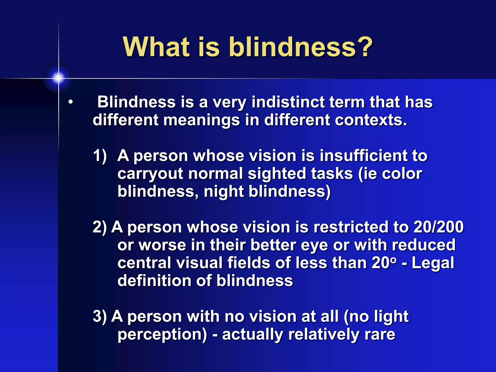
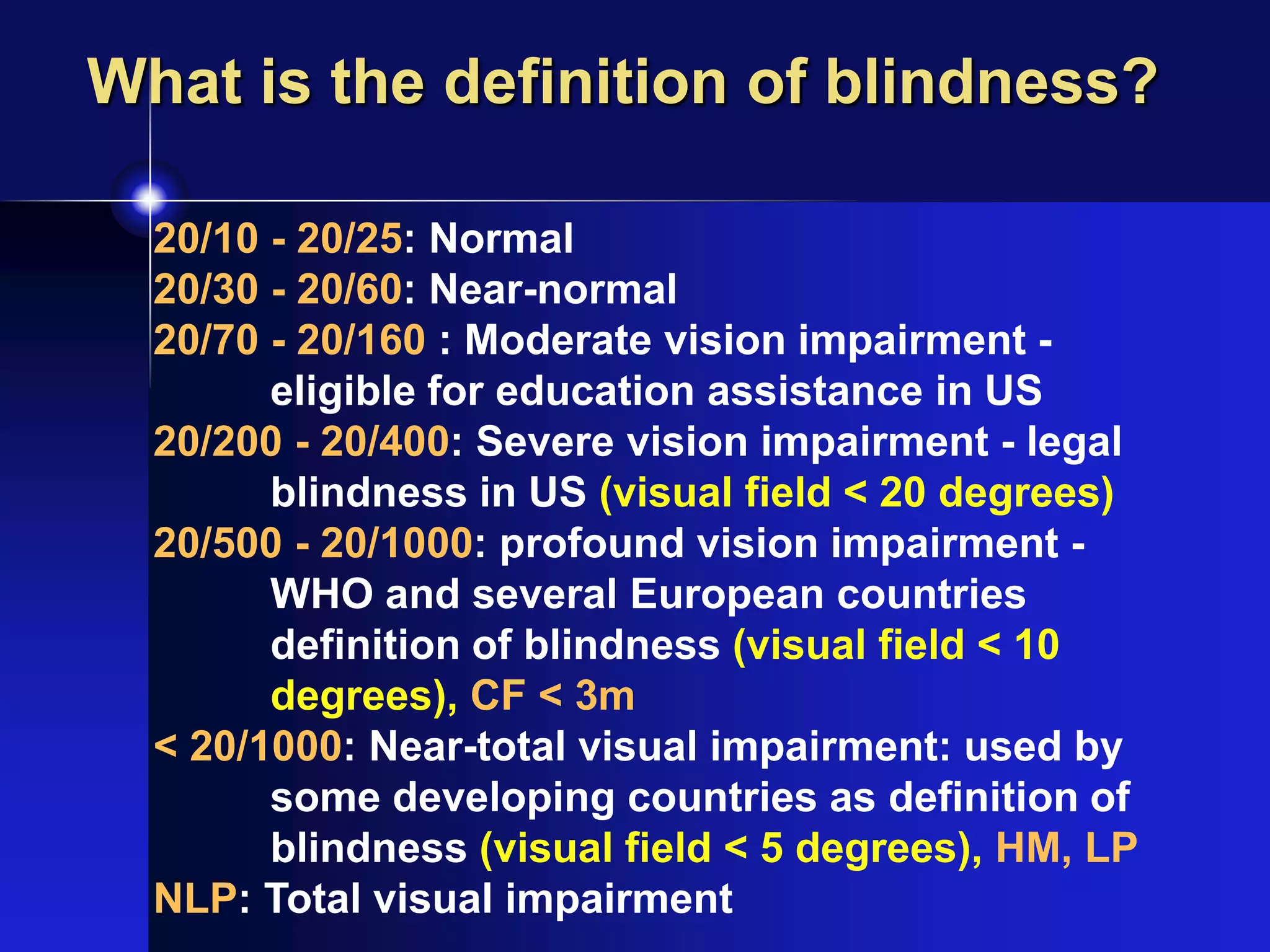
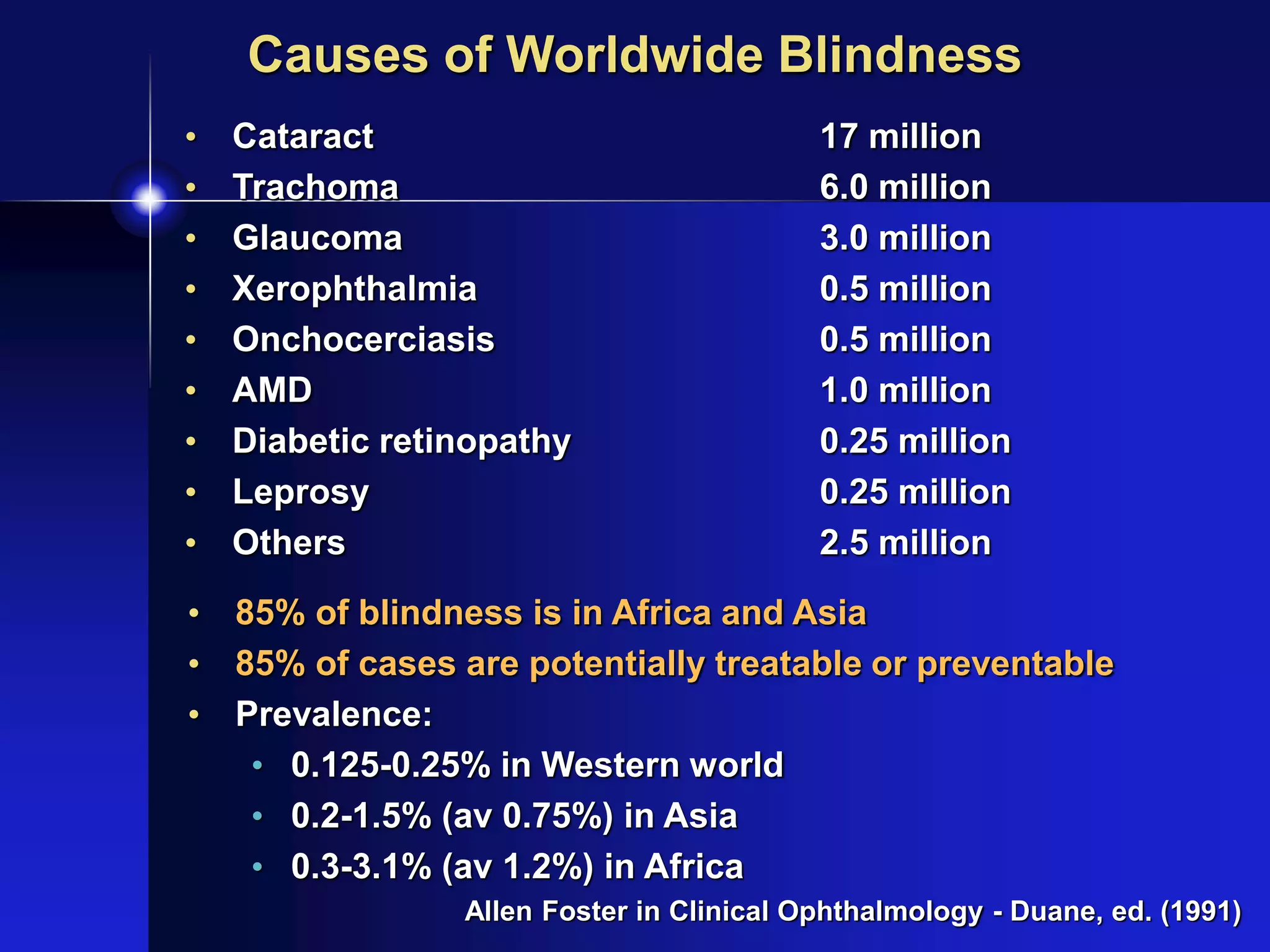
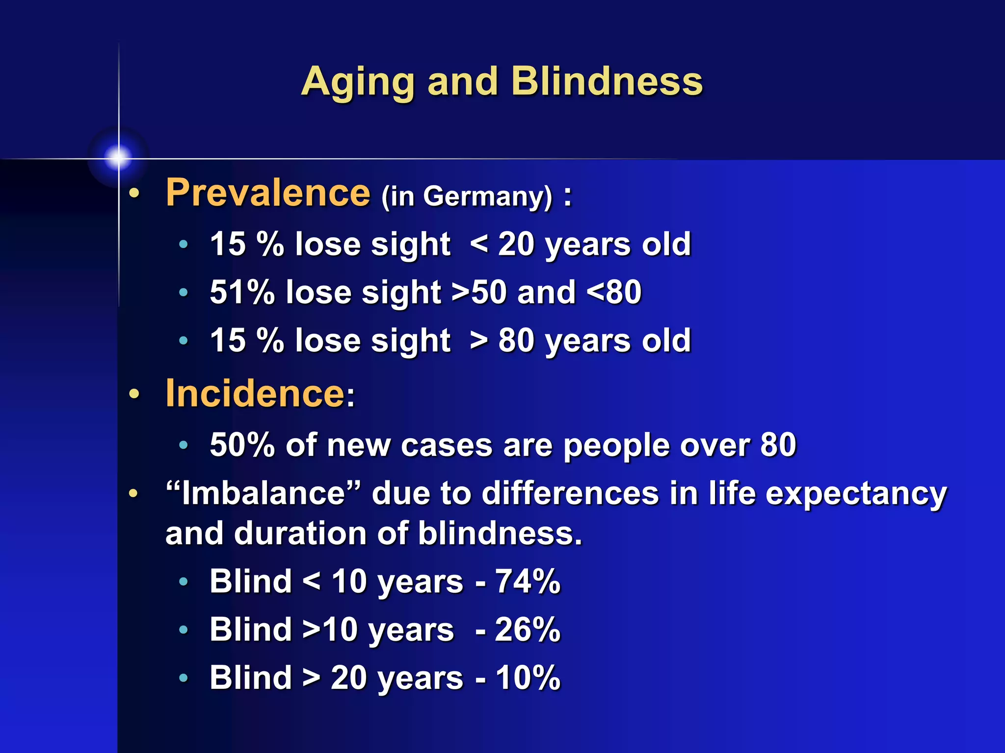
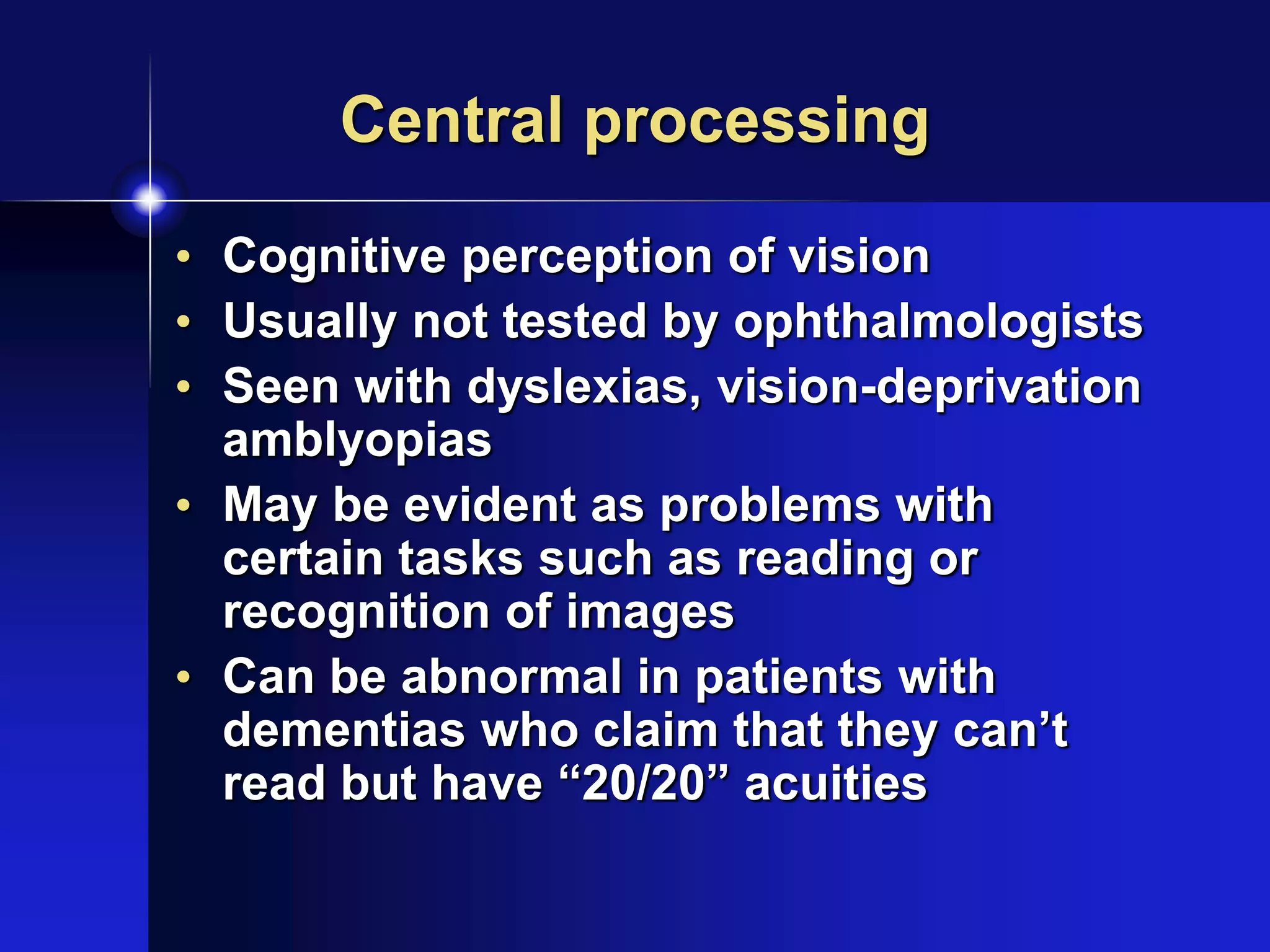
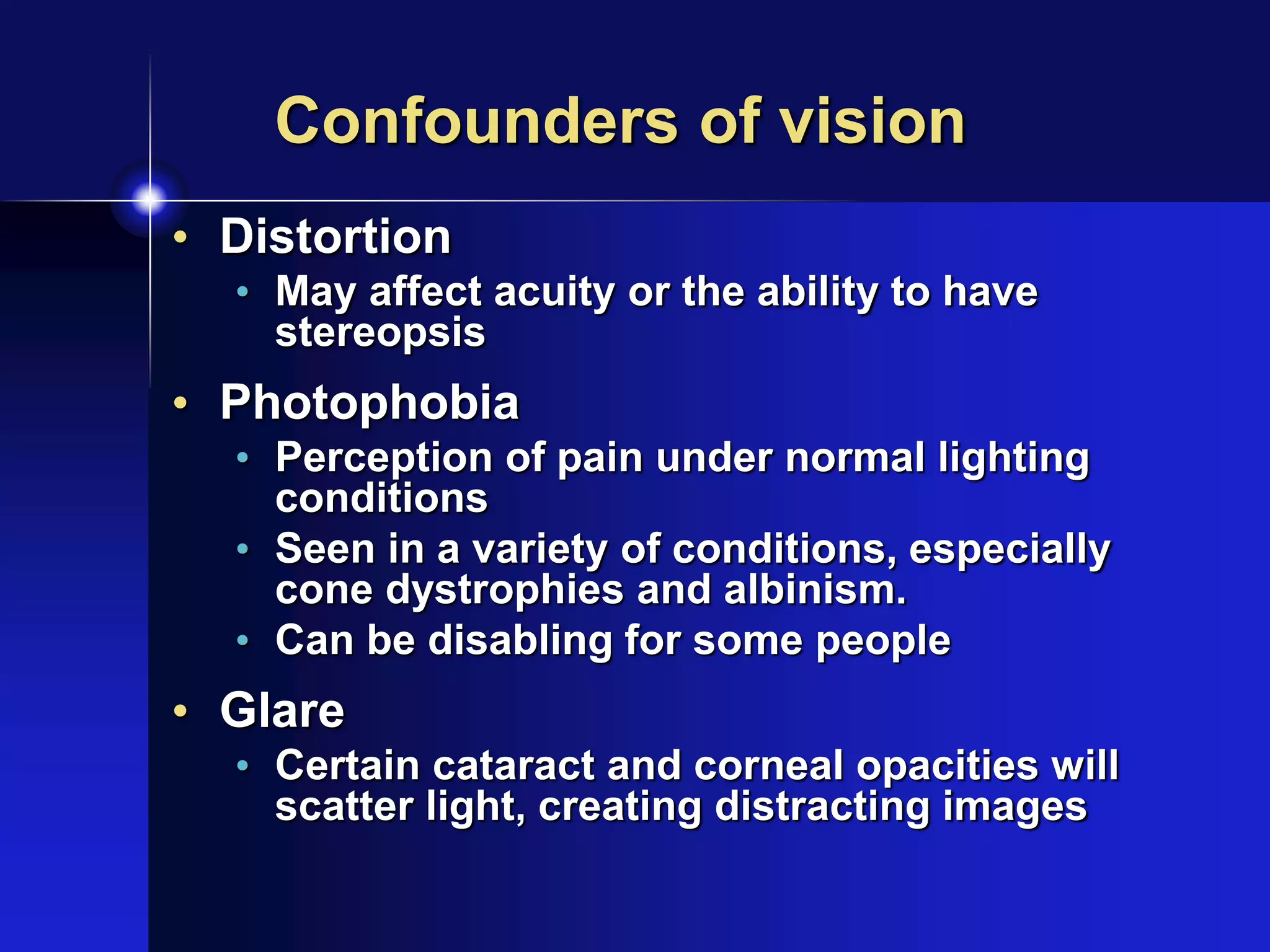
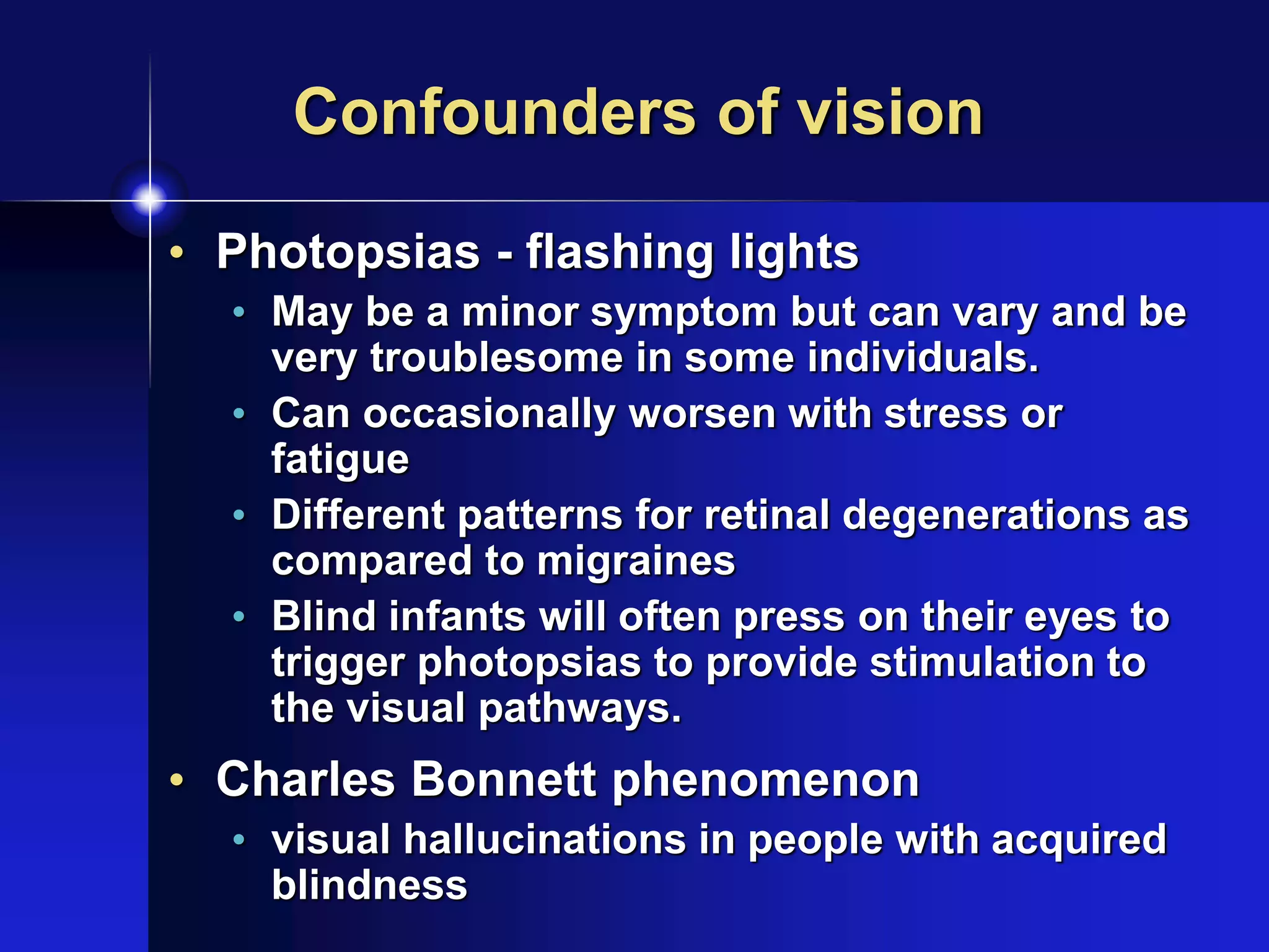
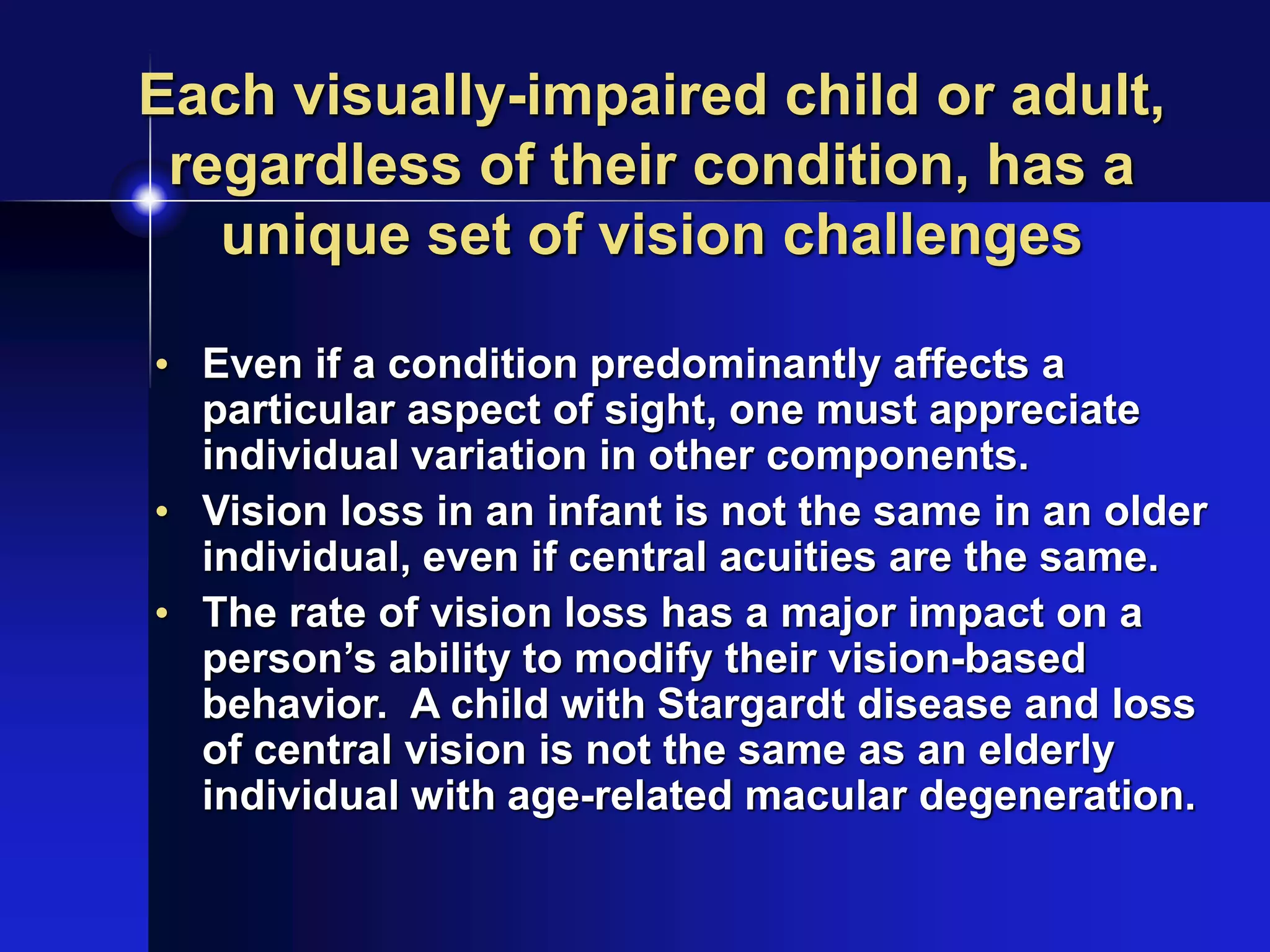
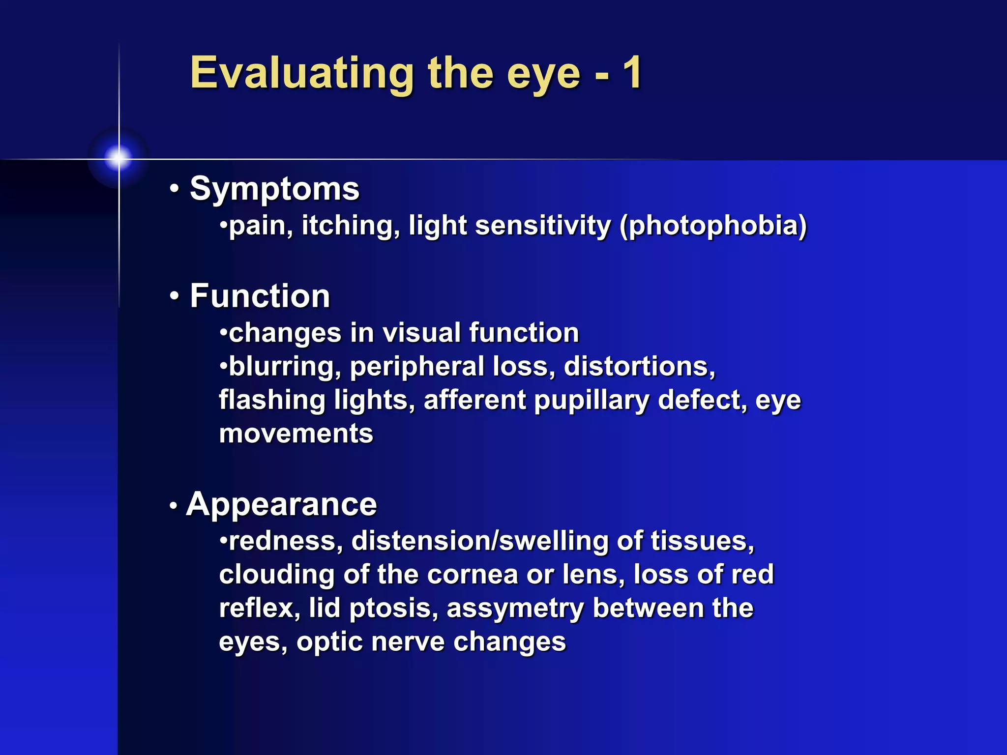
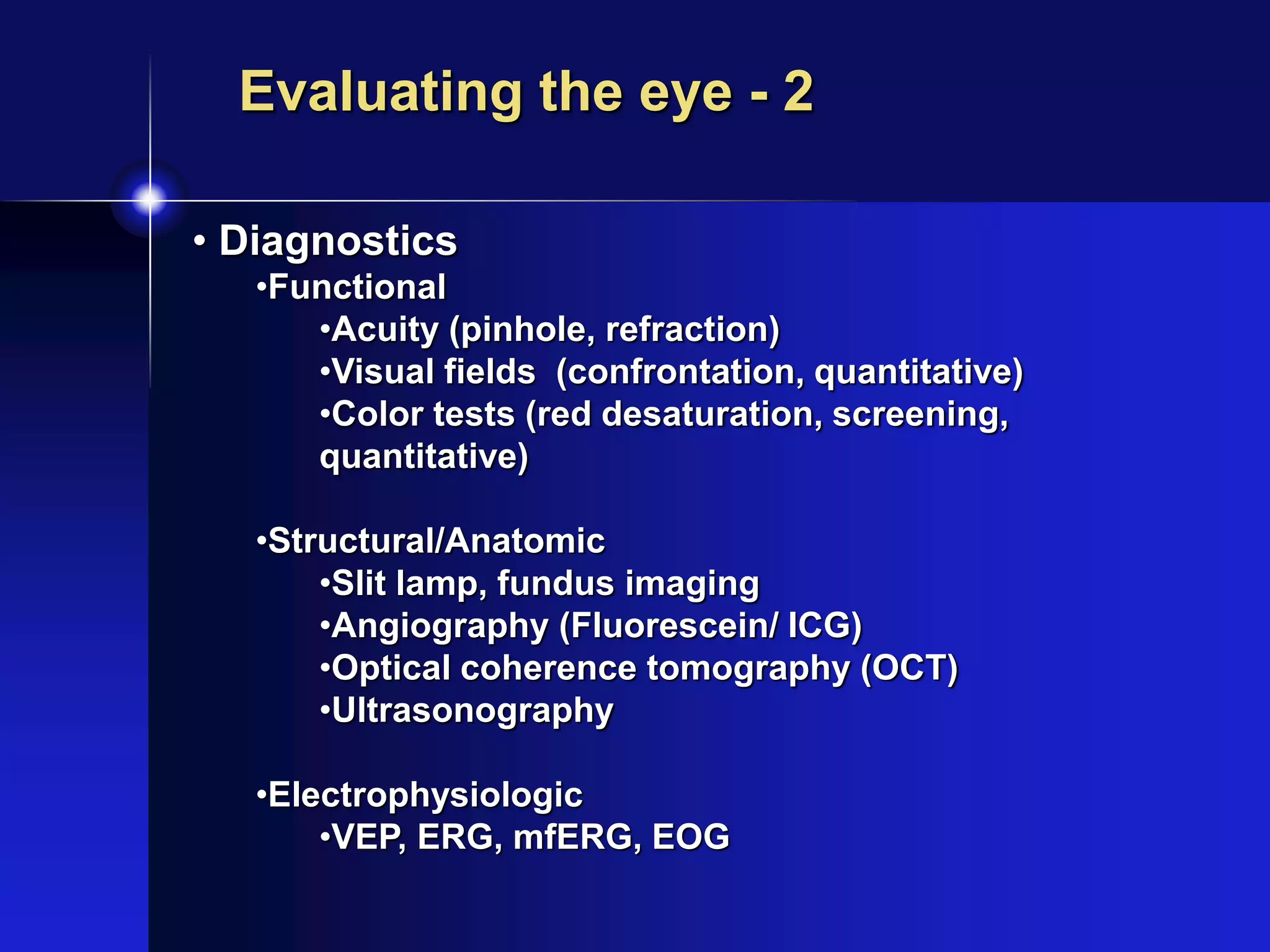
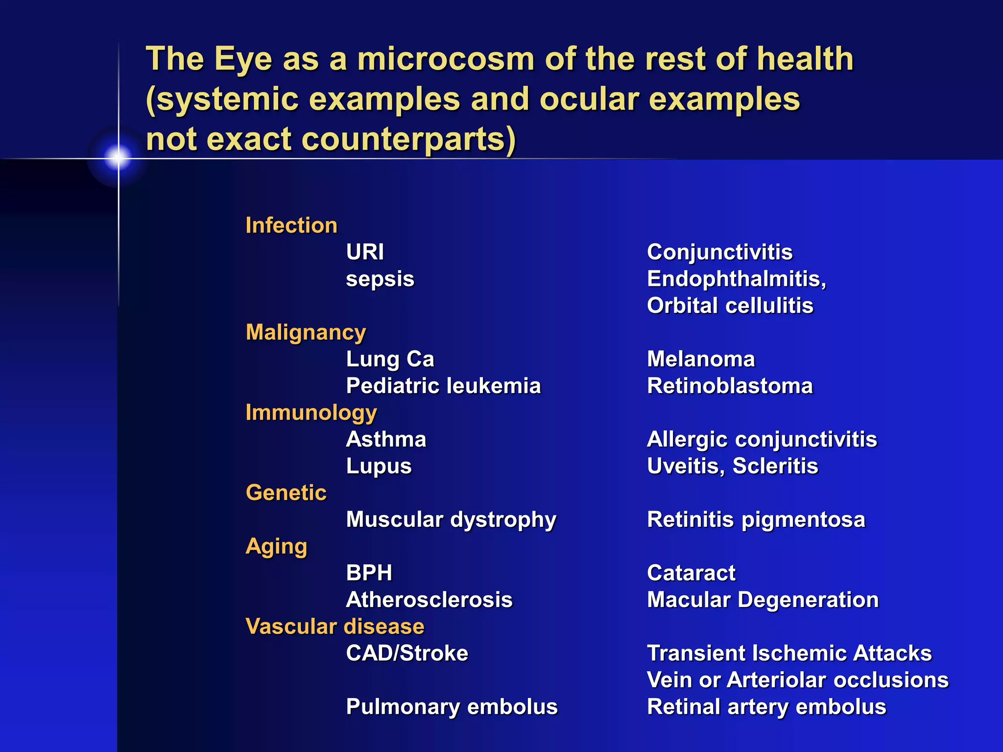
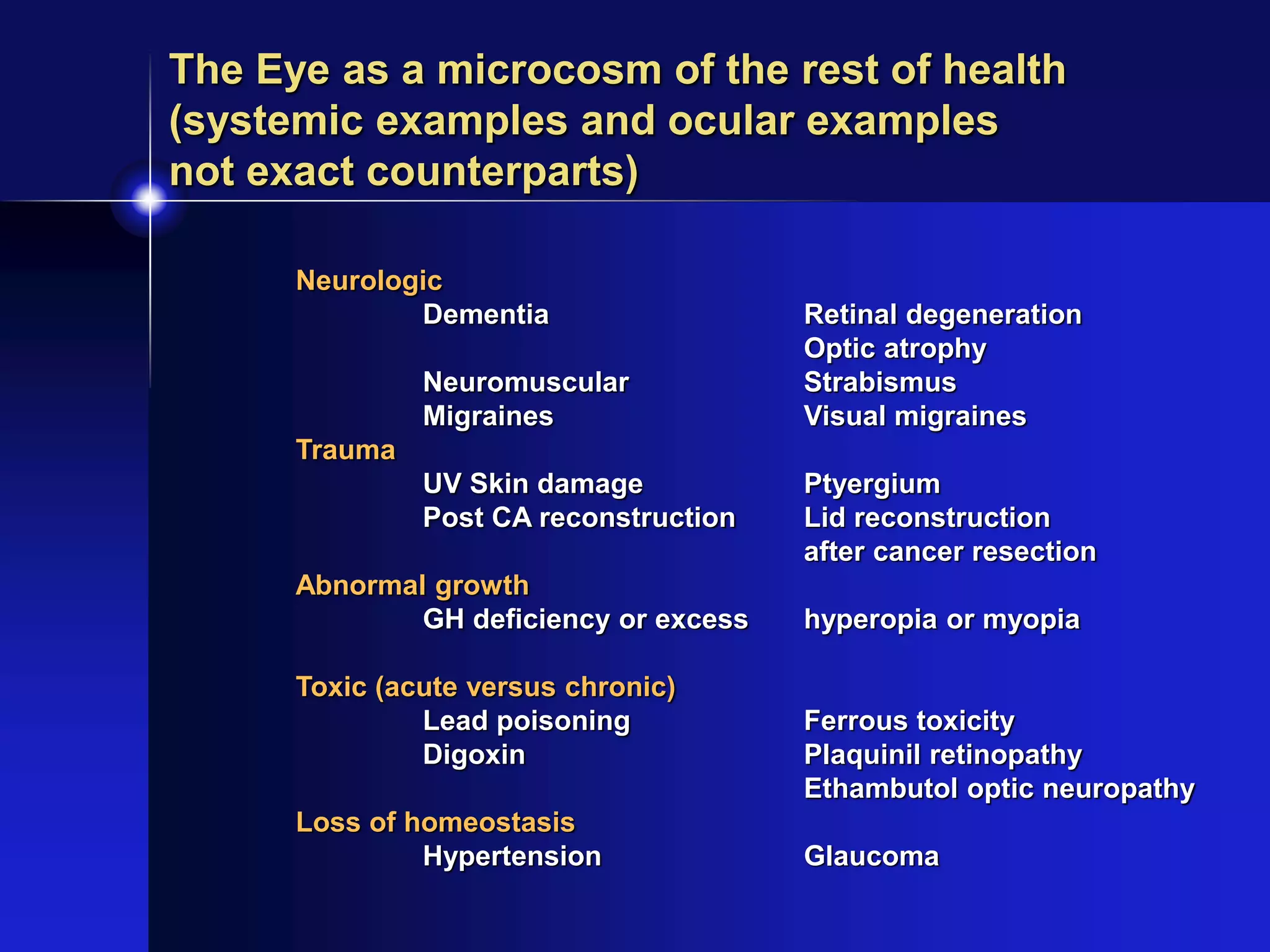
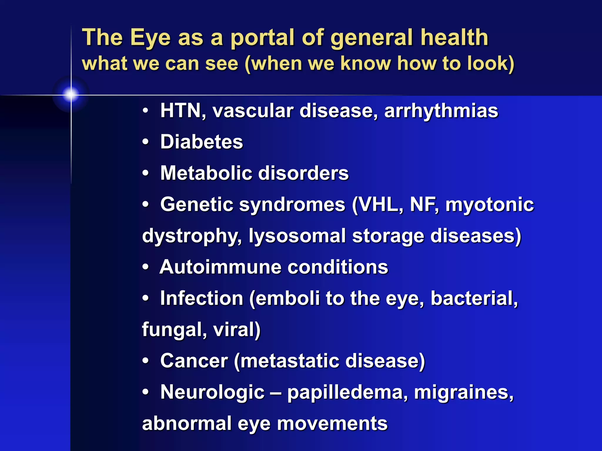
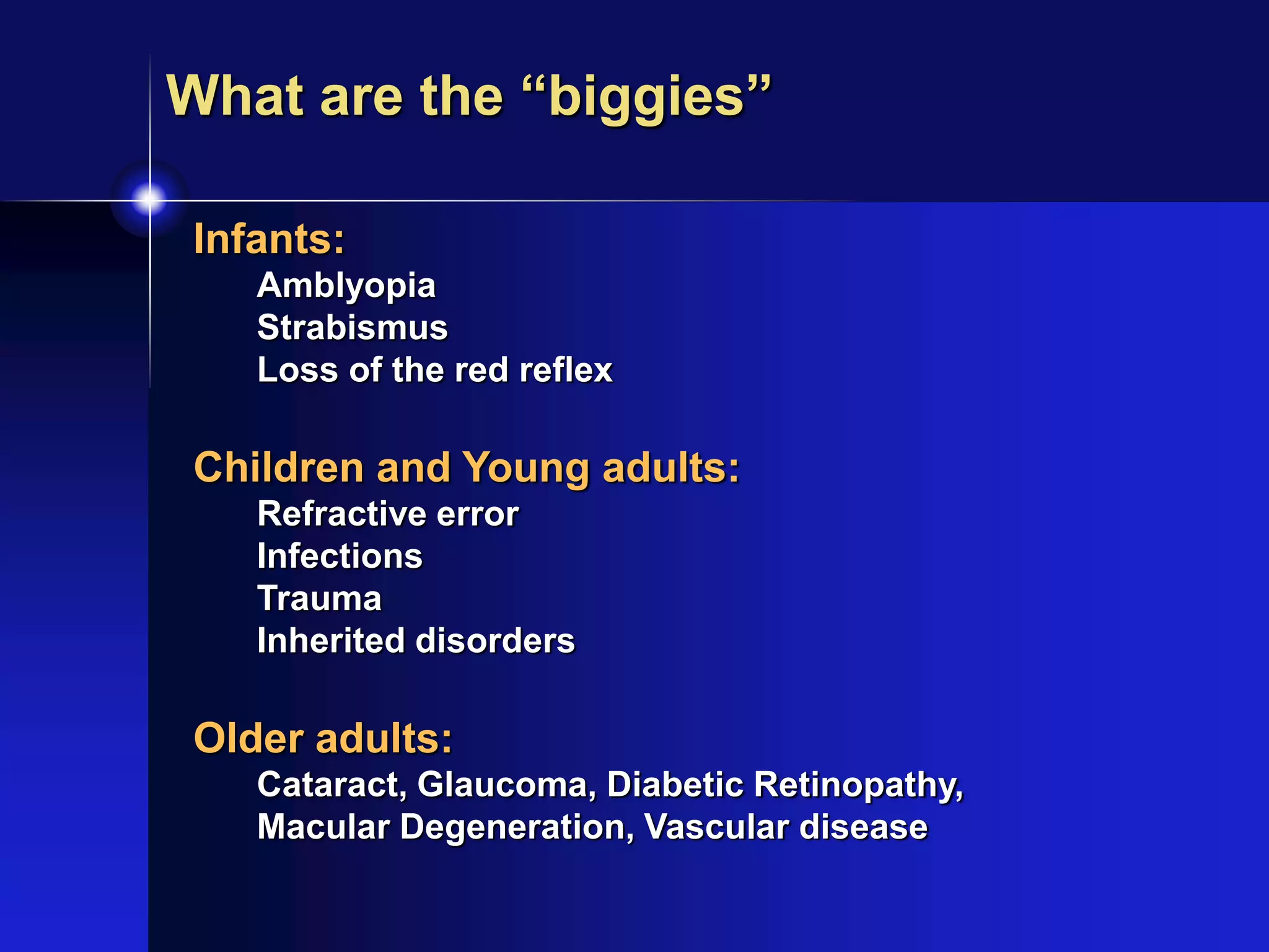
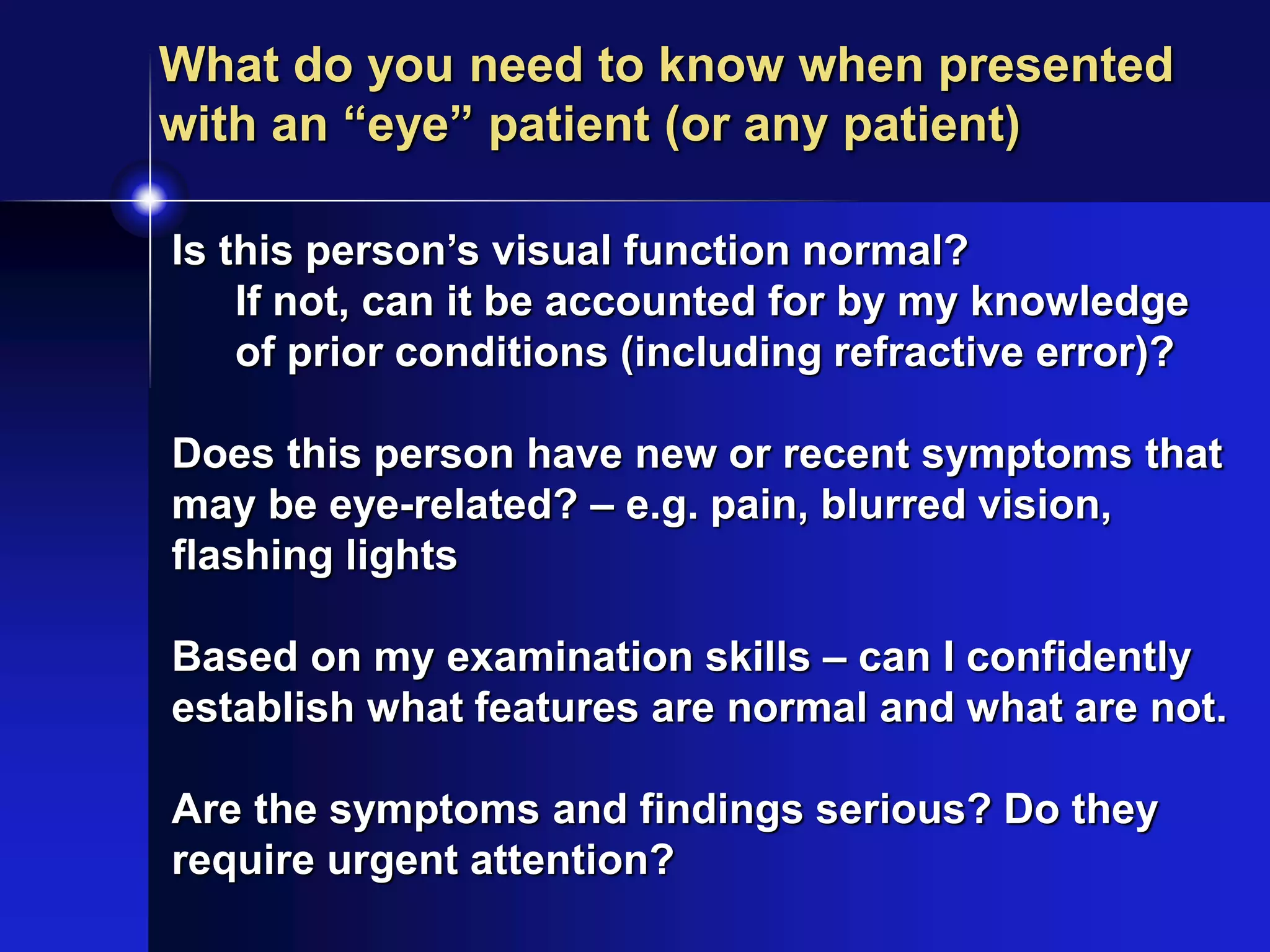
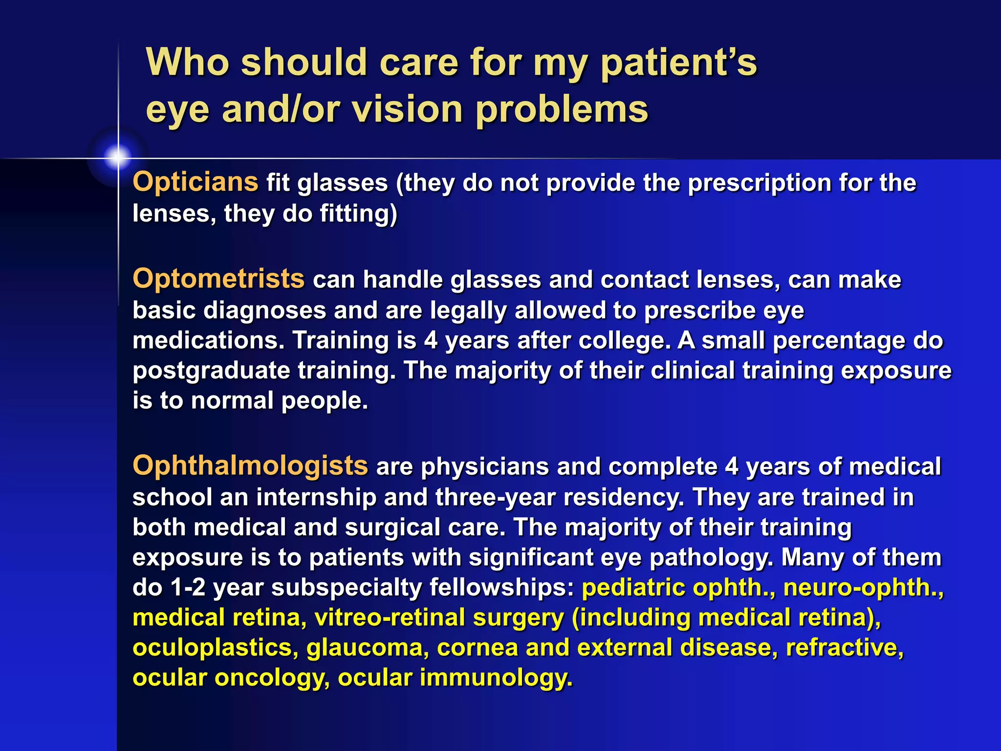
What every physician should
know about the eye
Michael B. Gorin, MD PhD
Professor of Ophthalmology
Jules Stein Eye Institute - UCLA
What every physician should
think about vision and the eye
Goals of this talk
• What is blindness
• Understand basic concepts regarding
vision and its assessment
• How do we evaluate the eye
• Appreciating the diversity of ocular
disease
• The eye with respect to general health
and systemic disease
• The “biggies”
• What do you do with a patient with
possible vision or eye problems.
“Non-goals” of this talk
• Detailed discussion of ocular anatomy,
physiology, biochemistry and genetics
• You should have already had this in prior
sections
• There is a website in your handout to review
this material and you will have a quiz on the
morning of the first day to ensure that you
have the necessary information for the
clerkship.
• Eye examination skills for the general
physician
• This was covered in the prior workshop (2nd
year)
“Non-goals” of this talk
• Recognition of the findings of different
types of eye disorders
• Management and treatment of major or
common eye conditions
• These are the primary goals of this 3rd year
clinical clerkship experience.
• Convincing you that ophthalmology is the
best specialty in medicine
What is blindness?
• Blindness is a very indistinct term that has
different meanings in different contexts.
1) A person whose vision is insufficient to
carryout normal sighted tasks (ie color
blindness, night blindness)
2) A person whose vision is restricted to 20/200
or worse in their better eye or with reduced
central visual fields of less than 20o - Legal
definition of blindness
3) A person with no vision at all (no light
perception) - actually relatively rare
What is the definition of blindness?
20/10 - 20/25: Normal
20/30 - 20/60: Near-normal
20/70 - 20/160 : Moderate vision impairment -
eligible for education assistance in US
20/200 - 20/400: Severe vision impairment - legal
blindness in US (visual field < 20 degrees)
20/500 - 20/1000: profound vision impairment -
WHO and several European countries
definition of blindness (visual field < 10
degrees), CF < 3m
< 20/1000: Near-total visual impairment: used by
some developing countries as definition of
blindness (visual field < 5 degrees), HM, LP
NLP: Total visual impairment
Causes of Worldwide Blindness
• Cataract 17 million
• Trachoma 6.0 million
• Glaucoma 3.0 million
• Xerophthalmia 0.5 million
• Onchocerciasis 0.5 million
• AMD 1.0 million
• Diabetic retinopathy 0.25 million
• Leprosy 0.25 million
• Others 2.5 million
• 85% of blindness is in Africa and Asia
• 85% of cases are potentially treatable or preventable
• Prevalence:
• 0.125-0.25% in Western world
• 0.2-1.5% (av 0.75%) in Asia
• 0.3-3.1% (av 1.2%) in Africa
Allen Foster in Clinical Ophthalmology - Duane, ed. (1991)
Aging and Blindness
• Prevalence (in Germany) :
• 15 % lose sight < 20 years old
• 51% lose sight >50 and <80
• 15 % lose sight > 80 years old
• Incidence:
• 50% of new cases are people over 80
• “Imbalance” due to differences in life expectancy
and duration of blindness.
• Blind < 10 years - 74%
• Blind >10 years - 26%
• Blind > 20 years - 10%
Vision parameters
• Central visual acuity
• Contrast sensitivity
• Color
• Adaptation
• Peripheral vision
• Binocularity and stereopsis
• Central Processing
• Confounders - nightblindness,
photopsias, photophobia, scotomas,
distortions, glare
Central visual acuity
• Derives from the central 250 microns of the
retina
• Beyond 250 microns, central acuity declines
rapidly
• Measured by Snellen chart or ETDRS
Note: high contrast, high luminance
conditions
• Requires proper central (brain) development
Early vision impairment can prevent good
central vision even if problem is corrected -
amblyopia
• Uncorrected and
corrected
• Refractive error
Myopia
Hyperopia
Astigmatism
Presbyopia
• Snellen chart,
near card, and the
ETDRS chart
ETDRS Chart
Back-illuminated,
High luminance,
High contrast
Central
visual acuity
Normal View
Central loss with
paracentral
blurring
Fixation on head
Central loss with
paracentral
blurring
Fixation on paper
Contrast sensitivity
• Variations in vision with
different lighting conditions
• Different tests
• Pelli Robison, others
• Important to consider how
different lighting conditions
can affect functional acuity
Normal contrast
Reduced contrast Pelli-Robison Chart
Color
• Most common vision deficiency other
than refractive error.
• Most screening tests are for red-green
congenital colorblindness
• Color deficiencies are also seen in
progressive conditions such as cone and
cone-rod dystrophies
• Testing (CRT, plates and chips)
Ishihara Color
plates
Left - Normal
Right - Red green
deficiency
Farnsworth D-85
Color Testing
1: Normal
2: Red-green
3: Complete color
deficiency
1
2
3
Adaptation
• Adjustment to lighting changes
• Nightblindness (such as with RP,
Diabetics)
• Delayed recovery from photostress test
• (macular dystrophies, some stationary
conditions (fundus albipunctatus)
• Goldman Weekers adaptometer,
qualitative macular stress test
• Not routinely tested
Goldman Weekers
Adaptometer
Central panel:
poor light adaptation,
nightblindness
Peripheral vision
• Tested with visual fields
• Abnormal in RP, glaucoma as well as
other conditions
• When loss is gradual, patients adapt very
well until advanced disease
• Important to understand how the brain
builds a picture of the world.
• A constricted visual field is like painting a
large wall with a small brush. It takes more
time and effort. Harder for a person to
perceive a sudden change.
Normal Glaucoma
Visual Field TestAutomated Perimetry
http://my-vision-simulator.com/
Web site to play with simulations of
vision loss for different conditions
glaucoma, macular degeneration,
cataracts, retinitis pigmentosa,
diabetic retinopathy, myopia,
astigmatism, hyperopia
Fun to use
Not very accurate
Only shows limited aspects of vision loss
Binocularity and Stereopsis - 1
• Binocularity refers to the use of both eyes
to obtain a merged view of the world.
• One can have binocular vision without
stereopsis.
• Stereopsis is the perception of depth based
upon image disparities perceived by the
brain from the input from both eyes.
• One cannot have stereopsis without binocular
vision.
• One can have binocular vision without
stereopsis.
Binocularity and Stereopsis - 2
• Depth perception is the awareness that objects
are closer or farther from the subject and the
position of objects with respect to each other.
• One can have depth perception without stereopsis.
• Often lost in strabismus and amblyopia
• May be diminished with poor central vision
• Can be lost over time if person loses too much
vision to allow for fusion
• Generally not critical to distance perception.
• Not essential for driving and most tasks
• Testing is done with polarizing glasses or special
examination devices.
Central processing
• Cognitive perception of vision
• Usually not tested by ophthalmologists
• Seen with dyslexias, vision-deprivation
amblyopias
• May be evident as problems with
certain tasks such as reading or
recognition of images
• Can be abnormal in patients with
dementias who claim that they can’t
read but have “20/20” acuities
Confounders of vision
• Distortion
• May affect acuity or the ability to have
stereopsis
• Photophobia
• Perception of pain under normal lighting
conditions
• Seen in a variety of conditions, especially
cone dystrophies and albinism.
• Can be disabling for some people
• Glare
• Certain cataract and corneal opacities will
scatter light, creating distracting images
Confounders of vision
• Photopsias - flashing lights
• May be a minor symptom but can vary and be
very troublesome in some individuals.
• Can occasionally worsen with stress or
fatigue
• Different patterns for retinal degenerations as
compared to migraines
• Blind infants will often press on their eyes to
trigger photopsias to provide stimulation to
the visual pathways.
• Charles Bonnett phenomenon
• visual hallucinations in people with acquired
blindness
Each visually-impaired child or adult,
regardless of their condition, has a
unique set of vision challenges
• Even if a condition predominantly affects a
particular aspect of sight, one must appreciate
individual variation in other components.
• Vision loss in an infant is not the same in an older
individual, even if central acuities are the same.
• The rate of vision loss has a major impact on a
person’s ability to modify their vision-based
behavior. A child with Stargardt disease and loss
of central vision is not the same as an elderly
individual with age-related macular degeneration.
• Symptoms
•pain, itching, light sensitivity (photophobia)
• Function
•changes in visual function
•blurring, peripheral loss, distortions,
flashing lights, afferent pupillary defect, eye
movements
• Appearance
•redness, distension/swelling of tissues,
clouding of the cornea or lens, loss of red
reflex, lid ptosis, assymetry between the
eyes, optic nerve changes
Evaluating the eye - 1
• Diagnostics
•Functional
•Acuity (pinhole, refraction)
•Visual fields (confrontation, quantitative)
•Color tests (red desaturation, screening,
quantitative)
•Structural/Anatomic
•Slit lamp, fundus imaging
•Angiography (Fluorescein/ ICG)
•Optical coherence tomography (OCT)
•Ultrasonography
•Electrophysiologic
•VEP, ERG, mfERG, EOG
Evaluating the eye - 2
The Eye as a microcosm of the rest of health
(systemic examples and ocular examples
not exact counterparts)
Infection
URI Conjunctivitis
sepsis Endophthalmitis,
Orbital cellulitis
Malignancy
Lung Ca Melanoma
Pediatric leukemia Retinoblastoma
Immunology
Asthma Allergic conjunctivitis
Lupus Uveitis, Scleritis
Genetic
Muscular dystrophy Retinitis pigmentosa
Aging
BPH Cataract
Atherosclerosis Macular Degeneration
Vascular disease
CAD/Stroke Transient Ischemic Attacks
Vein or Arteriolar occlusions
Pulmonary embolus Retinal artery embolus
The Eye as a microcosm of the rest of health
(systemic examples and ocular examples
not exact counterparts)
Neurologic
Dementia Retinal degeneration
Optic atrophy
Neuromuscular Strabismus
Migraines Visual migraines
Trauma
UV Skin damage Ptyergium
Post CA reconstruction Lid reconstruction
after cancer resection
Abnormal growth
GH deficiency or excess hyperopia or myopia
Toxic (acute versus chronic)
Lead poisoning Ferrous toxicity
Digoxin Plaquinil retinopathy
Ethambutol optic neuropathy
Loss of homeostasis
Hypertension Glaucoma
• HTN, vascular disease, arrhythmias
• Diabetes
• Metabolic disorders
• Genetic syndromes (VHL, NF, myotonic
dystrophy, lysosomal storage diseases)
• Autoimmune conditions
• Infection (emboli to the eye, bacterial,
fungal, viral)
• Cancer (metastatic disease)
• Neurologic – papilledema, migraines,
abnormal eye movements
The Eye as a portal of general health
what we can see (when we know how to look)
Infants:
Amblyopia
Strabismus
Loss of the red reflex
Children and Young adults:
Refractive error
Infections
Trauma
Inherited disorders
Older adults:
Cataract, Glaucoma, Diabetic Retinopathy,
Macular Degeneration, Vascular disease
What are the “biggies”
Is this person’s visual function normal?
If not, can it be accounted for by my knowledge
of prior conditions (including refractive error)?
Does this person have new or recent symptoms that
may be eye-related? – e.g. pain, blurred vision,
flashing lights
Based on my examination skills – can I confidently
establish what features are normal and what are not.
Are the symptoms and findings serious? Do they
require urgent attention?
What do you need to know when presented
with an “eye” patient (or any patient)
Opticians fit glasses (they do not provide the prescription for the
lenses, they do fitting)
Optometrists can handle glasses and contact lenses, can make
basic diagnoses and are legally allowed to prescribe eye
medications. Training is 4 years after college. A small percentage do
postgraduate training. The majority of their clinical training exposure
is to normal people.
Ophthalmologists are physicians and complete 4 years of medical
school an internship and three-year residency. They are trained in
both medical and surgical care. The majority of their training
exposure is to patients with significant eye pathology. Many of them
do 1-2 year subspecialty fellowships: pediatric ophth., neuro-ophth.,
medical retina, vitreo-retinal surgery (including medical retina),
oculoplastics, glaucoma, cornea and external disease, refractive,
ocular oncology, ocular immunology.
Who should care for my patient’s
eye and/or vision problems

What every md should know about the eye

  • 1.
    What every physicianshould know about the eye Michael B. Gorin, MD PhD Professor of Ophthalmology Jules Stein Eye Institute - UCLA What every physician should think about vision and the eye
  • 2.
    Goals of thistalk • What is blindness • Understand basic concepts regarding vision and its assessment • How do we evaluate the eye • Appreciating the diversity of ocular disease • The eye with respect to general health and systemic disease • The “biggies” • What do you do with a patient with possible vision or eye problems.
  • 3.
    “Non-goals” of thistalk • Detailed discussion of ocular anatomy, physiology, biochemistry and genetics • You should have already had this in prior sections • There is a website in your handout to review this material and you will have a quiz on the morning of the first day to ensure that you have the necessary information for the clerkship. • Eye examination skills for the general physician • This was covered in the prior workshop (2nd year)
  • 4.
    “Non-goals” of thistalk • Recognition of the findings of different types of eye disorders • Management and treatment of major or common eye conditions • These are the primary goals of this 3rd year clinical clerkship experience. • Convincing you that ophthalmology is the best specialty in medicine
  • 5.
    What is blindness? •Blindness is a very indistinct term that has different meanings in different contexts. 1) A person whose vision is insufficient to carryout normal sighted tasks (ie color blindness, night blindness) 2) A person whose vision is restricted to 20/200 or worse in their better eye or with reduced central visual fields of less than 20o - Legal definition of blindness 3) A person with no vision at all (no light perception) - actually relatively rare
  • 6.
    What is thedefinition of blindness? 20/10 - 20/25: Normal 20/30 - 20/60: Near-normal 20/70 - 20/160 : Moderate vision impairment - eligible for education assistance in US 20/200 - 20/400: Severe vision impairment - legal blindness in US (visual field < 20 degrees) 20/500 - 20/1000: profound vision impairment - WHO and several European countries definition of blindness (visual field < 10 degrees), CF < 3m < 20/1000: Near-total visual impairment: used by some developing countries as definition of blindness (visual field < 5 degrees), HM, LP NLP: Total visual impairment
  • 7.
    Causes of WorldwideBlindness • Cataract 17 million • Trachoma 6.0 million • Glaucoma 3.0 million • Xerophthalmia 0.5 million • Onchocerciasis 0.5 million • AMD 1.0 million • Diabetic retinopathy 0.25 million • Leprosy 0.25 million • Others 2.5 million • 85% of blindness is in Africa and Asia • 85% of cases are potentially treatable or preventable • Prevalence: • 0.125-0.25% in Western world • 0.2-1.5% (av 0.75%) in Asia • 0.3-3.1% (av 1.2%) in Africa Allen Foster in Clinical Ophthalmology - Duane, ed. (1991)
  • 8.
    Aging and Blindness •Prevalence (in Germany) : • 15 % lose sight < 20 years old • 51% lose sight >50 and <80 • 15 % lose sight > 80 years old • Incidence: • 50% of new cases are people over 80 • “Imbalance” due to differences in life expectancy and duration of blindness. • Blind < 10 years - 74% • Blind >10 years - 26% • Blind > 20 years - 10%
  • 9.
    Vision parameters • Centralvisual acuity • Contrast sensitivity • Color • Adaptation • Peripheral vision • Binocularity and stereopsis • Central Processing • Confounders - nightblindness, photopsias, photophobia, scotomas, distortions, glare
  • 10.
    Central visual acuity •Derives from the central 250 microns of the retina • Beyond 250 microns, central acuity declines rapidly • Measured by Snellen chart or ETDRS Note: high contrast, high luminance conditions • Requires proper central (brain) development Early vision impairment can prevent good central vision even if problem is corrected - amblyopia
  • 11.
    • Uncorrected and corrected •Refractive error Myopia Hyperopia Astigmatism Presbyopia • Snellen chart, near card, and the ETDRS chart ETDRS Chart Back-illuminated, High luminance, High contrast Central visual acuity
  • 12.
    Normal View Central losswith paracentral blurring Fixation on head Central loss with paracentral blurring Fixation on paper
  • 13.
    Contrast sensitivity • Variationsin vision with different lighting conditions • Different tests • Pelli Robison, others • Important to consider how different lighting conditions can affect functional acuity
  • 14.
  • 15.
    Color • Most commonvision deficiency other than refractive error. • Most screening tests are for red-green congenital colorblindness • Color deficiencies are also seen in progressive conditions such as cone and cone-rod dystrophies • Testing (CRT, plates and chips)
  • 16.
    Ishihara Color plates Left -Normal Right - Red green deficiency
  • 17.
    Farnsworth D-85 Color Testing 1:Normal 2: Red-green 3: Complete color deficiency 1 2 3
  • 18.
    Adaptation • Adjustment tolighting changes • Nightblindness (such as with RP, Diabetics) • Delayed recovery from photostress test • (macular dystrophies, some stationary conditions (fundus albipunctatus) • Goldman Weekers adaptometer, qualitative macular stress test • Not routinely tested
  • 19.
    Goldman Weekers Adaptometer Central panel: poorlight adaptation, nightblindness
  • 20.
    Peripheral vision • Testedwith visual fields • Abnormal in RP, glaucoma as well as other conditions • When loss is gradual, patients adapt very well until advanced disease • Important to understand how the brain builds a picture of the world. • A constricted visual field is like painting a large wall with a small brush. It takes more time and effort. Harder for a person to perceive a sudden change.
  • 21.
    Normal Glaucoma Visual FieldTestAutomated Perimetry
  • 22.
    http://my-vision-simulator.com/ Web site toplay with simulations of vision loss for different conditions glaucoma, macular degeneration, cataracts, retinitis pigmentosa, diabetic retinopathy, myopia, astigmatism, hyperopia Fun to use Not very accurate Only shows limited aspects of vision loss
  • 23.
    Binocularity and Stereopsis- 1 • Binocularity refers to the use of both eyes to obtain a merged view of the world. • One can have binocular vision without stereopsis. • Stereopsis is the perception of depth based upon image disparities perceived by the brain from the input from both eyes. • One cannot have stereopsis without binocular vision. • One can have binocular vision without stereopsis.
  • 24.
    Binocularity and Stereopsis- 2 • Depth perception is the awareness that objects are closer or farther from the subject and the position of objects with respect to each other. • One can have depth perception without stereopsis. • Often lost in strabismus and amblyopia • May be diminished with poor central vision • Can be lost over time if person loses too much vision to allow for fusion • Generally not critical to distance perception. • Not essential for driving and most tasks • Testing is done with polarizing glasses or special examination devices.
  • 25.
    Central processing • Cognitiveperception of vision • Usually not tested by ophthalmologists • Seen with dyslexias, vision-deprivation amblyopias • May be evident as problems with certain tasks such as reading or recognition of images • Can be abnormal in patients with dementias who claim that they can’t read but have “20/20” acuities
  • 26.
    Confounders of vision •Distortion • May affect acuity or the ability to have stereopsis • Photophobia • Perception of pain under normal lighting conditions • Seen in a variety of conditions, especially cone dystrophies and albinism. • Can be disabling for some people • Glare • Certain cataract and corneal opacities will scatter light, creating distracting images
  • 27.
    Confounders of vision •Photopsias - flashing lights • May be a minor symptom but can vary and be very troublesome in some individuals. • Can occasionally worsen with stress or fatigue • Different patterns for retinal degenerations as compared to migraines • Blind infants will often press on their eyes to trigger photopsias to provide stimulation to the visual pathways. • Charles Bonnett phenomenon • visual hallucinations in people with acquired blindness
  • 28.
    Each visually-impaired childor adult, regardless of their condition, has a unique set of vision challenges • Even if a condition predominantly affects a particular aspect of sight, one must appreciate individual variation in other components. • Vision loss in an infant is not the same in an older individual, even if central acuities are the same. • The rate of vision loss has a major impact on a person’s ability to modify their vision-based behavior. A child with Stargardt disease and loss of central vision is not the same as an elderly individual with age-related macular degeneration.
  • 29.
    • Symptoms •pain, itching,light sensitivity (photophobia) • Function •changes in visual function •blurring, peripheral loss, distortions, flashing lights, afferent pupillary defect, eye movements • Appearance •redness, distension/swelling of tissues, clouding of the cornea or lens, loss of red reflex, lid ptosis, assymetry between the eyes, optic nerve changes Evaluating the eye - 1
  • 30.
    • Diagnostics •Functional •Acuity (pinhole,refraction) •Visual fields (confrontation, quantitative) •Color tests (red desaturation, screening, quantitative) •Structural/Anatomic •Slit lamp, fundus imaging •Angiography (Fluorescein/ ICG) •Optical coherence tomography (OCT) •Ultrasonography •Electrophysiologic •VEP, ERG, mfERG, EOG Evaluating the eye - 2
  • 31.
    The Eye asa microcosm of the rest of health (systemic examples and ocular examples not exact counterparts) Infection URI Conjunctivitis sepsis Endophthalmitis, Orbital cellulitis Malignancy Lung Ca Melanoma Pediatric leukemia Retinoblastoma Immunology Asthma Allergic conjunctivitis Lupus Uveitis, Scleritis Genetic Muscular dystrophy Retinitis pigmentosa Aging BPH Cataract Atherosclerosis Macular Degeneration Vascular disease CAD/Stroke Transient Ischemic Attacks Vein or Arteriolar occlusions Pulmonary embolus Retinal artery embolus
  • 32.
    The Eye asa microcosm of the rest of health (systemic examples and ocular examples not exact counterparts) Neurologic Dementia Retinal degeneration Optic atrophy Neuromuscular Strabismus Migraines Visual migraines Trauma UV Skin damage Ptyergium Post CA reconstruction Lid reconstruction after cancer resection Abnormal growth GH deficiency or excess hyperopia or myopia Toxic (acute versus chronic) Lead poisoning Ferrous toxicity Digoxin Plaquinil retinopathy Ethambutol optic neuropathy Loss of homeostasis Hypertension Glaucoma
  • 33.
    • HTN, vasculardisease, arrhythmias • Diabetes • Metabolic disorders • Genetic syndromes (VHL, NF, myotonic dystrophy, lysosomal storage diseases) • Autoimmune conditions • Infection (emboli to the eye, bacterial, fungal, viral) • Cancer (metastatic disease) • Neurologic – papilledema, migraines, abnormal eye movements The Eye as a portal of general health what we can see (when we know how to look)
  • 34.
    Infants: Amblyopia Strabismus Loss of thered reflex Children and Young adults: Refractive error Infections Trauma Inherited disorders Older adults: Cataract, Glaucoma, Diabetic Retinopathy, Macular Degeneration, Vascular disease What are the “biggies”
  • 35.
    Is this person’svisual function normal? If not, can it be accounted for by my knowledge of prior conditions (including refractive error)? Does this person have new or recent symptoms that may be eye-related? – e.g. pain, blurred vision, flashing lights Based on my examination skills – can I confidently establish what features are normal and what are not. Are the symptoms and findings serious? Do they require urgent attention? What do you need to know when presented with an “eye” patient (or any patient)
  • 36.
    Opticians fit glasses(they do not provide the prescription for the lenses, they do fitting) Optometrists can handle glasses and contact lenses, can make basic diagnoses and are legally allowed to prescribe eye medications. Training is 4 years after college. A small percentage do postgraduate training. The majority of their clinical training exposure is to normal people. Ophthalmologists are physicians and complete 4 years of medical school an internship and three-year residency. They are trained in both medical and surgical care. The majority of their training exposure is to patients with significant eye pathology. Many of them do 1-2 year subspecialty fellowships: pediatric ophth., neuro-ophth., medical retina, vitreo-retinal surgery (including medical retina), oculoplastics, glaucoma, cornea and external disease, refractive, ocular oncology, ocular immunology. Who should care for my patient’s eye and/or vision problems