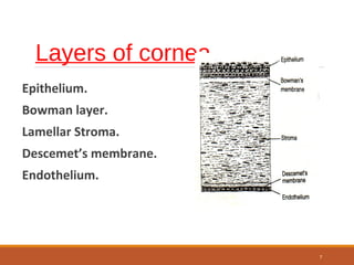
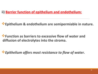
The cornea is a transparent tissue that forms the outer protective layer of the eye. It has five layers - epithelium, Bowman's layer, stroma, Descemet's membrane, and endothelium. The stroma makes up most of the corneal thickness and contains regularly arranged collagen fibrils that prevent light scattering through optical interference. The corneal endothelium actively pumps fluid out of the stroma to maintain corneal dehydration and transparency. Any disruption to the regular arrangement of collagen fibrils or the endothelial pump can compromise corneal transparency.
The aqueous humour is a transparent, watery fluid similar to plasma, but containing low protein concentrations. It is secreted from the ciliary epithelium, a structure supporting the lens
This presentation gives a brief idea about angle of anterior chamber along with its structures and diagnostic methods to grade and visualize the structures.
Cornea is the clear front surface of the eye. It lies directly in front of the iris and pupil, and it allows light to enter the eye.
Cornea forms the transparent and anterior 1/6th of the external fibrous coat of the globe of the eyeball.
The cornea is the eye's most powerful structure for focusing light that provides approximately 65 to 75 percent of the focusing power of the eye.
The cornea has unmyelinated nerve endings sensitive to touch, temperature and chemicals; a touch of the cornea causes an involuntary reflex to close the eyelid.
Main physiologic function of cornea is to act as a major refracting medium, so that a clear retinal image is formed. • Normal corneal transparency is result of • 1.anatomical factor such as uniform and regular arrangement of corneal epithelium, peculiar arrangement of corneal lamella and corneal vascularity 2.Physiological factor [ie] relative state of corneal dehydration.
3. • Therefore, any process which upsets the anatomy or physiology of cornea will cause LOSS OF TRANSPARENCY to some degree.
4. FACTORS AFFECTING CORNEAL TRANSPARENCY • CORNEAL EPITHELIUM &TEAR FLIM • ARRANGEMENT OF STROMAL LAMELLA • CORNEAL VASCULARIZATION • CORNEAL HYDRATION • CELLULAR FACTORS AFFECTING TRANSPARENCY
This lecture includes anatomy and Physiology of Cornea, if u like it kindly share it with colleagues and like it. I will share more lectures related to eye anatomy and optometry.
Thank You.
A brief presentation on corneal physiology (Functions ,cell shapes, histology ,biochemical compositions, transparency, drug permeability and cell turnover and wound healing )
Title: Sense of Smell
Presenter: Dr. Faiza, Assistant Professor of Physiology
Qualifications:
MBBS (Best Graduate, AIMC Lahore)
FCPS Physiology
ICMT, CHPE, DHPE (STMU)
MPH (GC University, Faisalabad)
MBA (Virtual University of Pakistan)
Learning Objectives:
Describe the primary categories of smells and the concept of odor blindness.
Explain the structure and location of the olfactory membrane and mucosa, including the types and roles of cells involved in olfaction.
Describe the pathway and mechanisms of olfactory signal transmission from the olfactory receptors to the brain.
Illustrate the biochemical cascade triggered by odorant binding to olfactory receptors, including the role of G-proteins and second messengers in generating an action potential.
Identify different types of olfactory disorders such as anosmia, hyposmia, hyperosmia, and dysosmia, including their potential causes.
Key Topics:
Olfactory Genes:
3% of the human genome accounts for olfactory genes.
400 genes for odorant receptors.
Olfactory Membrane:
Located in the superior part of the nasal cavity.
Medially: Folds downward along the superior septum.
Laterally: Folds over the superior turbinate and upper surface of the middle turbinate.
Total surface area: 5-10 square centimeters.
Olfactory Mucosa:
Olfactory Cells: Bipolar nerve cells derived from the CNS (100 million), with 4-25 olfactory cilia per cell.
Sustentacular Cells: Produce mucus and maintain ionic and molecular environment.
Basal Cells: Replace worn-out olfactory cells with an average lifespan of 1-2 months.
Bowman’s Gland: Secretes mucus.
Stimulation of Olfactory Cells:
Odorant dissolves in mucus and attaches to receptors on olfactory cilia.
Involves a cascade effect through G-proteins and second messengers, leading to depolarization and action potential generation in the olfactory nerve.
Quality of a Good Odorant:
Small (3-20 Carbon atoms), volatile, water-soluble, and lipid-soluble.
Facilitated by odorant-binding proteins in mucus.
Membrane Potential and Action Potential:
Resting membrane potential: -55mV.
Action potential frequency in the olfactory nerve increases with odorant strength.
Adaptation Towards the Sense of Smell:
Rapid adaptation within the first second, with further slow adaptation.
Psychological adaptation greater than receptor adaptation, involving feedback inhibition from the central nervous system.
Primary Sensations of Smell:
Camphoraceous, Musky, Floral, Pepperminty, Ethereal, Pungent, Putrid.
Odor Detection Threshold:
Examples: Hydrogen sulfide (0.0005 ppm), Methyl-mercaptan (0.002 ppm).
Some toxic substances are odorless at lethal concentrations.
Characteristics of Smell:
Odor blindness for single substances due to lack of appropriate receptor protein.
Behavioral and emotional influences of smell.
Transmission of Olfactory Signals:
From olfactory cells to glomeruli in the olfactory bulb, involving lateral inhibition.
Primitive, less old, and new olfactory systems with different path
Ozempic: Preoperative Management of Patients on GLP-1 Receptor Agonists Saeid Safari
Preoperative Management of Patients on GLP-1 Receptor Agonists like Ozempic and Semiglutide
ASA GUIDELINE
NYSORA Guideline
2 Case Reports of Gastric Ultrasound
Recomendações da OMS sobre cuidados maternos e neonatais para uma experiência pós-natal positiva.
Em consonância com os ODS – Objetivos do Desenvolvimento Sustentável e a Estratégia Global para a Saúde das Mulheres, Crianças e Adolescentes, e aplicando uma abordagem baseada nos direitos humanos, os esforços de cuidados pós-natais devem expandir-se para além da cobertura e da simples sobrevivência, de modo a incluir cuidados de qualidade.
Estas diretrizes visam melhorar a qualidade dos cuidados pós-natais essenciais e de rotina prestados às mulheres e aos recém-nascidos, com o objetivo final de melhorar a saúde e o bem-estar materno e neonatal.
Uma “experiência pós-natal positiva” é um resultado importante para todas as mulheres que dão à luz e para os seus recém-nascidos, estabelecendo as bases para a melhoria da saúde e do bem-estar a curto e longo prazo. Uma experiência pós-natal positiva é definida como aquela em que as mulheres, pessoas que gestam, os recém-nascidos, os casais, os pais, os cuidadores e as famílias recebem informação consistente, garantia e apoio de profissionais de saúde motivados; e onde um sistema de saúde flexível e com recursos reconheça as necessidades das mulheres e dos bebês e respeite o seu contexto cultural.
Estas diretrizes consolidadas apresentam algumas recomendações novas e já bem fundamentadas sobre cuidados pós-natais de rotina para mulheres e neonatos que recebem cuidados no pós-parto em unidades de saúde ou na comunidade, independentemente dos recursos disponíveis.
É fornecido um conjunto abrangente de recomendações para cuidados durante o período puerperal, com ênfase nos cuidados essenciais que todas as mulheres e recém-nascidos devem receber, e com a devida atenção à qualidade dos cuidados; isto é, a entrega e a experiência do cuidado recebido. Estas diretrizes atualizam e ampliam as recomendações da OMS de 2014 sobre cuidados pós-natais da mãe e do recém-nascido e complementam as atuais diretrizes da OMS sobre a gestão de complicações pós-natais.
O estabelecimento da amamentação e o manejo das principais intercorrências é contemplada.
Recomendamos muito.
Vamos discutir essas recomendações no nosso curso de pós-graduação em Aleitamento no Instituto Ciclos.
Esta publicação só está disponível em inglês até o momento.
Prof. Marcus Renato de Carvalho
www.agostodourado.com
Lung Cancer: Artificial Intelligence, Synergetics, Complex System Analysis, S...Oleg Kshivets
RESULTS: Overall life span (LS) was 2252.1±1742.5 days and cumulative 5-year survival (5YS) reached 73.2%, 10 years – 64.8%, 20 years – 42.5%. 513 LCP lived more than 5 years (LS=3124.6±1525.6 days), 148 LCP – more than 10 years (LS=5054.4±1504.1 days).199 LCP died because of LC (LS=562.7±374.5 days). 5YS of LCP after bi/lobectomies was significantly superior in comparison with LCP after pneumonectomies (78.1% vs.63.7%, P=0.00001 by log-rank test). AT significantly improved 5YS (66.3% vs. 34.8%) (P=0.00000 by log-rank test) only for LCP with N1-2. Cox modeling displayed that 5YS of LCP significantly depended on: phase transition (PT) early-invasive LC in terms of synergetics, PT N0—N12, cell ratio factors (ratio between cancer cells- CC and blood cells subpopulations), G1-3, histology, glucose, AT, blood cell circuit, prothrombin index, heparin tolerance, recalcification time (P=0.000-0.038). Neural networks, genetic algorithm selection and bootstrap simulation revealed relationships between 5YS and PT early-invasive LC (rank=1), PT N0—N12 (rank=2), thrombocytes/CC (3), erythrocytes/CC (4), eosinophils/CC (5), healthy cells/CC (6), lymphocytes/CC (7), segmented neutrophils/CC (8), stick neutrophils/CC (9), monocytes/CC (10); leucocytes/CC (11). Correct prediction of 5YS was 100% by neural networks computing (area under ROC curve=1.0; error=0.0).
CONCLUSIONS: 5YS of LCP after radical procedures significantly depended on: 1) PT early-invasive cancer; 2) PT N0--N12; 3) cell ratio factors; 4) blood cell circuit; 5) biochemical factors; 6) hemostasis system; 7) AT; 8) LC characteristics; 9) LC cell dynamics; 10) surgery type: lobectomy/pneumonectomy; 11) anthropometric data. Optimal diagnosis and treatment strategies for LC are: 1) screening and early detection of LC; 2) availability of experienced thoracic surgeons because of complexity of radical procedures; 3) aggressive en block surgery and adequate lymph node dissection for completeness; 4) precise prediction; 5) adjuvant chemoimmunoradiotherapy for LCP with unfavorable prognosis.
Knee anatomy and clinical tests 2024.pdfvimalpl1234
This includes all relevant anatomy and clinical tests compiled from standard textbooks, Campbell,netter etc..It is comprehensive and best suited for orthopaedicians and orthopaedic residents.
Couples presenting to the infertility clinic- Do they really have infertility...Sujoy Dasgupta
Dr Sujoy Dasgupta presented the study on "Couples presenting to the infertility clinic- Do they really have infertility? – The unexplored stories of non-consummation" in the 13th Congress of the Asia Pacific Initiative on Reproduction (ASPIRE 2024) at Manila on 24 May, 2024.
Title: Sense of Taste
Presenter: Dr. Faiza, Assistant Professor of Physiology
Qualifications:
MBBS (Best Graduate, AIMC Lahore)
FCPS Physiology
ICMT, CHPE, DHPE (STMU)
MPH (GC University, Faisalabad)
MBA (Virtual University of Pakistan)
Learning Objectives:
Describe the structure and function of taste buds.
Describe the relationship between the taste threshold and taste index of common substances.
Explain the chemical basis and signal transduction of taste perception for each type of primary taste sensation.
Recognize different abnormalities of taste perception and their causes.
Key Topics:
Significance of Taste Sensation:
Differentiation between pleasant and harmful food
Influence on behavior
Selection of food based on metabolic needs
Receptors of Taste:
Taste buds on the tongue
Influence of sense of smell, texture of food, and pain stimulation (e.g., by pepper)
Primary and Secondary Taste Sensations:
Primary taste sensations: Sweet, Sour, Salty, Bitter, Umami
Chemical basis and signal transduction mechanisms for each taste
Taste Threshold and Index:
Taste threshold values for Sweet (sucrose), Salty (NaCl), Sour (HCl), and Bitter (Quinine)
Taste index relationship: Inversely proportional to taste threshold
Taste Blindness:
Inability to taste certain substances, particularly thiourea compounds
Example: Phenylthiocarbamide
Structure and Function of Taste Buds:
Composition: Epithelial cells, Sustentacular/Supporting cells, Taste cells, Basal cells
Features: Taste pores, Taste hairs/microvilli, and Taste nerve fibers
Location of Taste Buds:
Found in papillae of the tongue (Fungiform, Circumvallate, Foliate)
Also present on the palate, tonsillar pillars, epiglottis, and proximal esophagus
Mechanism of Taste Stimulation:
Interaction of taste substances with receptors on microvilli
Signal transduction pathways for Umami, Sweet, Bitter, Sour, and Salty tastes
Taste Sensitivity and Adaptation:
Decrease in sensitivity with age
Rapid adaptation of taste sensation
Role of Saliva in Taste:
Dissolution of tastants to reach receptors
Washing away the stimulus
Taste Preferences and Aversions:
Mechanisms behind taste preference and aversion
Influence of receptors and neural pathways
Impact of Sensory Nerve Damage:
Degeneration of taste buds if the sensory nerve fiber is cut
Abnormalities of Taste Detection:
Conditions: Ageusia, Hypogeusia, Dysgeusia (parageusia)
Causes: Nerve damage, neurological disorders, infections, poor oral hygiene, adverse drug effects, deficiencies, aging, tobacco use, altered neurotransmitter levels
Neurotransmitters and Taste Threshold:
Effects of serotonin (5-HT) and norepinephrine (NE) on taste sensitivity
Supertasters:
25% of the population with heightened sensitivity to taste, especially bitterness
Increased number of fungiform papillae
- Video recording of this lecture in English language: https://youtu.be/lK81BzxMqdo
- Video recording of this lecture in Arabic language: https://youtu.be/Ve4P0COk9OI
- Link to download the book free: https://nephrotube.blogspot.com/p/nephrotube-nephrology-books.html
- Link to NephroTube website: www.NephroTube.com
- Link to NephroTube social media accounts: https://nephrotube.blogspot.com/p/join-nephrotube-on-social-media.html
ARTIFICIAL INTELLIGENCE IN HEALTHCARE.pdfAnujkumaranit
Artificial intelligence (AI) refers to the simulation of human intelligence processes by machines, especially computer systems. It encompasses tasks such as learning, reasoning, problem-solving, perception, and language understanding. AI technologies are revolutionizing various fields, from healthcare to finance, by enabling machines to perform tasks that typically require human intelligence.
Prix Galien International 2024 Forum ProgramLevi Shapiro
June 20, 2024, Prix Galien International and Jerusalem Ethics Forum in ROME. Detailed agenda including panels:
- ADVANCES IN CARDIOLOGY: A NEW PARADIGM IS COMING
- WOMEN’S HEALTH: FERTILITY PRESERVATION
- WHAT’S NEW IN THE TREATMENT OF INFECTIOUS,
ONCOLOGICAL AND INFLAMMATORY SKIN DISEASES?
- ARTIFICIAL INTELLIGENCE AND ETHICS
- GENE THERAPY
- BEYOND BORDERS: GLOBAL INITIATIVES FOR DEMOCRATIZING LIFE SCIENCE TECHNOLOGIES AND PROMOTING ACCESS TO HEALTHCARE
- ETHICAL CHALLENGES IN LIFE SCIENCES
- Prix Galien International Awards Ceremony
Pulmonary Thromboembolism - etilogy, types, medical- Surgical and nursing man...VarunMahajani
Disruption of blood supply to lung alveoli due to blockage of one or more pulmonary blood vessels is called as Pulmonary thromboembolism. In this presentation we will discuss its causes, types and its management in depth.
Explore natural remedies for syphilis treatment in Singapore. Discover alternative therapies, herbal remedies, and lifestyle changes that may complement conventional treatments. Learn about holistic approaches to managing syphilis symptoms and supporting overall health.
Acute scrotum is a general term referring to an emergency condition affecting the contents or the wall of the scrotum.
There are a number of conditions that present acutely, predominantly with pain and/or swelling
A careful and detailed history and examination, and in some cases, investigations allow differentiation between these diagnoses. A prompt diagnosis is essential as the patient may require urgent surgical intervention
Testicular torsion refers to twisting of the spermatic cord, causing ischaemia of the testicle.
Testicular torsion results from inadequate fixation of the testis to the tunica vaginalis producing ischemia from reduced arterial inflow and venous outflow obstruction.
The prevalence of testicular torsion in adult patients hospitalized with acute scrotal pain is approximately 25 to 50 percent
2. Transparency is the quality or state of
transmitting light without appreciable
scattering so that bodies lying beyond are
entirely visible .
2
3. The cornea is an exquisite layered composite
material with a structure ideally suited to its
function as the transparent window in the tough
outer tunic of the eye ball through which the
outside world is viewed.
3
4. The Cornea is a transparent avascular tissue with
smooth outer convex surface and concave inner
surface which resembles a small watch glass.
Forms anterior one –sixth of the fibrous
coat of the eye.
4
5. Corneal Dimension
Horizontal Vertical
Anterior surface-- 11.7mm 10.6mm
Posterior surface-11.7mm 11.7mm
Radius of curvature
Ant surface-7.8mm Post surface-6.5mm
Corneal thickness
Central-0.5-0.6mm Peripheral-1-1.2mm
5
6. Functions of Cornea
Optical : It forms the principal refractive surface
accounting for some 70%(40-45 D) of total refractive
power(60D).
Barrier: Cornea provides a protective interference with
the outer environment and also contain intraocular
pressure.
6
8. 1. Epithelium (50-90 um)
3 types of cell :-
i) A single layer basal columnar cells stand as paliside like manner in
perfect alignment on the basement membrane.
Attached by hemidesmosomes to the epithelial basement membrane.
ii)2- 3 rows of wing cells.
iii) 2 layers of flattened surface cells
- microplicae and microvilli
- excellent ability to regenerate
- attached by desmosomes , and zonulae occludens
8
10. BIOCHEMICAL COMPOSITION OF
EPITHELIUM
Water (70% of wet weight) .
Protein synthesis is 5 times higher than stroma.
Lipid (Phospholipid and cholesterol).
Enzymes.
ATP,glycogen,glutathione,Ascorbic acid.
Acetylecholine, cholinesterase.
Electrolytes (K+,Na+,and Cl– ).
10
11. 2. Bowman’s layer (8-14 um):
Acellular mass of condensed collagen fibril.
It is not true elastic membrane but simply continuation
of stroma.
Shows considerable resistance to infection and injury
Does not regenerate.
11
12. 3.3. Lamellar Stroma (0.5mm):(0.5mm):
Consists collagen fibrils (lamellae) (200-250)and cells embedded in matrix
(proteoglycans).
Mean diameter of collagen fibril between
22.5&32nm,predominantly type I collagen.
Fibril within the lamellae are parallel to each other & also parallel to corneal
plane
Fibril within adjacent lamellae make various angles with respect to one other.
12
13. Stromal cells are keratocytes, wandering
macrophages, histocytes etc.
Keratocytes are fibroblast which produce ground
substance (keratin & chondrotin sulfate) and collagen
fibril.
The highly negatively charged keratin sulfate around
collagen fibril maintain spatial relationship between
them.
13
14. BIOCHEMICAL COMPOSITION OF STROMA :
-Water(78%)
- Collagen (15%)
- other protein (5%)
- Glycosaminoglycan
# Keratan sulfate(0.7%)
# Chondroitin sulfate(0.3)
# Chondroitin
- Salts
14
15. 4.4. Descemet’s membrane (3-40um):(3-40um):
Homogeneous layer.
Made up of collagen & glycoprotein.
Resistant to chemical agent, trauma infection and
pathological process .
Can regenerate.
15
16. 5.5. Endothelium ::
-- Single layer polygonal cells
- Attached to Descemet’s membrane by
hemidesmosomes and laterally to each other
by tight junctional complex .
- barrier function is calcium dependent
-Contains active pump mechanism
-Involved in active secretion and protein
synthesis
16
17. 17
FACTORS AFFECTING CORNEAL TRANSPERANCY
Anatomical factors:
* Uniform & regular arrangement of corneal
epithelium .
* Peculiar arrangement of corneal stromal lamellae.
* Corneal avascularity.
Physiological factors:
Relative state of corneal dehydration.
19. 1. Corneal epithelium
Epithelial cells are closely packed.
Uniformity & regularity in arrangement.
Homogenicity in refractive index.
Tight intercellular junction.
19
20. 2. Tear film
Keeps epithelial surface smooth.
Provides high quality optical surface.
20
21. 3.3. Arrangement of stromal lamellae:
Maurice Theory:
Collagen fibrils of uniform diameter (275-
350Å)are packed regularly and thus
creates a lattice pattern.
The fibril axis are located at the position
of a perfect crystalline lattice.
Interfibrillar spacing is about 50-60nm
and is smaller than wavelength of light
(400-700nm).
Scattered light is destroyed by mutual or
destructive interference
21
22. Interference:
Interference is the phenomenon by virtue of which
there is a modification in the distribution of energy due
to superimposition of two or more waves.
‘ Principle of superimposition’. This can be stated as: “
Whenever two or more sets of waves pass through and
cross one another in the same medium, they behave
independent of each other and net displacement of a
particle , at any instant , is equal to the algebraic sum of
the individual displacements due to all the waves.”
22
23. Interference may be ‘constructive’ or ‘destructive’
In constructive interference the crest of one wave coincides with
the crest of another and the net amplitude is equal to the sum of
individual amplitudes.
In destructive interference the crest of one wave coincides with the
trough of another and the net amplitude is equal to the difference
between the individual amplitudes.
Scattered rays - destructive interference
Rays on the line of incident
light- Constructive interference
23
Destructive interference
25. Findings contradictory to Maurice theory
i) The cornea is not perfectly transparent, otherwise an
ophthalmologist could not view cornea in the slit lamp..
ii) Shark’s cornea with regions of disorganised fibres and
random distribution of interfibrillar distances is also quite
transparent.
.
25
26. Hard core cylinder model of Twersky:
Twersky proposed a model in which the fibrils had a composite
structure consisting of an inner core, composed of collagen fibrils
and outer coating of a material that matches the refractive index
of the ground substance. The coating would increase correlations
but not affect the light scattering properties of a fibril.
The loss of transparency observed when the cornea swells would
be explained in the hard core model on the basis of increased
area available per fibril.
26
27. Theory of Goldman & Benedek :
Fibrils are small in relationship to
wavelength of light and do not interfere
with light transmission unless they are larger
then one-half of a wavelength of light
(2000Å).
27
28. 4. Endotheliumis transparent because it is
* Single layer.
* Homogeneous.
* Closely packed cells .
* It has deturgescence function.
28
29. 5. Corneal Vascularisation:
Except for capillary palisade of limbus, normal cornea is
avascular.
Corneal avascularity factors are not known.
When vessels present are due to corneal pathology.
Vascularisation -Loss of transparency.
29
30. Mechanical Theory : According to Cogan (1948)
Blood vessel can not invade normal cornea.
Loosening of compactness of corneal tissue
due to oedema is a must for neovsacularisation.
Interstitial keratitis where oedema always precedes
vascularisation.
Some vesostimulatory factor may be needed along with
corneal oedema for neovascularisation to occur.
30
31. Chemical theory:
Role of vasoinhibitory factors(VIF)
Sulfate ester of hyaluronic acidSulfate ester of hyaluronic acid
Role of vasostimulatory factors (VSF)
Low mol wt. AminesLow mol wt. Amines
Corneal hypoxia→VSF stimulation→ Neovascularisation
31
33. Corneal hydration :
Normal cornea maintains itself in a state of relative dehydration (80% water
content ) which is essential for corneal transparency.
It is kept constant by---
1) Factors which draw water in the cornea, like
-Stromal swelling pressure (SP)
-Intraocular pressure (IOP)
2 ) Factors which prevent flow of water in the cornea
- Mechanical barrier function of epithelium &
endothelium.
3) Factors which draw water out of cornea
-active pumping action of endothelium.
33
34. FACTORS AFFECTING CORNEAL HYDRATION:
i) STROMAL SWELLING PRESSURE (SP, 60 mmHg):
Pressure exerted by glycosaminoglycans(GAGs) of
the corneal stroma which act like a sponge.
Electrostatic repulsion of the anionic charges on
the GAG molecule expands the tissue, sucking in the
fluid with equal but negative pressure called,
imbibition pressure (IP).
34
35. In vitro, IP=SP
In vivo, IP is reduced by values
equivalent to IOP.
i.e. IP=IOP- SP
i.e. IP=17-60= - 43mmHg.
Negative imbibation pressure
draws out water from stroma.
35
36. ii) Barrier function of epithelium and endothelium:
Epithelium & endothelium are semipermiable in nature.
Function as barriers to excessive flow of water and
diffusion of electrolytes into the stroma.
Epithelium offers most resistance to flow of water.
36
37. iii) Hydration control by active pump mechanism:
a) Na+
/K+
ATPase pump system:
•Endothelium is more active than &epithelium,
•Pumps are located in basolateral membrane of endothelial cell.
•Stromal transparency develops 13-20 days after birth due to greatest increase in
pump sites/cell.
37
38. •Enzyme “Na+
/K+
activated ATPase” mediate pump causes extrusion of the Na+
& water from the stroma and thus maintain corneal transparency.
•Corneal hydration depend on extent to which endothelial barrier and pump
function can be reestablished.
38
39. b) Bicarbonate dependent ATPase present in endothelium are also reported to have
role in
fluid /ion balance in the cornea.
c) Carbonic enhydrase enzyme catalyzes the conversion of CO2and water
into HCO3
-
and H+
, thus provides important source for HCO3
-
for endothelial
pump.
d) Na+
/H+
pump has also been postulated.
39
41. Corneal thickness is increased and transparency is
decreased when there is endothelial damage and to a
lesser extend when epithelium is damaged.
41
42. iv) Passive ion movement, like
- K+
, Cl-
and HCO3
-
ions diffuse into
aqueous humor.
- Na+
, Cl-
and HCO3
-
diffusion in
contra lateral direction.
42
43. v) Hypoxia, pH and changes in the temperature
can alter the metabolic activity of the cornea and
thereby may cause alteration in corneal thickness
and loss of transparency.
43
44. vi) Intraocular pressure (IOP) :
As we know ,
I P = IOP - SP, i.e. 17- 60= - 43 mmHg,
i.e. I P is a negative pressure.
When IOP exceeds SP, i.eWhen IOP exceeds SP, i.e. when I Pwhen I P becomes positive , corneal oedemabecomes positive , corneal oedema resul
It can occur when there is
- high IOP and normal SP,as in acute glaucoma,
- Normal IOP and low SP, as in endothelial dystrophy.
44
46. Corneal Swelling:
Electron micrograph of a swollen cornea shows fibril
distribution with region completely devoid of fibrils.
These voids are called lakes, which have larger dimensions &
spoils the interference that is critical to transparency.
46
47. Photorefractive keratectomy using argon fluoride excimer
laser:
. Despite the excimer laser’s ability to ablate corneal tissue
with good precision and with little collateral damage, the
treatments commonly result in the development of increased
subepithelial light scattering that gives the cornea a hazy
appearance in the treated area.
increased numbers of activated keratocytes and vacuoles within
and around keratocytes adjacent to the treated area. Activated
keratocytes may have different effective refractive indices than
normal keratocytes, thus increasing their contribution to
scattering. Vacuoles within and around keratocytes may act like
the lakes seen in swollen cornea.
47
48. vii) Evaporation of water from corneal surface:
Evaporation of water from the pre-corneal tear film fluid →
increase in osmolarity relative to cornea→hyper tonicity of
tear film → flow of water from the cornea.
48
49. Corneal transparency results from-----
Interference among the residual waves scattered
by different fibrils.
The inefficiency of the fibrils as scatters.
Avascularity of the cornea.
The thinness of cornea which is maintained by a
complex series of metabolically dependent reaction
in the corneal endothelium and epithelium.
49