SlideShare a Scribd company logo
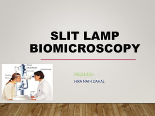
SLIT LAMP
BIOMICROSCOPY
PRESENTER-
HIRA NATH DAHAL
PRESENTATION LAYOUT
• Introduction
• Optical principle
• Components
• Examination procedures
• Illumination types
• Uses
INTRODUCTION
Slit lamp:
• instrument designed specifically to examine the eye and
adnexa
• Operational components consists of a binocular
microscope and a light source
• provides light in the form of slit to observe various ocular
structures
• Provides stereoscopic view of external adnexa, external
eye, AC, iris, lens and anterior vitreous
HISTORY
• Alvar Gullstrand( Stockholm)
• Nobel Prize in medicine and
physiology(1911)
• Vogt(1919)- first to describe use
of specular microscopy
• Mawas(1925)- biomicroscopy
• Various modification by
Kohler, Goldmann
OPTICAL PRINCIPLE
• Two systems- illumination and observation
• Mounted on a movable trolley about a common centre
and vertical axis
• Foci on the same plane
• These two systems coupled around the same centre of
rotation to ensure par-focus of microscope and slit beam
• Coplanar, coaxial and copivotal
• Works on the same principle of compund
microscope
• Objective lens(+22D) and eye piece(10-14D)
• Adjustable illumination system
• A narrow "slit" beam of very bright light
by lamp. This beam is focused on to the eye
which is then viewed under magnification with
microscope
TYPES BASED ON ILLUMINATION
SYSTEM
Zeiss slit lamp biomicroscope-
 Light source at the bottom
Haag streit slit lamp
biomicroscope-
 Light source at the top
HAAG STREIT ZIESS
OPTICAL PRINCIPLE OF HAAG-STREIT TYPE
Vertical illumination system
OPTICAL PRINCIPLE OF ZEISS TYPE
Horizontal prism reflected light source
PARTS OF SLIT LAMP
Mechanical support
Forehead rest
Chin rest
Fixation target
Power supply unit
Locking controls
Joystick
Observation system
Binocular eyepieces
Camera/video adapter
Observation tube
Magnification changer
ILLUMINATION SYSTEM
• Light source-halogen, xenon, W lamps(200000-
400000lux)
• Condenser lens system- 2 planoconvex lens,
convexities in apposition
• Slit and other diaphragms-stenopaic slits
• Filters(Neutral density filter, cobalt blue, red free filter)
• Projection lens- small diameter
• Reflecting mirror/prism
TYPICAL SLIT
LAMP
ILLUMINATION
SYSTEM
• The slit within illumination system must have sharply
demarcated edges and be adjustable
• Slit width and height must be adjustable such that any
shaped patch from a slit to circle may be projected-
increase illumination methods
• Graduated slit width- size of lesion
• Ability to rotate lamp housing- if a protractor scale
included
• Must have the facility to displaced of offset
sideways(decoupled)
THE LIGHT BEAM IS CONTROLLED BY KNOBS OR LEVERS
FILTERS
• Green(red free)-
• Increase contrast when
looking for corneal and
iris neovascularization
• Increase the visibility of rose bengal staining
• ND filters-
• Reduce beam brightness and increase comfort for the pt
• Polarizing filters-
• Reduce unwanted specular reflection and enhance visibility of
subtle defects
FILTERS
• Cobalt blue-
• Fluorescein staining
• Keratoconus- fleischer’s ring
• Kodak Wratten No.12(Yellow)
• Barrier filter placed in front of viewing system
• Enhancing green staining
GRATICULE
•Measurement and lens fit
•Pupil size, HVID, etc
OBJECTIVE SYSTEM
• The resolution of image is governed by NA of
microscope dependant upon-
• The diameter of objective
• The working distance
• The refractive index of medium between objective lens
and eye
• The wavelength of light
BIOMICROSCOPE
• Objective(2 planoconvex lens=22D), eye piece(+10D), enlarged image of
near object
• Tubes converged at 10-15° for good stereopsis
• A pair of prisms to re-invert the image
• Range of accomodation- ×6 to ×40
• Czapskiscope with rotating objectives- Haag Streit, B&L, Thorpe
• Littman Galilean telescopic system- Zeiss, Rodenstock, American optical
• Zoom system- Nikon
BIOMICROSCOPE
• Variable magnification
• Low 7x-10x general eye
• Medium 20x-25x structure layer
• High 30x-40x details
• Optics of compund microscope
• Two types-
– The Grenough type
– The Galilean changer type
MECHANICAL PARTS
PROCEDURE
• Position the patient
• Adjust the chin rest height so that the outer
canthus of the patient is at the level of the
mark given
• Forehead on the head rest.
• Turn the switch on – begin with minimum
illumination
• Use the focusing rod to adjust the focus of
the eyepiece
• Now start ur observation.
ORDER OF EXAMINATION
• Tears
• Lid margins/Lashes
• Conjunctiva
• Cornea
• Anterior chamber
• Iris
• Lens
• Anterior vitreous
HAND HELD SLIT LAMP
• A portable slit lamp
• Used to examine the pt
in supine position
• Fits into lightweight case
• Wider interpupillary dioptic
range and field of view.
ILLUMINATION TYPES:
• Diffuse
• Direct
 Wide beam, optic, parallelopiped, conical
 Specular
 Tangential
• Indirect
 Proximal
 Retro
 Sclerotic scatter
DIFFUSE ILLUMINATION
• light is spread evenly over the entire observed surface
• most often used in slit lamp photography
• 45 degree angle and fully open slit
• If no ND filter(diffuser), decrease intensity
• least amount of magnification (6X or 10X).
• The cobalt blue and red-free filters also act as diffusers, but white light
is generally used
•Observe: eyelids, lashes, conjunctiva, sclera, pattern of
redness, iris, pupil, gross pathology, and media opacities
•CL fit
DIRECT ILLUMINATION
• Observation and illumination system focus at the same point
• Vary angle of illumination
• Variable magnification
• Variable width and height of light
• Types:
 Broad beam
 Optic section
 Parallelopiped
 conical
BROAD BEAM ILLUMINATION
To examine large area
OPTIC SECTION
• Narrow focused light(<0.25mm wide)
• Indicate depths
• Localize:
• Nerve fibre,
• blood vessels,
• infiltrates,
• Cataract
• Anterior chamber angle
OPTIC SECTION
Used to evaluate the structural layers of the cornea and lens
Good judgement of the depth of corneal foreign body or position
of cataract
PARALLELOPIPED
• Broader view, illuminated block of cornea
• Angle between two systems 40 - 50 deg.
• Slit width: 1 to 2 mm
• Provides a layered view of the cornea and the lens
• Higher magnification than the wide beam to evaluate both the depth
and extent of corneal abrasion, scar of FB
Observe corneal stroma, epithelial breakdown, lens surface and
endothelium
Punctate keratitis, corneal nerve fibres in stroma, water clefts
CONICAL ILLUMINATION
• Produced by reducing the height of a parallelopiped
• Square spot of light, darkened room
• Used to examine AC cells and flares
INDIRECT
(PROXIMAL)ILLUMINATION
• Observation and illumination system are not focused at the same point
• Vary angle of illumination
• Slit beam is off set
• Low to high magnification
• Beam is focused on an area adjacent to the area to be
observed
Iris pathology, iris sphinters, epithelial vesicles and erosions
RETRO ILLUMINATION
• Object of regard is illuminated by
reflected light
• Vary angle of illumination
• Moderately wide beam
• Slit beam is off set
• Medium to high magnification
• Reflected light from iris or fundus
TYPES
• Direct
 Prism or mirror is used so that light reflected from lens or iris is directly aligned
with area under observation
 Pathology is seen against light background
• Indirect
 Prism is offset so that area under observation is between focal light beam and
light reflected from iris/lens
 Pathology is seen against dark background
• Marginal
 Prism is offset so that light reflected from lens/iris at pupil margin is aligned with
area under observation
Vascularization, epithelial edema, microcysts,
vacuoles, dystrophies, lens opacities and CL deposits
SCLEROTIC SCATTER
• A tall, wide beam is directed onto the limbal area, light undergoes TIR
and comes out from the limbus of next side
• The illuminator should be slightly offset for this technique and directed
from a moderate angle.
• 10X magnification, with the microscope directed straight ahead.
• Normal portion of cornea looks dark and any opacities on the path of
light show grey reflex.
Central corneal epithelial edema, corneal abrasions,
corneal nebula and macula, FB in cornea
SPECULAR REFLECTION
• Produced by separating the microscope and slit beam by
equal angles from the normal to the cornea
• Separation of 50 deg produces the best specular reflection
• The area of high reflection --- zone of specular reflection
• Small zone of reflection is seen at one time , so we should
instruct the patient to change gaze so that large area can be
examined.
• High magnification is required.(40 times)
• Endothelial cells can be counted and pathology in them can
be viewed.
Endothelial cell layer, tear film debris,
tear film lipid layer thickness
TANGENTIAL ILLUMINATION
• Angle between the slit and microscope 70 – 80 deg
• Used to see iris freckles and tumors, general integrity
of cornea and iris
OSCILLATORY ILLUMINATION
• Quick to and fro movement
• Minutes objects in AC
CLINICAL USES
• Diagnostic
– Anterior segment Evaluation
– Goldmann Applanation Tonometry
– TBUT test
– Staining (Fluorescein, Rose Bengal etc.)
– Visiometry
– Gonioscopy
– FFA and Clinical Photography
• Therapeutic
–Epilation
–Foreign Body Removal
–Contact Lens (fitting and post-wear
evaluation)
–Corneal epithelial debridement
(herpetic keratitis)
–Insertion of punctal plugs
REFERENCES
• Schmidt A.F.T.Slit Lamp Microscopy
• Bhatt S S. Basics of slit lamp microscopy
• IACLE contact lens modules
• Grosvenor T.Primary Care Optometry
• Clinical procedures in optometry

More Related Content

What's hot (20)

corneal Pachymetry
 corneal Pachymetry corneal Pachymetry
corneal Pachymetry
 
Corneal Topography
Corneal TopographyCorneal Topography
Corneal Topography
 
Slit lamp (methods of illumination)
Slit lamp (methods of illumination)Slit lamp (methods of illumination)
Slit lamp (methods of illumination)
 
The Slit lamp Biomicroscope
The Slit lamp BiomicroscopeThe Slit lamp Biomicroscope
The Slit lamp Biomicroscope
 
Photostress recovery time
Photostress recovery timePhotostress recovery time
Photostress recovery time
 
Measuring interpupillary distance
Measuring interpupillary distanceMeasuring interpupillary distance
Measuring interpupillary distance
 
Retinoscopy
RetinoscopyRetinoscopy
Retinoscopy
 
Slit Lamp Illumination Techniques
Slit Lamp Illumination TechniquesSlit Lamp Illumination Techniques
Slit Lamp Illumination Techniques
 
Vertex distance
Vertex distanceVertex distance
Vertex distance
 
JCC -Jackson Cross Cylinder
JCC -Jackson Cross CylinderJCC -Jackson Cross Cylinder
JCC -Jackson Cross Cylinder
 
A scan ultrasonography
A scan ultrasonographyA scan ultrasonography
A scan ultrasonography
 
Keratometry
KeratometryKeratometry
Keratometry
 
Ophthalmic prisms
Ophthalmic prismsOphthalmic prisms
Ophthalmic prisms
 
FUNDUS FLUORESCEIN ANGIOGRAPHY
FUNDUS FLUORESCEIN ANGIOGRAPHYFUNDUS FLUORESCEIN ANGIOGRAPHY
FUNDUS FLUORESCEIN ANGIOGRAPHY
 
Humphrey visual field analyser (HVFA)
Humphrey visual field analyser (HVFA)Humphrey visual field analyser (HVFA)
Humphrey visual field analyser (HVFA)
 
Trial box
Trial boxTrial box
Trial box
 
Binocular balancing
Binocular balancing Binocular balancing
Binocular balancing
 
Maddox rod
Maddox rodMaddox rod
Maddox rod
 
Potential acuity meter
Potential acuity meterPotential acuity meter
Potential acuity meter
 
Acaratio
AcaratioAcaratio
Acaratio
 

Similar to Slit lamp biomicroscopy

Slit lamp techniques.pptx
Slit lamp techniques.pptxSlit lamp techniques.pptx
Slit lamp techniques.pptxRaju Kaiti
 
SLIT LAMP BIOMICROSCOPE MOPB 021.pptx
SLIT LAMP BIOMICROSCOPE MOPB 021.pptxSLIT LAMP BIOMICROSCOPE MOPB 021.pptx
SLIT LAMP BIOMICROSCOPE MOPB 021.pptxbakanangemmahpholoan
 
SLIT LAMP AND ITS DIFFERENT ILLUMINATION TECHNIQUES.pptx
SLIT LAMP AND ITS DIFFERENT ILLUMINATION TECHNIQUES.pptxSLIT LAMP AND ITS DIFFERENT ILLUMINATION TECHNIQUES.pptx
SLIT LAMP AND ITS DIFFERENT ILLUMINATION TECHNIQUES.pptxAbhishek Kashyap
 
Slitlamp bimicroscopy
Slitlamp bimicroscopy Slitlamp bimicroscopy
Slitlamp bimicroscopy Obehi Osoata
 
Slit lamp biomicroscopy
Slit lamp biomicroscopySlit lamp biomicroscopy
Slit lamp biomicroscopynafiz mahmood
 
Slit Lamp Biomicroscopy.
Slit Lamp Biomicroscopy.Slit Lamp Biomicroscopy.
Slit Lamp Biomicroscopy.Mystic Dider
 
Slit lamp biomicroscopy and illumination techniques
Slit lamp biomicroscopy and illumination techniquesSlit lamp biomicroscopy and illumination techniques
Slit lamp biomicroscopy and illumination techniquesLoknath Goswami
 
Magnification in endodontics
Magnification in endodonticsMagnification in endodontics
Magnification in endodonticsKarthik Shetty
 
Anterior segment examination
Anterior segment examinationAnterior segment examination
Anterior segment examinationUdbuddha Dutta
 
Slit lamp examination lecture
Slit lamp examination lectureSlit lamp examination lecture
Slit lamp examination lecturePrashant Patel
 
Indirect ophthalmoscopy and fundus drawing
Indirect ophthalmoscopy and fundus drawingIndirect ophthalmoscopy and fundus drawing
Indirect ophthalmoscopy and fundus drawingSonali Singh
 
Direct & indirect ophthalmoscopy
Direct & indirect ophthalmoscopyDirect & indirect ophthalmoscopy
Direct & indirect ophthalmoscopyDR. ROHIT AGRAWAL
 
Slit lamp evaluation
Slit lamp evaluationSlit lamp evaluation
Slit lamp evaluationManasiNayak
 

Similar to Slit lamp biomicroscopy (20)

Slit lamp techniques.pptx
Slit lamp techniques.pptxSlit lamp techniques.pptx
Slit lamp techniques.pptx
 
SLIT LAMP BIOMICROSCOPE MOPB 021.pptx
SLIT LAMP BIOMICROSCOPE MOPB 021.pptxSLIT LAMP BIOMICROSCOPE MOPB 021.pptx
SLIT LAMP BIOMICROSCOPE MOPB 021.pptx
 
Slit lamp examination
Slit lamp examinationSlit lamp examination
Slit lamp examination
 
Ophthalmoscopy
OphthalmoscopyOphthalmoscopy
Ophthalmoscopy
 
SLIT LAMP AND ITS DIFFERENT ILLUMINATION TECHNIQUES.pptx
SLIT LAMP AND ITS DIFFERENT ILLUMINATION TECHNIQUES.pptxSLIT LAMP AND ITS DIFFERENT ILLUMINATION TECHNIQUES.pptx
SLIT LAMP AND ITS DIFFERENT ILLUMINATION TECHNIQUES.pptx
 
Slitlamp bimicroscopy
Slitlamp bimicroscopy Slitlamp bimicroscopy
Slitlamp bimicroscopy
 
Slit lamp biomicroscopy
Slit lamp biomicroscopySlit lamp biomicroscopy
Slit lamp biomicroscopy
 
Slit Lamp Biomicroscopy.
Slit Lamp Biomicroscopy.Slit Lamp Biomicroscopy.
Slit Lamp Biomicroscopy.
 
Slit lamp biomicroscopy and illumination techniques
Slit lamp biomicroscopy and illumination techniquesSlit lamp biomicroscopy and illumination techniques
Slit lamp biomicroscopy and illumination techniques
 
Slit lamp
Slit lampSlit lamp
Slit lamp
 
Magnification in endodontics
Magnification in endodonticsMagnification in endodontics
Magnification in endodontics
 
Anterior segment examination
Anterior segment examinationAnterior segment examination
Anterior segment examination
 
Slit lamp
Slit lamp Slit lamp
Slit lamp
 
Slit lamp biomicroscope
Slit lamp biomicroscopeSlit lamp biomicroscope
Slit lamp biomicroscope
 
Slit lamp examination lecture
Slit lamp examination lectureSlit lamp examination lecture
Slit lamp examination lecture
 
INDIRECT OPHTHALMOSCOPY ppt
INDIRECT OPHTHALMOSCOPY pptINDIRECT OPHTHALMOSCOPY ppt
INDIRECT OPHTHALMOSCOPY ppt
 
Indirect ophthalmoscopy and fundus drawing
Indirect ophthalmoscopy and fundus drawingIndirect ophthalmoscopy and fundus drawing
Indirect ophthalmoscopy and fundus drawing
 
Direct & indirect ophthalmoscopy
Direct & indirect ophthalmoscopyDirect & indirect ophthalmoscopy
Direct & indirect ophthalmoscopy
 
Slit lamp evaluation
Slit lamp evaluationSlit lamp evaluation
Slit lamp evaluation
 
Presentation mopb.pptx
Presentation mopb.pptxPresentation mopb.pptx
Presentation mopb.pptx
 

More from Hira Dahal

Common binocular vision disorder neglected
Common binocular vision disorder neglectedCommon binocular vision disorder neglected
Common binocular vision disorder neglectedHira Dahal
 
Gonioscopy presentation
Gonioscopy presentationGonioscopy presentation
Gonioscopy presentationHira Dahal
 
Glare testing and dark adaptation
Glare testing and dark adaptationGlare testing and dark adaptation
Glare testing and dark adaptationHira Dahal
 
Embryology and anatomy of human lens
Embryology and anatomy of human lensEmbryology and anatomy of human lens
Embryology and anatomy of human lensHira Dahal
 
Corneal transparency
Corneal transparencyCorneal transparency
Corneal transparencyHira Dahal
 
Electrophysiology
ElectrophysiologyElectrophysiology
ElectrophysiologyHira Dahal
 
Anatomic and physiological ocular changes with age final
Anatomic and physiological ocular changes with age finalAnatomic and physiological ocular changes with age final
Anatomic and physiological ocular changes with age finalHira Dahal
 
Visual field testing and interpretation
Visual field testing and interpretationVisual field testing and interpretation
Visual field testing and interpretationHira Dahal
 
Cyclorefraction
CyclorefractionCyclorefraction
CyclorefractionHira Dahal
 
Colour vision examination
Colour vision examinationColour vision examination
Colour vision examinationHira Dahal
 
Prisms in optometry practice
Prisms in optometry practicePrisms in optometry practice
Prisms in optometry practiceHira Dahal
 
Therapeutic contact lens
Therapeutic contact lensTherapeutic contact lens
Therapeutic contact lensHira Dahal
 
Effects of radiation and glare in eye
Effects of radiation and glare in eyeEffects of radiation and glare in eye
Effects of radiation and glare in eyeHira Dahal
 
General eye overview
General eye overviewGeneral eye overview
General eye overviewHira Dahal
 
Frame slection
Frame slectionFrame slection
Frame slectionHira Dahal
 
Circadian cycle
Circadian cycleCircadian cycle
Circadian cycleHira Dahal
 
RGP Complications
RGP ComplicationsRGP Complications
RGP ComplicationsHira Dahal
 
Low vision rehabilitation
Low vision rehabilitationLow vision rehabilitation
Low vision rehabilitationHira Dahal
 
Contact lens options in keratoconus hira
Contact lens options in keratoconus hiraContact lens options in keratoconus hira
Contact lens options in keratoconus hiraHira Dahal
 

More from Hira Dahal (20)

Common binocular vision disorder neglected
Common binocular vision disorder neglectedCommon binocular vision disorder neglected
Common binocular vision disorder neglected
 
Gonioscopy presentation
Gonioscopy presentationGonioscopy presentation
Gonioscopy presentation
 
Glare testing and dark adaptation
Glare testing and dark adaptationGlare testing and dark adaptation
Glare testing and dark adaptation
 
Embryology and anatomy of human lens
Embryology and anatomy of human lensEmbryology and anatomy of human lens
Embryology and anatomy of human lens
 
Vitamins a
Vitamins aVitamins a
Vitamins a
 
Corneal transparency
Corneal transparencyCorneal transparency
Corneal transparency
 
Electrophysiology
ElectrophysiologyElectrophysiology
Electrophysiology
 
Anatomic and physiological ocular changes with age final
Anatomic and physiological ocular changes with age finalAnatomic and physiological ocular changes with age final
Anatomic and physiological ocular changes with age final
 
Visual field testing and interpretation
Visual field testing and interpretationVisual field testing and interpretation
Visual field testing and interpretation
 
Cyclorefraction
CyclorefractionCyclorefraction
Cyclorefraction
 
Colour vision examination
Colour vision examinationColour vision examination
Colour vision examination
 
Prisms in optometry practice
Prisms in optometry practicePrisms in optometry practice
Prisms in optometry practice
 
Therapeutic contact lens
Therapeutic contact lensTherapeutic contact lens
Therapeutic contact lens
 
Effects of radiation and glare in eye
Effects of radiation and glare in eyeEffects of radiation and glare in eye
Effects of radiation and glare in eye
 
General eye overview
General eye overviewGeneral eye overview
General eye overview
 
Frame slection
Frame slectionFrame slection
Frame slection
 
Circadian cycle
Circadian cycleCircadian cycle
Circadian cycle
 
RGP Complications
RGP ComplicationsRGP Complications
RGP Complications
 
Low vision rehabilitation
Low vision rehabilitationLow vision rehabilitation
Low vision rehabilitation
 
Contact lens options in keratoconus hira
Contact lens options in keratoconus hiraContact lens options in keratoconus hira
Contact lens options in keratoconus hira
 

Recently uploaded

PT MANAGEMENT OF URINARY INCONTINENCE.pptx
PT MANAGEMENT OF URINARY INCONTINENCE.pptxPT MANAGEMENT OF URINARY INCONTINENCE.pptx
PT MANAGEMENT OF URINARY INCONTINENCE.pptxdrtabassum4
 
"Central Hypertension"‚ in China: Towards the nation-wide use of SphygmoCor t...
"Central Hypertension"‚ in China: Towards the nation-wide use of SphygmoCor t..."Central Hypertension"‚ in China: Towards the nation-wide use of SphygmoCor t...
"Central Hypertension"‚ in China: Towards the nation-wide use of SphygmoCor t...Catherine Liao
 
TEST BANK For Advanced Practice Nursing in the Care of Older Adults, 2nd Edit...
TEST BANK For Advanced Practice Nursing in the Care of Older Adults, 2nd Edit...TEST BANK For Advanced Practice Nursing in the Care of Older Adults, 2nd Edit...
TEST BANK For Advanced Practice Nursing in the Care of Older Adults, 2nd Edit...kevinkariuki227
 
Introducing VarSeq Dx as a Medical Device in the European Union
Introducing VarSeq Dx as a Medical Device in the European UnionIntroducing VarSeq Dx as a Medical Device in the European Union
Introducing VarSeq Dx as a Medical Device in the European UnionGolden Helix
 
TEST BANK For Wong’s Essentials of Pediatric Nursing, 11th Edition by Marilyn...
TEST BANK For Wong’s Essentials of Pediatric Nursing, 11th Edition by Marilyn...TEST BANK For Wong’s Essentials of Pediatric Nursing, 11th Edition by Marilyn...
TEST BANK For Wong’s Essentials of Pediatric Nursing, 11th Edition by Marilyn...kevinkariuki227
 
Is preeclampsia and spontaneous preterm delivery associate with vascular and ...
Is preeclampsia and spontaneous preterm delivery associate with vascular and ...Is preeclampsia and spontaneous preterm delivery associate with vascular and ...
Is preeclampsia and spontaneous preterm delivery associate with vascular and ...Catherine Liao
 
Cardiac Impulse: Rhythmical Excitation and Conduction in the Heart
Cardiac Impulse: Rhythmical Excitation and Conduction in the HeartCardiac Impulse: Rhythmical Excitation and Conduction in the Heart
Cardiac Impulse: Rhythmical Excitation and Conduction in the HeartMedicoseAcademics
 
Young at heart: Cardiovascular health stations to empower healthy lifestyle b...
Young at heart: Cardiovascular health stations to empower healthy lifestyle b...Young at heart: Cardiovascular health stations to empower healthy lifestyle b...
Young at heart: Cardiovascular health stations to empower healthy lifestyle b...Catherine Liao
 
𝕔ALL #𝕘IRLS Service in Jaipur %(8901183002)# Jaipur #𝕔ALL #𝕘IRLS
𝕔ALL #𝕘IRLS Service in Jaipur %(8901183002)#  Jaipur #𝕔ALL #𝕘IRLS𝕔ALL #𝕘IRLS Service in Jaipur %(8901183002)#  Jaipur #𝕔ALL #𝕘IRLS
𝕔ALL #𝕘IRLS Service in Jaipur %(8901183002)# Jaipur #𝕔ALL #𝕘IRLSlakehe2738
 
Scientificity and feasibility study of non-invasive central arterial pressure...
Scientificity and feasibility study of non-invasive central arterial pressure...Scientificity and feasibility study of non-invasive central arterial pressure...
Scientificity and feasibility study of non-invasive central arterial pressure...Catherine Liao
 
Factors Affecting child behavior in Pediatric Dentistry
Factors Affecting child behavior in Pediatric DentistryFactors Affecting child behavior in Pediatric Dentistry
Factors Affecting child behavior in Pediatric DentistryDr Simran Deepak Vangani
 
A thorough review of supernormal conduction.pptx
A thorough review of supernormal conduction.pptxA thorough review of supernormal conduction.pptx
A thorough review of supernormal conduction.pptxSergio Pinski
 
Impact of cancers therapies on the loss in cardiac function, myocardial fffic...
Impact of cancers therapies on the loss in cardiac function, myocardial fffic...Impact of cancers therapies on the loss in cardiac function, myocardial fffic...
Impact of cancers therapies on the loss in cardiac function, myocardial fffic...Catherine Liao
 
Book Trailer: PGMEE in a Nutshell (CEE MD/MS PG Entrance Examination)
Book Trailer: PGMEE in a Nutshell (CEE MD/MS PG Entrance Examination)Book Trailer: PGMEE in a Nutshell (CEE MD/MS PG Entrance Examination)
Book Trailer: PGMEE in a Nutshell (CEE MD/MS PG Entrance Examination)Dr. Aryan (Anish Dhakal)
 
Aptopadesha Pramana / Pariksha: The Verbal Testimony
Aptopadesha Pramana / Pariksha: The Verbal TestimonyAptopadesha Pramana / Pariksha: The Verbal Testimony
Aptopadesha Pramana / Pariksha: The Verbal TestimonyDr KHALID B.M
 
180-hour Power Capsules For Men In Ghana
180-hour Power Capsules For Men In Ghana180-hour Power Capsules For Men In Ghana
180-hour Power Capsules For Men In Ghanahealthwatchghana
 
Gauri Gawande(9) Constipation Final.pptx
Gauri Gawande(9) Constipation Final.pptxGauri Gawande(9) Constipation Final.pptx
Gauri Gawande(9) Constipation Final.pptxgauripg8
 
ANATOMY OF THE LOWER URINARY TRACT AND MALE [Autosaved] [Autosaved].pptx
ANATOMY OF THE LOWER URINARY TRACT AND MALE [Autosaved] [Autosaved].pptxANATOMY OF THE LOWER URINARY TRACT AND MALE [Autosaved] [Autosaved].pptx
ANATOMY OF THE LOWER URINARY TRACT AND MALE [Autosaved] [Autosaved].pptxBright Chipili
 
Hemodialysis: Chapter 2, Extracorporeal Blood Circuit - Dr.Gawad
Hemodialysis: Chapter 2, Extracorporeal Blood Circuit - Dr.GawadHemodialysis: Chapter 2, Extracorporeal Blood Circuit - Dr.Gawad
Hemodialysis: Chapter 2, Extracorporeal Blood Circuit - Dr.GawadNephroTube - Dr.Gawad
 
Non-Invasive assessment of arterial stiffness in advanced heart failure patie...
Non-Invasive assessment of arterial stiffness in advanced heart failure patie...Non-Invasive assessment of arterial stiffness in advanced heart failure patie...
Non-Invasive assessment of arterial stiffness in advanced heart failure patie...Catherine Liao
 

Recently uploaded (20)

PT MANAGEMENT OF URINARY INCONTINENCE.pptx
PT MANAGEMENT OF URINARY INCONTINENCE.pptxPT MANAGEMENT OF URINARY INCONTINENCE.pptx
PT MANAGEMENT OF URINARY INCONTINENCE.pptx
 
"Central Hypertension"‚ in China: Towards the nation-wide use of SphygmoCor t...
"Central Hypertension"‚ in China: Towards the nation-wide use of SphygmoCor t..."Central Hypertension"‚ in China: Towards the nation-wide use of SphygmoCor t...
"Central Hypertension"‚ in China: Towards the nation-wide use of SphygmoCor t...
 
TEST BANK For Advanced Practice Nursing in the Care of Older Adults, 2nd Edit...
TEST BANK For Advanced Practice Nursing in the Care of Older Adults, 2nd Edit...TEST BANK For Advanced Practice Nursing in the Care of Older Adults, 2nd Edit...
TEST BANK For Advanced Practice Nursing in the Care of Older Adults, 2nd Edit...
 
Introducing VarSeq Dx as a Medical Device in the European Union
Introducing VarSeq Dx as a Medical Device in the European UnionIntroducing VarSeq Dx as a Medical Device in the European Union
Introducing VarSeq Dx as a Medical Device in the European Union
 
TEST BANK For Wong’s Essentials of Pediatric Nursing, 11th Edition by Marilyn...
TEST BANK For Wong’s Essentials of Pediatric Nursing, 11th Edition by Marilyn...TEST BANK For Wong’s Essentials of Pediatric Nursing, 11th Edition by Marilyn...
TEST BANK For Wong’s Essentials of Pediatric Nursing, 11th Edition by Marilyn...
 
Is preeclampsia and spontaneous preterm delivery associate with vascular and ...
Is preeclampsia and spontaneous preterm delivery associate with vascular and ...Is preeclampsia and spontaneous preterm delivery associate with vascular and ...
Is preeclampsia and spontaneous preterm delivery associate with vascular and ...
 
Cardiac Impulse: Rhythmical Excitation and Conduction in the Heart
Cardiac Impulse: Rhythmical Excitation and Conduction in the HeartCardiac Impulse: Rhythmical Excitation and Conduction in the Heart
Cardiac Impulse: Rhythmical Excitation and Conduction in the Heart
 
Young at heart: Cardiovascular health stations to empower healthy lifestyle b...
Young at heart: Cardiovascular health stations to empower healthy lifestyle b...Young at heart: Cardiovascular health stations to empower healthy lifestyle b...
Young at heart: Cardiovascular health stations to empower healthy lifestyle b...
 
𝕔ALL #𝕘IRLS Service in Jaipur %(8901183002)# Jaipur #𝕔ALL #𝕘IRLS
𝕔ALL #𝕘IRLS Service in Jaipur %(8901183002)#  Jaipur #𝕔ALL #𝕘IRLS𝕔ALL #𝕘IRLS Service in Jaipur %(8901183002)#  Jaipur #𝕔ALL #𝕘IRLS
𝕔ALL #𝕘IRLS Service in Jaipur %(8901183002)# Jaipur #𝕔ALL #𝕘IRLS
 
Scientificity and feasibility study of non-invasive central arterial pressure...
Scientificity and feasibility study of non-invasive central arterial pressure...Scientificity and feasibility study of non-invasive central arterial pressure...
Scientificity and feasibility study of non-invasive central arterial pressure...
 
Factors Affecting child behavior in Pediatric Dentistry
Factors Affecting child behavior in Pediatric DentistryFactors Affecting child behavior in Pediatric Dentistry
Factors Affecting child behavior in Pediatric Dentistry
 
A thorough review of supernormal conduction.pptx
A thorough review of supernormal conduction.pptxA thorough review of supernormal conduction.pptx
A thorough review of supernormal conduction.pptx
 
Impact of cancers therapies on the loss in cardiac function, myocardial fffic...
Impact of cancers therapies on the loss in cardiac function, myocardial fffic...Impact of cancers therapies on the loss in cardiac function, myocardial fffic...
Impact of cancers therapies on the loss in cardiac function, myocardial fffic...
 
Book Trailer: PGMEE in a Nutshell (CEE MD/MS PG Entrance Examination)
Book Trailer: PGMEE in a Nutshell (CEE MD/MS PG Entrance Examination)Book Trailer: PGMEE in a Nutshell (CEE MD/MS PG Entrance Examination)
Book Trailer: PGMEE in a Nutshell (CEE MD/MS PG Entrance Examination)
 
Aptopadesha Pramana / Pariksha: The Verbal Testimony
Aptopadesha Pramana / Pariksha: The Verbal TestimonyAptopadesha Pramana / Pariksha: The Verbal Testimony
Aptopadesha Pramana / Pariksha: The Verbal Testimony
 
180-hour Power Capsules For Men In Ghana
180-hour Power Capsules For Men In Ghana180-hour Power Capsules For Men In Ghana
180-hour Power Capsules For Men In Ghana
 
Gauri Gawande(9) Constipation Final.pptx
Gauri Gawande(9) Constipation Final.pptxGauri Gawande(9) Constipation Final.pptx
Gauri Gawande(9) Constipation Final.pptx
 
ANATOMY OF THE LOWER URINARY TRACT AND MALE [Autosaved] [Autosaved].pptx
ANATOMY OF THE LOWER URINARY TRACT AND MALE [Autosaved] [Autosaved].pptxANATOMY OF THE LOWER URINARY TRACT AND MALE [Autosaved] [Autosaved].pptx
ANATOMY OF THE LOWER URINARY TRACT AND MALE [Autosaved] [Autosaved].pptx
 
Hemodialysis: Chapter 2, Extracorporeal Blood Circuit - Dr.Gawad
Hemodialysis: Chapter 2, Extracorporeal Blood Circuit - Dr.GawadHemodialysis: Chapter 2, Extracorporeal Blood Circuit - Dr.Gawad
Hemodialysis: Chapter 2, Extracorporeal Blood Circuit - Dr.Gawad
 
Non-Invasive assessment of arterial stiffness in advanced heart failure patie...
Non-Invasive assessment of arterial stiffness in advanced heart failure patie...Non-Invasive assessment of arterial stiffness in advanced heart failure patie...
Non-Invasive assessment of arterial stiffness in advanced heart failure patie...
 

Slit lamp biomicroscopy

  • 2. PRESENTATION LAYOUT • Introduction • Optical principle • Components • Examination procedures • Illumination types • Uses
  • 3. INTRODUCTION Slit lamp: • instrument designed specifically to examine the eye and adnexa • Operational components consists of a binocular microscope and a light source • provides light in the form of slit to observe various ocular structures • Provides stereoscopic view of external adnexa, external eye, AC, iris, lens and anterior vitreous
  • 4. HISTORY • Alvar Gullstrand( Stockholm) • Nobel Prize in medicine and physiology(1911) • Vogt(1919)- first to describe use of specular microscopy • Mawas(1925)- biomicroscopy • Various modification by Kohler, Goldmann
  • 5. OPTICAL PRINCIPLE • Two systems- illumination and observation • Mounted on a movable trolley about a common centre and vertical axis • Foci on the same plane • These two systems coupled around the same centre of rotation to ensure par-focus of microscope and slit beam • Coplanar, coaxial and copivotal
  • 6. • Works on the same principle of compund microscope • Objective lens(+22D) and eye piece(10-14D) • Adjustable illumination system • A narrow "slit" beam of very bright light by lamp. This beam is focused on to the eye which is then viewed under magnification with microscope
  • 7. TYPES BASED ON ILLUMINATION SYSTEM Zeiss slit lamp biomicroscope-  Light source at the bottom Haag streit slit lamp biomicroscope-  Light source at the top
  • 9. OPTICAL PRINCIPLE OF HAAG-STREIT TYPE Vertical illumination system
  • 10. OPTICAL PRINCIPLE OF ZEISS TYPE Horizontal prism reflected light source
  • 11. PARTS OF SLIT LAMP Mechanical support Forehead rest Chin rest Fixation target Power supply unit Locking controls Joystick Observation system Binocular eyepieces Camera/video adapter Observation tube Magnification changer
  • 12. ILLUMINATION SYSTEM • Light source-halogen, xenon, W lamps(200000- 400000lux) • Condenser lens system- 2 planoconvex lens, convexities in apposition • Slit and other diaphragms-stenopaic slits • Filters(Neutral density filter, cobalt blue, red free filter) • Projection lens- small diameter • Reflecting mirror/prism
  • 14.
  • 15. • The slit within illumination system must have sharply demarcated edges and be adjustable • Slit width and height must be adjustable such that any shaped patch from a slit to circle may be projected- increase illumination methods • Graduated slit width- size of lesion • Ability to rotate lamp housing- if a protractor scale included • Must have the facility to displaced of offset sideways(decoupled)
  • 16. THE LIGHT BEAM IS CONTROLLED BY KNOBS OR LEVERS
  • 17.
  • 18. FILTERS • Green(red free)- • Increase contrast when looking for corneal and iris neovascularization • Increase the visibility of rose bengal staining • ND filters- • Reduce beam brightness and increase comfort for the pt • Polarizing filters- • Reduce unwanted specular reflection and enhance visibility of subtle defects
  • 19. FILTERS • Cobalt blue- • Fluorescein staining • Keratoconus- fleischer’s ring • Kodak Wratten No.12(Yellow) • Barrier filter placed in front of viewing system • Enhancing green staining
  • 20. GRATICULE •Measurement and lens fit •Pupil size, HVID, etc
  • 21. OBJECTIVE SYSTEM • The resolution of image is governed by NA of microscope dependant upon- • The diameter of objective • The working distance • The refractive index of medium between objective lens and eye • The wavelength of light
  • 22. BIOMICROSCOPE • Objective(2 planoconvex lens=22D), eye piece(+10D), enlarged image of near object • Tubes converged at 10-15° for good stereopsis • A pair of prisms to re-invert the image • Range of accomodation- ×6 to ×40 • Czapskiscope with rotating objectives- Haag Streit, B&L, Thorpe • Littman Galilean telescopic system- Zeiss, Rodenstock, American optical • Zoom system- Nikon
  • 23. BIOMICROSCOPE • Variable magnification • Low 7x-10x general eye • Medium 20x-25x structure layer • High 30x-40x details • Optics of compund microscope • Two types- – The Grenough type – The Galilean changer type
  • 24.
  • 25.
  • 26.
  • 27.
  • 29. PROCEDURE • Position the patient • Adjust the chin rest height so that the outer canthus of the patient is at the level of the mark given • Forehead on the head rest. • Turn the switch on – begin with minimum illumination • Use the focusing rod to adjust the focus of the eyepiece • Now start ur observation.
  • 30.
  • 31. ORDER OF EXAMINATION • Tears • Lid margins/Lashes • Conjunctiva • Cornea • Anterior chamber • Iris • Lens • Anterior vitreous
  • 32. HAND HELD SLIT LAMP • A portable slit lamp • Used to examine the pt in supine position • Fits into lightweight case • Wider interpupillary dioptic range and field of view.
  • 33. ILLUMINATION TYPES: • Diffuse • Direct  Wide beam, optic, parallelopiped, conical  Specular  Tangential • Indirect  Proximal  Retro  Sclerotic scatter
  • 34. DIFFUSE ILLUMINATION • light is spread evenly over the entire observed surface • most often used in slit lamp photography • 45 degree angle and fully open slit • If no ND filter(diffuser), decrease intensity • least amount of magnification (6X or 10X). • The cobalt blue and red-free filters also act as diffusers, but white light is generally used
  • 35. •Observe: eyelids, lashes, conjunctiva, sclera, pattern of redness, iris, pupil, gross pathology, and media opacities •CL fit
  • 36. DIRECT ILLUMINATION • Observation and illumination system focus at the same point • Vary angle of illumination • Variable magnification • Variable width and height of light • Types:  Broad beam  Optic section  Parallelopiped  conical
  • 37. BROAD BEAM ILLUMINATION To examine large area
  • 38. OPTIC SECTION • Narrow focused light(<0.25mm wide) • Indicate depths • Localize: • Nerve fibre, • blood vessels, • infiltrates, • Cataract • Anterior chamber angle
  • 39. OPTIC SECTION Used to evaluate the structural layers of the cornea and lens Good judgement of the depth of corneal foreign body or position of cataract
  • 40. PARALLELOPIPED • Broader view, illuminated block of cornea • Angle between two systems 40 - 50 deg. • Slit width: 1 to 2 mm • Provides a layered view of the cornea and the lens • Higher magnification than the wide beam to evaluate both the depth and extent of corneal abrasion, scar of FB
  • 41. Observe corneal stroma, epithelial breakdown, lens surface and endothelium Punctate keratitis, corneal nerve fibres in stroma, water clefts
  • 42. CONICAL ILLUMINATION • Produced by reducing the height of a parallelopiped • Square spot of light, darkened room • Used to examine AC cells and flares
  • 43. INDIRECT (PROXIMAL)ILLUMINATION • Observation and illumination system are not focused at the same point • Vary angle of illumination • Slit beam is off set • Low to high magnification • Beam is focused on an area adjacent to the area to be observed
  • 44. Iris pathology, iris sphinters, epithelial vesicles and erosions
  • 45. RETRO ILLUMINATION • Object of regard is illuminated by reflected light • Vary angle of illumination • Moderately wide beam • Slit beam is off set • Medium to high magnification • Reflected light from iris or fundus
  • 46. TYPES • Direct  Prism or mirror is used so that light reflected from lens or iris is directly aligned with area under observation  Pathology is seen against light background • Indirect  Prism is offset so that area under observation is between focal light beam and light reflected from iris/lens  Pathology is seen against dark background • Marginal  Prism is offset so that light reflected from lens/iris at pupil margin is aligned with area under observation
  • 47. Vascularization, epithelial edema, microcysts, vacuoles, dystrophies, lens opacities and CL deposits
  • 48. SCLEROTIC SCATTER • A tall, wide beam is directed onto the limbal area, light undergoes TIR and comes out from the limbus of next side • The illuminator should be slightly offset for this technique and directed from a moderate angle. • 10X magnification, with the microscope directed straight ahead. • Normal portion of cornea looks dark and any opacities on the path of light show grey reflex.
  • 49. Central corneal epithelial edema, corneal abrasions, corneal nebula and macula, FB in cornea
  • 50. SPECULAR REFLECTION • Produced by separating the microscope and slit beam by equal angles from the normal to the cornea • Separation of 50 deg produces the best specular reflection • The area of high reflection --- zone of specular reflection • Small zone of reflection is seen at one time , so we should instruct the patient to change gaze so that large area can be examined. • High magnification is required.(40 times) • Endothelial cells can be counted and pathology in them can be viewed.
  • 51. Endothelial cell layer, tear film debris, tear film lipid layer thickness
  • 52. TANGENTIAL ILLUMINATION • Angle between the slit and microscope 70 – 80 deg • Used to see iris freckles and tumors, general integrity of cornea and iris
  • 53. OSCILLATORY ILLUMINATION • Quick to and fro movement • Minutes objects in AC
  • 54. CLINICAL USES • Diagnostic – Anterior segment Evaluation – Goldmann Applanation Tonometry – TBUT test – Staining (Fluorescein, Rose Bengal etc.) – Visiometry – Gonioscopy – FFA and Clinical Photography
  • 55. • Therapeutic –Epilation –Foreign Body Removal –Contact Lens (fitting and post-wear evaluation) –Corneal epithelial debridement (herpetic keratitis) –Insertion of punctal plugs
  • 56. REFERENCES • Schmidt A.F.T.Slit Lamp Microscopy • Bhatt S S. Basics of slit lamp microscopy • IACLE contact lens modules • Grosvenor T.Primary Care Optometry • Clinical procedures in optometry