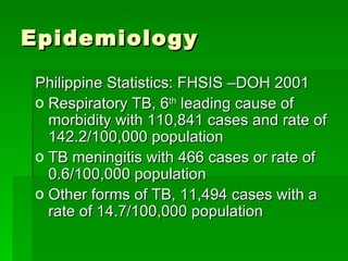
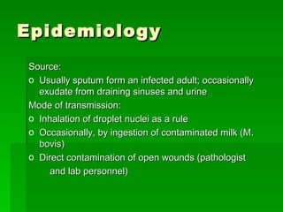
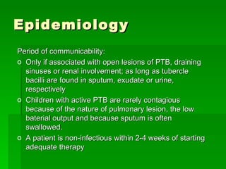
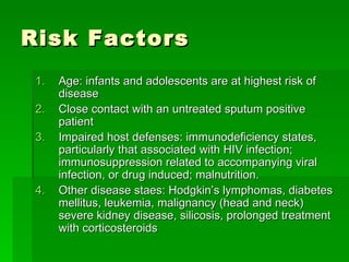
Pulmonary Tuberculosis http://crisbertcualteros.page.tl
Pulmonary Tuberculosis Etiology: 1.1  Mycobacterium tuberculosis 1.2  Mycobacterium bovis , rarely
Epidemiology Philippine Statistics: FHSIS –DOH 2001 Respiratory TB, 6 th  leading cause of morbidity with 110,841 cases and rate of 142.2/100,000 population  TB meningitis with 466 cases or rate of 0.6/100,000 population Other forms of TB, 11,494 cases with a rate of 14.7/100,000 population
Epidemiology Source: Usually sputum form an infected adult; occasionally exudate from draining sinuses and urine Mode of transmission: Inhalation of droplet nuclei as a rule Occasionally, by ingestion of contaminated milk (M. bovis) Direct contamination of open wounds (pathologist and lab personnel)
Epidemiology Period of communicability: Only if associated with open lesions of PTB, draining sinuses or renal involvement; as long as tubercle bacilli are found in sputum, exudate or urine, respectively Children with active PTB are rarely contagious because of the nature of pulmonary lesion, the low baterial output and because sputum is often swallowed. A patient is non-infectious within 2-4 weeks of starting adequate therapy
Risk Factors Age: infants and adolescents are at highest risk of disease Close contact with an untreated sputum positive patient Impaired host defenses: immunodeficiency states, particularly that associated with HIV infection; immunosuppression related to accompanying viral infection, or drug induced; malnutrition. Other disease staes: Hodgkin’s lymphomas, diabetes mellitus, leukemia, malignancy (head and neck) severe kidney disease, silicosis, prolonged treatment with corticosteroids
Risk Factors 5. Persons whose tuberculin skin test results converted to (+) in the past 1-2 years 6. Persons who have CXR suggestive of old TB
Portal of Entry Usually respiratory tract (inhalation of aerosolized particles containing 1-3 tubercle bacilli); rarely, skin, gastrointestinaltract, mucous membrane, transplacentally from mother to fetus or via infected amniotic fluid
Incubation Period From 3 to 8 weeks
CLASSIFICATION Class I (TB exposure) (+) exposure (-) Mantoux tuberculin test (-) signs and symptoms suggestive of TB (-) chest radiograph
CLASSIFICATION Class II (TB infection) (±) exposure (+) Mantoux tuberculin test (-) signs and symptoms suggestive of TB (-) chest radiograph
CLASSIFICATION Class III (TB disease) Has three or more of the ff. criteria (+) history of exposure to an adult/adolescent with active TB disease (+) Mantoux tuberculin test (+) signs and symptoms suggestive of TB Cough/wheezing > 2 weeks; fever > 2 weeks Painless cervical and/or other lymphadenopathy Poor weight gain; failure to make a quick return to normal after an infection (measles, tonsillitis, whooping cough) or failure to respond to approriate antibiotic therapy (pneumonia, otitis media) Abnormal Chest radiograph Laboratory findings suggestive of TB (histological, cytological, biochemical, immunological or molecular)
CLASSIFICATION Class IV (TB inactive) A child/adolescent with or without history of previous TB and any of the ff: (±) previous chemotherapy (+) radiographic evidence of healed/calcified TB (+) Mantoux tuberculin test (-) signs and symptoms suggestive of TB (-) smear/culture for M. tuberculosis
Clinical Forms of Tuberculosis Pulmonary/endothoracic Asymptomatic or Latent TB infection Primary TB/primary complex Primary focus, lymphangitis and regional lymphadenitis Most common clinical symptoms Non-productive cough Mild dyspnea Cervical lymphadenopathies
Clinical Forms of Tuberculosis Pleurisy with effusion Accompanies primary focus Considered a component of the primary complex Onset is usually abrupt Fever, chest pain, shortness of breath Dullness to flatness and diminished breath sounds Obliteration of costophrenic sulcus on CXR (minimal) Layering of fluid density (moderate effusion) Occupy one hemithorax (massive effusion)
Clinical Forms of Tuberculosis Progressive primary tuberculosis  More severe fever, malaise, cough, weight loss Classical signs of cavitation Crepitant rales, diminished breath sounds, lymphadenopathy Endobronchial TB Bronchial obstruction due to enlargement of peribronchial lymph nodes Sudden death by asphyxia Emphysema Atelectasis
Clinical Forms of Tuberculosis Miliary TB  Generalized hematogenous tuberculosis due to massive invasion of the blood stream by the tubercle bacilli Arises from a discharge of a caseous focus often from a lymph node into the blood vessel (pulmonary vein) High fever, cough, dyspnea Crepitant rales, splenomagly, hepatomegaly, signs of menigeal irritation
Clinical Forms of Tuberculosis Chronic TB Reinfection or adult TB Apical or infraclavicular infiltrates often with cavitation and no hilar lymphadenopathy Persistent cough, prlonged fever, chest pain, hemoptysis and supraclavicular adenitis Tuberculoma Pericardial TB
Clinical Forms of Tuberculosis Extrapulmonary TB TB of the cervical lymph nodes/Scrofula Involved LN are painless, firm, discrete, movable becoming adherent to each other and anchored to the surrounding tissues and skin as they enlarge Scofuloderma (when left untreated and ruptures resulting in a draining sinus tract TB of the CNS TB meningitis TB abscess
Clinical Forms of Tuberculosis Skeletal TB TB of the bones and joints TB of the spine or Pott’s GI TB TB enteritis TB peritonitis Hepatobiliary TB TB of the pancreas Cutaneous TB Ocular TB GUT TB TB of the Middle Ear
Diagnostic Tests Mantoux Testing/Tuberculin skin test Most widely used method to determine latent TB infection  Standard method for screening positive if ≥ 8 mm induration size A dose of 0.1 ml of 2-TU PPD-RT23 or 0.1 ml of 5-TU PPD-S Provides a general measure of a person’s cellular response
Diagnostic Tests Mantoux Testing/Tuberculin skin test Features of reaction  Delayed course reaching a peak of more than 24h after injection of antigen Indurated character Occasional vesiculation and necrosis A pale wheal of 6 to 10mm in diameter should be evident after injection Read within 48-72hrs from the time of administration
Diagnostic Tests Mantoux Testing/Tuberculin skin test False positive Nontuberculous mycobacteria BCG vaccination  Reaction develops 6-12 weeks after vaccination Wanes after 5 years from immunization False negative Anergy Very young age (< 6 months) Recent TB infection or overwhelming TB disease Live-virus vaccination  postpone for at least 4 – 6 weeks after immunization or do it on the same day of vaccination
Diagnostic Tests AFB smears (microscopic examination) Provides presumptive diagnosis of active TB Gives a quantitative estimation of the number of bacilli on the smear  Implies infectiousness of the patient Low sensitivity (51.8 – 53.1%) High specificity (97.5 – 99.8%) 10 4  bacilli per ml of sputum : lowest concentration that can be detected
Diagnostic Tests Culture : gold std.  Solid media: 4-6 weeks for isolation and  another 2-4 weeks for susceptibility testing Middlebrook  7H-11 7H-10  (agar-based) Lowenstein-Jensen (egg-based) Liquid media Bactec : as few as 7 to 10 days; carbon-14 (marker of bacterial growth) Middlebrook broth Septi-check AFB BBL mycobacteria growth incubator tube
Diagnostic Tests Specimens collected for demonstration of tubercle bacilli Sputum  for older children able to expectorate  Series of three early morning specimens on different days before starting chemotherapy Make sure brought up from the lungs
Diagnostic Tests Specimens collected for demonstration of tubercle bacilli Gastric aspirate For infants and children who cannot expectorate even with aerosol inhalation 5-10 ml of gastric contents aspirated early in the morning after the person has fasted for at least 8 – 10 hours preferably before the child arises and peristalsis empties the stomach of respiratory secretions swallowed overnight
Diagnostic Tests Specimens collected for demonstration of tubercle bacilli Bronchial washings Urine First morning-voided midstream specimen Other body fluids and tissues Bone marrow, lung and liver biopsy in patients with hematogenous spread/disseminated disease must be considered
Diagnostic Tests Radiographic Findings No pathognomonic findings in childhood TB Lateral projections are important wherein partially calcified mediastinal nodes may be visible Most common cause of calcification in children  Uniform stippling of both lungs found in miliary tuberculosis Lobar or lobular consolidations Common findings: Enlarged retrocardiac lymphadenopathy (70%), hilar adenopathy with pulmonary infiltrates (20%), and pleural effusion
Initial Empiric Therapy of Tuberculosis in Infants, Children and Adolescents Immediately prophylaxis controversial for those 5 years, but is recommended by some experts specially if with risk factors e.g. malnutrition, immunocom-promised states 3 months INH Class I TB Exposure <5 years 5 years Remarks Regimen Category
Initial Empiric Therapy of Tuberculosis in Infants, Children and Adolescents Corticosteroids (usually prednisone at 1 mkday for 6-8 weeks with gradual tapering) beneficial for the following: meningitis, pericarditis, pleuritis, endobronchial TB, miliary TB 2 months HRZ + E or S ffd by 10 months HR ± E/S given once daily or as DOT 3x weekly  Same regimen as pulmonary disease Extrapulmonary Severe, life-threatening disease: disseminated/ miliary, meningitis, bone/joint disease Other extrapulmonary sites
Initial Empiric Therapy of Tuberculosis in Infants, Children and Adolescents In the presence of primary INH resistance, use Rifampicin 9 months INH 9 months INH 9 months INH Class II TB infection PPD conversion  within past 1-2 years, (-) CXR PPD (+) not due to BCG,(-)CXR, (-) previous treatment PPD (+) with stable/ healed lesion,  (-) previous treatment
Initial Empiric Therapy of Tuberculosis in Infants, Children and Adolescents 1-2 mos For the duration of immunosup-pression 12  Months INH Class II TB infection PPD (+) with stable / healed lesion, (+) previous treatment, at risk of reactivation due to: Measles, pertussis, etc Conditions/drugs inducing immunosuppression (IDDM leukemia chronicdialysis) HIV infection/ persons at risk for infection but HIV status unknown
Initial Empiric Therapy of Tuberculosis in Infants, Children and Adolescents Streptomycin preferred in children < 6 years of age, where visual acuity/color perception cannot be monitored reliably In immunocompromised patients, continuation phase extended to 7 months (total duration of therapy:9 months) or for at least 6 months after sputum conversion (if applicable) whichever is longer. If susceptibility results anavailable, continue E/S for the entire duration of therapy 2 months HRZ once daily, ffd by 4 months HR given once daily or as DOT 3x weekly 2 months HRZ plus E or S once daily, ffd by 4 months HR ± E/S given once daily or as DOT 3x weekly Class IIIB TB Disease Pulmonary Fully susceptible: based on culture results of index case, (-) previous treatment, <10% local prevalence of primary INH resistance (b) Susceptibility unknown or initial drug resistance suspected because of big bacillary population, previous treatment (1 month), close contact with resistant source case, residence in area with >10% primary INH resistance
Algorithm for Preventive Therapy of Childhood Tuberculosis TB Exposure Class I yes <5years old   Start INH for 3 months   No Repeat Mantoux test  Yes  Radiologic findings  Yes  TB Disease After 3 months(+)  and /or, signs/symptoms  (Class III)   No   No   suggestive of TB   Multiple drug tx If no    Discontinue INH  No BCG scar,   If no BCG scar,  TB Infection Give BCG   give BCG   (Class II) Continue    6 INH

Pulmonary Tuberculosis

  • 1.
  • 2.
    Pulmonary Tuberculosis Etiology:1.1 Mycobacterium tuberculosis 1.2 Mycobacterium bovis , rarely
  • 3.
    Epidemiology Philippine Statistics:FHSIS –DOH 2001 Respiratory TB, 6 th leading cause of morbidity with 110,841 cases and rate of 142.2/100,000 population TB meningitis with 466 cases or rate of 0.6/100,000 population Other forms of TB, 11,494 cases with a rate of 14.7/100,000 population
  • 4.
    Epidemiology Source: Usuallysputum form an infected adult; occasionally exudate from draining sinuses and urine Mode of transmission: Inhalation of droplet nuclei as a rule Occasionally, by ingestion of contaminated milk (M. bovis) Direct contamination of open wounds (pathologist and lab personnel)
  • 5.
    Epidemiology Period ofcommunicability: Only if associated with open lesions of PTB, draining sinuses or renal involvement; as long as tubercle bacilli are found in sputum, exudate or urine, respectively Children with active PTB are rarely contagious because of the nature of pulmonary lesion, the low baterial output and because sputum is often swallowed. A patient is non-infectious within 2-4 weeks of starting adequate therapy
  • 6.
    Risk Factors Age:infants and adolescents are at highest risk of disease Close contact with an untreated sputum positive patient Impaired host defenses: immunodeficiency states, particularly that associated with HIV infection; immunosuppression related to accompanying viral infection, or drug induced; malnutrition. Other disease staes: Hodgkin’s lymphomas, diabetes mellitus, leukemia, malignancy (head and neck) severe kidney disease, silicosis, prolonged treatment with corticosteroids
  • 7.
    Risk Factors 5.Persons whose tuberculin skin test results converted to (+) in the past 1-2 years 6. Persons who have CXR suggestive of old TB
  • 8.
    Portal of EntryUsually respiratory tract (inhalation of aerosolized particles containing 1-3 tubercle bacilli); rarely, skin, gastrointestinaltract, mucous membrane, transplacentally from mother to fetus or via infected amniotic fluid
  • 9.
  • 10.
    CLASSIFICATION Class I(TB exposure) (+) exposure (-) Mantoux tuberculin test (-) signs and symptoms suggestive of TB (-) chest radiograph
  • 11.
    CLASSIFICATION Class II(TB infection) (±) exposure (+) Mantoux tuberculin test (-) signs and symptoms suggestive of TB (-) chest radiograph
  • 12.
    CLASSIFICATION Class III(TB disease) Has three or more of the ff. criteria (+) history of exposure to an adult/adolescent with active TB disease (+) Mantoux tuberculin test (+) signs and symptoms suggestive of TB Cough/wheezing > 2 weeks; fever > 2 weeks Painless cervical and/or other lymphadenopathy Poor weight gain; failure to make a quick return to normal after an infection (measles, tonsillitis, whooping cough) or failure to respond to approriate antibiotic therapy (pneumonia, otitis media) Abnormal Chest radiograph Laboratory findings suggestive of TB (histological, cytological, biochemical, immunological or molecular)
  • 13.
    CLASSIFICATION Class IV(TB inactive) A child/adolescent with or without history of previous TB and any of the ff: (±) previous chemotherapy (+) radiographic evidence of healed/calcified TB (+) Mantoux tuberculin test (-) signs and symptoms suggestive of TB (-) smear/culture for M. tuberculosis
  • 14.
    Clinical Forms ofTuberculosis Pulmonary/endothoracic Asymptomatic or Latent TB infection Primary TB/primary complex Primary focus, lymphangitis and regional lymphadenitis Most common clinical symptoms Non-productive cough Mild dyspnea Cervical lymphadenopathies
  • 15.
    Clinical Forms ofTuberculosis Pleurisy with effusion Accompanies primary focus Considered a component of the primary complex Onset is usually abrupt Fever, chest pain, shortness of breath Dullness to flatness and diminished breath sounds Obliteration of costophrenic sulcus on CXR (minimal) Layering of fluid density (moderate effusion) Occupy one hemithorax (massive effusion)
  • 16.
    Clinical Forms ofTuberculosis Progressive primary tuberculosis More severe fever, malaise, cough, weight loss Classical signs of cavitation Crepitant rales, diminished breath sounds, lymphadenopathy Endobronchial TB Bronchial obstruction due to enlargement of peribronchial lymph nodes Sudden death by asphyxia Emphysema Atelectasis
  • 17.
    Clinical Forms ofTuberculosis Miliary TB Generalized hematogenous tuberculosis due to massive invasion of the blood stream by the tubercle bacilli Arises from a discharge of a caseous focus often from a lymph node into the blood vessel (pulmonary vein) High fever, cough, dyspnea Crepitant rales, splenomagly, hepatomegaly, signs of menigeal irritation
  • 18.
    Clinical Forms ofTuberculosis Chronic TB Reinfection or adult TB Apical or infraclavicular infiltrates often with cavitation and no hilar lymphadenopathy Persistent cough, prlonged fever, chest pain, hemoptysis and supraclavicular adenitis Tuberculoma Pericardial TB
  • 19.
    Clinical Forms ofTuberculosis Extrapulmonary TB TB of the cervical lymph nodes/Scrofula Involved LN are painless, firm, discrete, movable becoming adherent to each other and anchored to the surrounding tissues and skin as they enlarge Scofuloderma (when left untreated and ruptures resulting in a draining sinus tract TB of the CNS TB meningitis TB abscess
  • 20.
    Clinical Forms ofTuberculosis Skeletal TB TB of the bones and joints TB of the spine or Pott’s GI TB TB enteritis TB peritonitis Hepatobiliary TB TB of the pancreas Cutaneous TB Ocular TB GUT TB TB of the Middle Ear
  • 21.
    Diagnostic Tests MantouxTesting/Tuberculin skin test Most widely used method to determine latent TB infection Standard method for screening positive if ≥ 8 mm induration size A dose of 0.1 ml of 2-TU PPD-RT23 or 0.1 ml of 5-TU PPD-S Provides a general measure of a person’s cellular response
  • 22.
    Diagnostic Tests MantouxTesting/Tuberculin skin test Features of reaction Delayed course reaching a peak of more than 24h after injection of antigen Indurated character Occasional vesiculation and necrosis A pale wheal of 6 to 10mm in diameter should be evident after injection Read within 48-72hrs from the time of administration
  • 23.
    Diagnostic Tests MantouxTesting/Tuberculin skin test False positive Nontuberculous mycobacteria BCG vaccination Reaction develops 6-12 weeks after vaccination Wanes after 5 years from immunization False negative Anergy Very young age (< 6 months) Recent TB infection or overwhelming TB disease Live-virus vaccination postpone for at least 4 – 6 weeks after immunization or do it on the same day of vaccination
  • 24.
    Diagnostic Tests AFBsmears (microscopic examination) Provides presumptive diagnosis of active TB Gives a quantitative estimation of the number of bacilli on the smear Implies infectiousness of the patient Low sensitivity (51.8 – 53.1%) High specificity (97.5 – 99.8%) 10 4 bacilli per ml of sputum : lowest concentration that can be detected
  • 25.
    Diagnostic Tests Culture: gold std. Solid media: 4-6 weeks for isolation and another 2-4 weeks for susceptibility testing Middlebrook 7H-11 7H-10 (agar-based) Lowenstein-Jensen (egg-based) Liquid media Bactec : as few as 7 to 10 days; carbon-14 (marker of bacterial growth) Middlebrook broth Septi-check AFB BBL mycobacteria growth incubator tube
  • 26.
    Diagnostic Tests Specimenscollected for demonstration of tubercle bacilli Sputum for older children able to expectorate Series of three early morning specimens on different days before starting chemotherapy Make sure brought up from the lungs
  • 27.
    Diagnostic Tests Specimenscollected for demonstration of tubercle bacilli Gastric aspirate For infants and children who cannot expectorate even with aerosol inhalation 5-10 ml of gastric contents aspirated early in the morning after the person has fasted for at least 8 – 10 hours preferably before the child arises and peristalsis empties the stomach of respiratory secretions swallowed overnight
  • 28.
    Diagnostic Tests Specimenscollected for demonstration of tubercle bacilli Bronchial washings Urine First morning-voided midstream specimen Other body fluids and tissues Bone marrow, lung and liver biopsy in patients with hematogenous spread/disseminated disease must be considered
  • 29.
    Diagnostic Tests RadiographicFindings No pathognomonic findings in childhood TB Lateral projections are important wherein partially calcified mediastinal nodes may be visible Most common cause of calcification in children Uniform stippling of both lungs found in miliary tuberculosis Lobar or lobular consolidations Common findings: Enlarged retrocardiac lymphadenopathy (70%), hilar adenopathy with pulmonary infiltrates (20%), and pleural effusion
  • 30.
    Initial Empiric Therapyof Tuberculosis in Infants, Children and Adolescents Immediately prophylaxis controversial for those 5 years, but is recommended by some experts specially if with risk factors e.g. malnutrition, immunocom-promised states 3 months INH Class I TB Exposure <5 years 5 years Remarks Regimen Category
  • 31.
    Initial Empiric Therapyof Tuberculosis in Infants, Children and Adolescents Corticosteroids (usually prednisone at 1 mkday for 6-8 weeks with gradual tapering) beneficial for the following: meningitis, pericarditis, pleuritis, endobronchial TB, miliary TB 2 months HRZ + E or S ffd by 10 months HR ± E/S given once daily or as DOT 3x weekly Same regimen as pulmonary disease Extrapulmonary Severe, life-threatening disease: disseminated/ miliary, meningitis, bone/joint disease Other extrapulmonary sites
  • 32.
    Initial Empiric Therapyof Tuberculosis in Infants, Children and Adolescents In the presence of primary INH resistance, use Rifampicin 9 months INH 9 months INH 9 months INH Class II TB infection PPD conversion within past 1-2 years, (-) CXR PPD (+) not due to BCG,(-)CXR, (-) previous treatment PPD (+) with stable/ healed lesion, (-) previous treatment
  • 33.
    Initial Empiric Therapyof Tuberculosis in Infants, Children and Adolescents 1-2 mos For the duration of immunosup-pression 12 Months INH Class II TB infection PPD (+) with stable / healed lesion, (+) previous treatment, at risk of reactivation due to: Measles, pertussis, etc Conditions/drugs inducing immunosuppression (IDDM leukemia chronicdialysis) HIV infection/ persons at risk for infection but HIV status unknown
  • 34.
    Initial Empiric Therapyof Tuberculosis in Infants, Children and Adolescents Streptomycin preferred in children < 6 years of age, where visual acuity/color perception cannot be monitored reliably In immunocompromised patients, continuation phase extended to 7 months (total duration of therapy:9 months) or for at least 6 months after sputum conversion (if applicable) whichever is longer. If susceptibility results anavailable, continue E/S for the entire duration of therapy 2 months HRZ once daily, ffd by 4 months HR given once daily or as DOT 3x weekly 2 months HRZ plus E or S once daily, ffd by 4 months HR ± E/S given once daily or as DOT 3x weekly Class IIIB TB Disease Pulmonary Fully susceptible: based on culture results of index case, (-) previous treatment, <10% local prevalence of primary INH resistance (b) Susceptibility unknown or initial drug resistance suspected because of big bacillary population, previous treatment (1 month), close contact with resistant source case, residence in area with >10% primary INH resistance
  • 35.
    Algorithm for PreventiveTherapy of Childhood Tuberculosis TB Exposure Class I yes <5years old Start INH for 3 months No Repeat Mantoux test Yes Radiologic findings Yes TB Disease After 3 months(+) and /or, signs/symptoms (Class III) No No suggestive of TB Multiple drug tx If no Discontinue INH No BCG scar, If no BCG scar, TB Infection Give BCG give BCG (Class II) Continue  6 INH