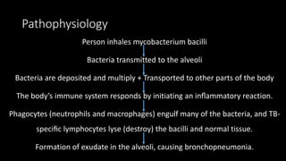
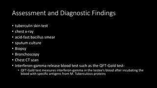
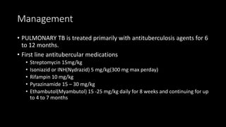
Pulmonary tuberculosis is caused by the bacterium Mycobacterium tuberculosis. It primarily affects the lung parenchyma but can spread to other organs. Risk factors include close contact with an active case, immunocompromised status, substance abuse, poor living conditions, and coming from a country with high TB rates. The bacteria are transmitted via airborne droplets when an infected person coughs or sneezes. Symptoms include cough, fever, night sweats and weight loss. Diagnosis involves tuberculin skin testing, chest x-ray, and sputum culture and analysis. Treatment consists of a multi-drug regimen for 6-12 months to prevent resistance and includes isoniazid, rifampin,
Pulmonary tuberculosis
The bacterium Mycobacterium tuberculosis causes tuberculosis (TB), a contagious, airborne infection that destroys body tissue. Pulmonary TB occurs when M. tuberculosis primarily attacks the lungs. However, it can spread from there to other organs.
New treatment regimen is mentioned here.
Pneumonia is an inflammatory condition of the lung affecting primarily the small air sacs known as alveoli. Typically symptoms include some combination of productive or dry cough, chest pain, fever, and trouble breathing. Severity is variable.
Pneumonia is usually caused by infection with viruses or bacteria and less commonly by other microorganisms, certain medications and conditions such as autoimmune diseases. Risk factors include cystic fibrosis, chronic obstructive pulmonary disease (COPD), asthma, diabetes, heart failure, a history of smoking, a poor ability to cough such as following a stroke, and a weak immune system. Diagnosis is often based on the symptoms and physical examination. Chest X-ray, blood tests, and culture of the sputum may help confirm the diagnosis. The disease may be classified by where it was acquired with community, hospital, or health care associated pneumonia.
Vaccines to prevent certain types of pneumonia are available. Other methods of prevention include handwashing and not smoking. Treatment depends on the underlying cause. Pneumonia believed to be due to bacteria is treated with antibiotics. If the pneumonia is severe, the affected person is generally hospitalized. Oxygen therapy may be used if oxygen levels are low.
Pneumonia affects approximately 450 million people globally (7% of the population) and results in about four million deaths per year. Pneumonia was regarded by William Osler in the 19th century as "the captain of the men of death". With the introduction of antibiotics and vaccines in the 20th century, survival improved. Nevertheless, in developing countries, and among the very old, the very young, and the chronically ill, pneumonia remains a leading cause of death. Pneumonia often shortens suffering among those already close to death and has thus been called "the old man's friend"
Pneumonia is an inflammation of the lung parenchyma caused by various microorganisms, including bacteria, mycobacteria, fungi, and viruses.
Pneumonitis is a more general term that describes the inflammatory process in the lung tissue that may predispose and Pneumonia is an inflammation of the lung parenchyma that is caused by a microbial agent.
place the patient at risk for microbial invasion.
Pneumonia is classified into four: community-acquired pneumonia (CAP) and hospital-acquired pneumonia (HAP), pneumonia in the immunocompromised host, and aspiration pneumonia.
These lecture notes were prepared by Dr. Hamdi Turkey- Pulmonologist- Department of internal medicine - Taiz university
Do Not Forget To Visit Our Pages On Facebook on the following Links:
https://www.facebook.com/groups/569435236444761/
AND
https://www.facebook.com/groups/690331650977113/
My Powerpoint on Tuberculosis, includes:
What is the incidence and prevalence?
What are the symptoms?
How is it diagnosed?
How is it treated?
What are the treatment guidelines?
Brief idea- tuberculosis, causative agent, epidemiology of disease in world and India, burden in HIV patients, Burden on Indian Economy, disease symptoms, control programmes implemented by government
This is about tuberculosis , features, diagnosis and management. With reference to Uganda Clinical Guidelines
By Okeke Gloria, Kasule Steven, Sengooba Dennis Nyanzi
Explore natural remedies for syphilis treatment in Singapore. Discover alternative therapies, herbal remedies, and lifestyle changes that may complement conventional treatments. Learn about holistic approaches to managing syphilis symptoms and supporting overall health.
HOT NEW PRODUCT! BIG SALES FAST SHIPPING NOW FROM CHINA!! EU KU DB BK substit...GL Anaacs
Contact us if you are interested:
Email / Skype : kefaya1771@gmail.com
Threema: PXHY5PDH
New BATCH Ku !!! MUCH IN DEMAND FAST SALE EVERY BATCH HAPPY GOOD EFFECT BIG BATCH !
Contact me on Threema or skype to start big business!!
Hot-sale products:
NEW HOT EUTYLONE WHITE CRYSTAL!!
5cl-adba precursor (semi finished )
5cl-adba raw materials
ADBB precursor (semi finished )
ADBB raw materials
APVP powder
5fadb/4f-adb
Jwh018 / Jwh210
Eutylone crystal
Protonitazene (hydrochloride) CAS: 119276-01-6
Flubrotizolam CAS: 57801-95-3
Metonitazene CAS: 14680-51-4
Payment terms: Western Union,MoneyGram,Bitcoin or USDT.
Deliver Time: Usually 7-15days
Shipping method: FedEx, TNT, DHL,UPS etc.Our deliveries are 100% safe, fast, reliable and discreet.
Samples will be sent for your evaluation!If you are interested in, please contact me, let's talk details.
We specializes in exporting high quality Research chemical, medical intermediate, Pharmaceutical chemicals and so on. Products are exported to USA, Canada, France, Korea, Japan,Russia, Southeast Asia and other countries.
Title: Sense of Taste
Presenter: Dr. Faiza, Assistant Professor of Physiology
Qualifications:
MBBS (Best Graduate, AIMC Lahore)
FCPS Physiology
ICMT, CHPE, DHPE (STMU)
MPH (GC University, Faisalabad)
MBA (Virtual University of Pakistan)
Learning Objectives:
Describe the structure and function of taste buds.
Describe the relationship between the taste threshold and taste index of common substances.
Explain the chemical basis and signal transduction of taste perception for each type of primary taste sensation.
Recognize different abnormalities of taste perception and their causes.
Key Topics:
Significance of Taste Sensation:
Differentiation between pleasant and harmful food
Influence on behavior
Selection of food based on metabolic needs
Receptors of Taste:
Taste buds on the tongue
Influence of sense of smell, texture of food, and pain stimulation (e.g., by pepper)
Primary and Secondary Taste Sensations:
Primary taste sensations: Sweet, Sour, Salty, Bitter, Umami
Chemical basis and signal transduction mechanisms for each taste
Taste Threshold and Index:
Taste threshold values for Sweet (sucrose), Salty (NaCl), Sour (HCl), and Bitter (Quinine)
Taste index relationship: Inversely proportional to taste threshold
Taste Blindness:
Inability to taste certain substances, particularly thiourea compounds
Example: Phenylthiocarbamide
Structure and Function of Taste Buds:
Composition: Epithelial cells, Sustentacular/Supporting cells, Taste cells, Basal cells
Features: Taste pores, Taste hairs/microvilli, and Taste nerve fibers
Location of Taste Buds:
Found in papillae of the tongue (Fungiform, Circumvallate, Foliate)
Also present on the palate, tonsillar pillars, epiglottis, and proximal esophagus
Mechanism of Taste Stimulation:
Interaction of taste substances with receptors on microvilli
Signal transduction pathways for Umami, Sweet, Bitter, Sour, and Salty tastes
Taste Sensitivity and Adaptation:
Decrease in sensitivity with age
Rapid adaptation of taste sensation
Role of Saliva in Taste:
Dissolution of tastants to reach receptors
Washing away the stimulus
Taste Preferences and Aversions:
Mechanisms behind taste preference and aversion
Influence of receptors and neural pathways
Impact of Sensory Nerve Damage:
Degeneration of taste buds if the sensory nerve fiber is cut
Abnormalities of Taste Detection:
Conditions: Ageusia, Hypogeusia, Dysgeusia (parageusia)
Causes: Nerve damage, neurological disorders, infections, poor oral hygiene, adverse drug effects, deficiencies, aging, tobacco use, altered neurotransmitter levels
Neurotransmitters and Taste Threshold:
Effects of serotonin (5-HT) and norepinephrine (NE) on taste sensitivity
Supertasters:
25% of the population with heightened sensitivity to taste, especially bitterness
Increased number of fungiform papillae
Lung Cancer: Artificial Intelligence, Synergetics, Complex System Analysis, S...Oleg Kshivets
RESULTS: Overall life span (LS) was 2252.1±1742.5 days and cumulative 5-year survival (5YS) reached 73.2%, 10 years – 64.8%, 20 years – 42.5%. 513 LCP lived more than 5 years (LS=3124.6±1525.6 days), 148 LCP – more than 10 years (LS=5054.4±1504.1 days).199 LCP died because of LC (LS=562.7±374.5 days). 5YS of LCP after bi/lobectomies was significantly superior in comparison with LCP after pneumonectomies (78.1% vs.63.7%, P=0.00001 by log-rank test). AT significantly improved 5YS (66.3% vs. 34.8%) (P=0.00000 by log-rank test) only for LCP with N1-2. Cox modeling displayed that 5YS of LCP significantly depended on: phase transition (PT) early-invasive LC in terms of synergetics, PT N0—N12, cell ratio factors (ratio between cancer cells- CC and blood cells subpopulations), G1-3, histology, glucose, AT, blood cell circuit, prothrombin index, heparin tolerance, recalcification time (P=0.000-0.038). Neural networks, genetic algorithm selection and bootstrap simulation revealed relationships between 5YS and PT early-invasive LC (rank=1), PT N0—N12 (rank=2), thrombocytes/CC (3), erythrocytes/CC (4), eosinophils/CC (5), healthy cells/CC (6), lymphocytes/CC (7), segmented neutrophils/CC (8), stick neutrophils/CC (9), monocytes/CC (10); leucocytes/CC (11). Correct prediction of 5YS was 100% by neural networks computing (area under ROC curve=1.0; error=0.0).
CONCLUSIONS: 5YS of LCP after radical procedures significantly depended on: 1) PT early-invasive cancer; 2) PT N0--N12; 3) cell ratio factors; 4) blood cell circuit; 5) biochemical factors; 6) hemostasis system; 7) AT; 8) LC characteristics; 9) LC cell dynamics; 10) surgery type: lobectomy/pneumonectomy; 11) anthropometric data. Optimal diagnosis and treatment strategies for LC are: 1) screening and early detection of LC; 2) availability of experienced thoracic surgeons because of complexity of radical procedures; 3) aggressive en block surgery and adequate lymph node dissection for completeness; 4) precise prediction; 5) adjuvant chemoimmunoradiotherapy for LCP with unfavorable prognosis.
Title: Sense of Smell
Presenter: Dr. Faiza, Assistant Professor of Physiology
Qualifications:
MBBS (Best Graduate, AIMC Lahore)
FCPS Physiology
ICMT, CHPE, DHPE (STMU)
MPH (GC University, Faisalabad)
MBA (Virtual University of Pakistan)
Learning Objectives:
Describe the primary categories of smells and the concept of odor blindness.
Explain the structure and location of the olfactory membrane and mucosa, including the types and roles of cells involved in olfaction.
Describe the pathway and mechanisms of olfactory signal transmission from the olfactory receptors to the brain.
Illustrate the biochemical cascade triggered by odorant binding to olfactory receptors, including the role of G-proteins and second messengers in generating an action potential.
Identify different types of olfactory disorders such as anosmia, hyposmia, hyperosmia, and dysosmia, including their potential causes.
Key Topics:
Olfactory Genes:
3% of the human genome accounts for olfactory genes.
400 genes for odorant receptors.
Olfactory Membrane:
Located in the superior part of the nasal cavity.
Medially: Folds downward along the superior septum.
Laterally: Folds over the superior turbinate and upper surface of the middle turbinate.
Total surface area: 5-10 square centimeters.
Olfactory Mucosa:
Olfactory Cells: Bipolar nerve cells derived from the CNS (100 million), with 4-25 olfactory cilia per cell.
Sustentacular Cells: Produce mucus and maintain ionic and molecular environment.
Basal Cells: Replace worn-out olfactory cells with an average lifespan of 1-2 months.
Bowman’s Gland: Secretes mucus.
Stimulation of Olfactory Cells:
Odorant dissolves in mucus and attaches to receptors on olfactory cilia.
Involves a cascade effect through G-proteins and second messengers, leading to depolarization and action potential generation in the olfactory nerve.
Quality of a Good Odorant:
Small (3-20 Carbon atoms), volatile, water-soluble, and lipid-soluble.
Facilitated by odorant-binding proteins in mucus.
Membrane Potential and Action Potential:
Resting membrane potential: -55mV.
Action potential frequency in the olfactory nerve increases with odorant strength.
Adaptation Towards the Sense of Smell:
Rapid adaptation within the first second, with further slow adaptation.
Psychological adaptation greater than receptor adaptation, involving feedback inhibition from the central nervous system.
Primary Sensations of Smell:
Camphoraceous, Musky, Floral, Pepperminty, Ethereal, Pungent, Putrid.
Odor Detection Threshold:
Examples: Hydrogen sulfide (0.0005 ppm), Methyl-mercaptan (0.002 ppm).
Some toxic substances are odorless at lethal concentrations.
Characteristics of Smell:
Odor blindness for single substances due to lack of appropriate receptor protein.
Behavioral and emotional influences of smell.
Transmission of Olfactory Signals:
From olfactory cells to glomeruli in the olfactory bulb, involving lateral inhibition.
Primitive, less old, and new olfactory systems with different path
MANAGEMENT OF ATRIOVENTRICULAR CONDUCTION BLOCK.pdfJim Jacob Roy
Cardiac conduction defects can occur due to various causes.
Atrioventricular conduction blocks ( AV blocks ) are classified into 3 types.
This document describes the acute management of AV block.
- Video recording of this lecture in English language: https://youtu.be/lK81BzxMqdo
- Video recording of this lecture in Arabic language: https://youtu.be/Ve4P0COk9OI
- Link to download the book free: https://nephrotube.blogspot.com/p/nephrotube-nephrology-books.html
- Link to NephroTube website: www.NephroTube.com
- Link to NephroTube social media accounts: https://nephrotube.blogspot.com/p/join-nephrotube-on-social-media.html
These lecture slides, by Dr Sidra Arshad, offer a quick overview of physiological basis of a normal electrocardiogram.
Learning objectives:
1. Define an electrocardiogram (ECG) and electrocardiography
2. Describe how dipoles generated by the heart produce the waveforms of the ECG
3. Describe the components of a normal electrocardiogram of a typical bipolar leads (limb II)
4. Differentiate between intervals and segments
5. Enlist some common indications for obtaining an ECG
Study Resources:
1. Chapter 11, Guyton and Hall Textbook of Medical Physiology, 14th edition
2. Chapter 9, Human Physiology - From Cells to Systems, Lauralee Sherwood, 9th edition
3. Chapter 29, Ganong’s Review of Medical Physiology, 26th edition
4. Electrocardiogram, StatPearls - https://www.ncbi.nlm.nih.gov/books/NBK549803/
5. ECG in Medical Practice by ABM Abdullah, 4th edition
6. ECG Basics, http://www.nataliescasebook.com/tag/e-c-g-basics
2. • Tuberculosis (TB) is an infectious disease that primarily affects the
lung parenchyma.
• It also may be transmitted to other parts of the body, including the
meninges, kidneys, bones, and lymph nodes.
3. Causative agent
• Mycobacterium tuberculosis
• Mycobacterium bovis and Mycobacterium avium have rarely been
associated with the development of a TB infection.
4. Risk factors
• Close contact with someone who has active TB.
• Immunocompromised status (eg, those with HIV infection, cancer,
transplanted organs, and prolonged high-dose corticosteroid therapy)
• Substance abuse (IV or injection drug users and alcoholics)
• Any person without adequate health care
• Preexisting medical conditions or special treatment (eg, diabetes,
chronic renal failure, malnourishment, selected malignancies,
hemodialysis, transplanted organ, gastrectomy, or jejunoileal bypass)
5. • Immigration from countries with a high prevalence of TB
• Institutionalization (eg, long-term care facilities, psychiatric
institutions, prisons)
• Living in overcrowded, substandard housing
• health care workers
6. Transmission
• TB spreads from person to person by airborne transmission
• An infected person releases droplet nuclei (generally particles 1 to
5micrometers in diameter) through talking, coughing, sneezing,
laughing, or singing. Larger droplets settle; smaller droplets remain
suspended in the air and are inhaled by the susceptible person.
7. Pathophysiology
Person inhales mycobacterium bacilli
Bacteria transmitted to the alveoli
Bacteria are deposited and multiply + Transported to other parts of the body
The body’s immune system responds by initiating an inflammatory reaction.
Phagocytes (neutrophils and macrophages) engulf many of the bacteria, and TB-
specific lymphocytes lyse (destroy) the bacilli and normal tissue.
Formation of exudate in the alveoli, causing bronchopneumonia.
8. • The initial infection usually occurs 2 to 10 weeks after exposure.
9. Granulomas form (new tissue masses of live and dead bacilli)
it is surrounded by macrophages, which form a protective wall
Granulomas are then transformed to a fibrous tissue mass, the central portion
of which is called a Ghon tubercle.
The material becomes necrotic, forming a cheesy mass.
This mass may become calcified and form a collagenous scar.
The bacteria become dormant, there is no further progression of active disease.
10. • After initial exposure and infection, the person may develop active
disease because of a compromised or inadequate immune system
response.
• Active disease also may occur with reinfection and activation of
dormant bacteria.
11. The Ghon tubercle ulcerates, releasing the cheesy material into the bronchi.
The bacteria become airborne, resulting in further spread of the disease.
The ulcerated tubercle heals and forms scar tissue.
The infected lung become more inflamed
Further development of bronchopneumonia and tubercle formation.
13. PULMONARY SYMPTOMS
• Dyspnea
• Non resolving bronchopneumonia
• Chest tightness
• Non productive cough
• Mucopurulent sputum with hemoptpysis
• Chest pain
14. Assessment and Diagnostic Findings
• tuberculin skin test
• chest x-ray
• acid-fast bacillus smear
• sputum culture
• Biopsy
• Bronchoscopy
• Chest CT scan
• Interferon-gamma release blood test such as the QFT-Gold test-
• QFT-Gold test measures interferon-gamma in the testee's blood after incubating the
blood with specific antigens from M. Tuberculosis proteins
15. CLASSIFICATION OF TB
• Class 0: no exposure; no infection
• Class 1: exposure; no evidence of infection
• Class 2: latent infection; no disease (eg, positive PPD reaction but no
clinical evidence of active TB)
• Class 3: disease; clinically active
• Class 4: disease; not clinically active
• Class 5: suspected disease; diagnosis pending
16. Management
• PULMONARY TB is treated primarily with antituberculosis agents for 6
to 12 months.
• First line antitubercular medications
• Streptomycin 15mg/kg
• Isoniazid or INH(Nydrazid) 5 mg/kg(300 mg max perday)
• Rifampin 10 mg/kg
• Pyrazinamide 15 – 30 mg/kg
• Ethambutol(Myambutol) 15 -25 mg/kg daily for 8 weeks and continuing for up
to 4 to 7 months
18. • Recommended treatment guidelines - multiple-medication regimen of INH,
rifampin, pyrazinamide, and either streptomycin or ethambutol.
• administered daily for 8 weeks.
• If cultures demonstrate that the organism is sensitive to the medications
before the 8 weeks of therapy have been completed, either ethambutol or
streptomycin can be discontinued.
• After 8 weeks of this medication regimen, pyrazinamide can be
discontinued and INH and rifampin are administered for an additional 4
months.
• A person is considered noninfectious after 2 to 3 weeks of continuous
medication therapy.
• Vitamin B (pyridoxine) is usually administered with INH to prevent INH-
associated peripheral neuropathy
19. Complications
• Bones - Spinal pain and joint destruction may result from TB (TB spine
or potss spine)
• Brain(meningitis)
• Liver or kidneys – military TB
• Heart(cardiac tamponade)
• Pleural effusion
• Tb pneumonia
• Serious reactions to drug therapy(hepato toxicity;hypersentivity)