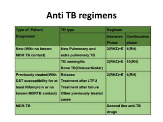
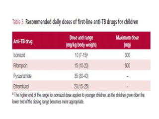
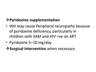
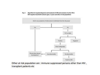
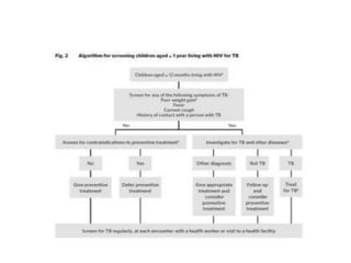
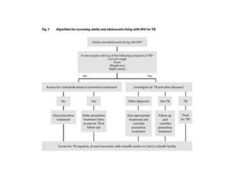
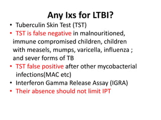
Tuberculosis
Zenawit.A( Assistant professor in
pediatics and child health)
Outline
• Key epidemiological data: Global, Ethiopia
• What do we mean by contact?
• Is cough important for TB transmission?
• Extent of transmission?
• Who is at risk?
• Diagnostic modalities
• TB-Prevention
• Treatment
Tuberculosis
• TB has been there for >4,000 yrs (studies of human
skeleton)
• First recognized as a clinical entity by Schönlein, who
used the term tuberculosis in 1830,tubercle means
lesion of consumption
• March 22, 1882, Dr.Robert Koch discovered
Mycobacterium Tuberculosis (World TB day)
• 1.7 billion of world population infected; 5-10% of
adults and up to 40% of children will develop the
disease.
Key facts
• 1.4 million died from Tb in 2019, including
• 208,000 HIV+ people
• 10 million people fell ill with TB
– 5.6 million men
– 3.2 million women
– 1.2 million children
• TB is one of the 10 top causes of deaths
Countries with high burden of TB
• Eight countries accounted for 2/3 of the global
burden: India (27%), China (9%) Indonesia (8%),
Philippines (6%), Pakistan (6%), Nigeria (4%),
Bangladesh (4%) and South Africa (3%).
• A total of 30 countries accounted for 87% of the
world’s cases.
• Ethiopia is among the first 20 high burden
countries
TB Classification
Classification based on anatomical site
• Pulmonary TB (PTB): TB involving the lung
parenchyma or the tracheobronchial tree.
• Miliary TB is classified as PTB.
• TB intrathoracic lymphadenopathy (mediastinal
and/or hilar) or pleural effusion, without CXR
abnormalities in the lungs, is a case of
extrapulmonary TB.
• A patient with both pulmonary and
extrapulmonaryTB should be classified as a case
of PTB.
• Extrapulmonary TB (EPTB): involving organs
other than the lungs, e.g. pleura, lymph
nodes, abdomen, GUT, skin, joints and bones,
meninges.
Classification based on Hx of previous
TB Tx
• New patients have never been treated for TB or
have taken anti-TB drugs for < 1 month.
• Previously treated patients: received anti TB ≥ 1
month further classified by the outcome as
follows:
– Relapse/re-infection patient diagnosed
with an episode of TB after declared cured
or Tx completed
_Treatment failure are those who have
previously been treated for TB and whose
Tx failed at the end of most recent Tx
_Treatment after loss to follow-up (LTFU)
patients: previously treated for TB and
were lost (previously known as Tx default )
– Other previously treated patients : treated
for TB but whose outcome after their
most recent Tx is unknown or
undocumented
Classification based on drug resistance
• Monoresistance: resistance to one first-line anti-
TB drug only .
• Multidrug resistance (MDR TB) : resistance to at
least both isoniazid and rifampicin.
• Polydrug resistance: resistance to >1 first-line
antiTB drug (other than both INH and rifampicin).
• Extensive drug resistance(XDR TB) : resistance to
any fluoroquinolone and to at least one of three
second-line inject able drugs (capreomycin,
kanamycin and amikacin), in addition to MDR.
… based on resistance
• Rifampicin resistance (RR TB) : resistance to
rifampicin with or without resistance to other
anti-TB drugs.(Mono-RR, MDR, PolyDR, XDR) .
• Patients found to have an RR-TB or MDR-TB :
should be started on second-line drug regimen
Lab Diagnosis
• Acid fast stains AFS : Detected > 100 yrs ago
* Ziehl–Neelsen stain
* Kinyoun stain does not need heating
* Auramine –rhodamine staining in the US
* 10,000 organisms/ml are required for
smear positivity
* Sensitivity of sputum AFS is 60% compared
with culture
Why collection of 3 sputum sample ?
• Detection of a single bacilli is highly suggestive
• Detection of 10 bacilli per slide is optimal
• Sensitivity increases:
* By 10% with the collection of the 2nd
sample
* By 2% with the 3rd sample
What fluids can be collected ?
• Any biologic fluid or material can be examined:
• Pleural fluid,
• Peritoneal fluid
• CSF
• Urine
• Gastric aspirate
• Sputum
• Pus aspirate etc
• thin fluids are best examined after sedimentation
by centrifugation.
Tb-Culture
• Gold standard
• Mycobacterium grow very slowly: rate 1/20
compared with the growth of conventional
bacteria
• Bacteria generation time 12-24 hrs
• Cultured on Löwenstein-Jensen culture media
• growth will take 3-6 wks( up to 12 wks)
Fluid samples vs Biopsies yield for Tb
culture
• Pleural fluid
• <30% are culture
• positive
• Pleural Biopsies
• 40-80% are culture
• positive
• Peritoneal fluid
• <20% are culture
• positive
• Peritoneal Biopsies
• >60% % are culture
• positive
Molecular tests
1. Rapid molecular test: Xpert MTB/RIF assay
* Provide result in 2 hrs
* Better accuracy than microscopy
* Positive when > 10 bacteria/ml
* Costly and needs trained Lab staffs
2. First line Probe assay(LPAs)(2008): tests
forRIF and INH
3. Second line probe assay(2016): tests for
flouroquinolones and injectable anti-Tbs
Transmission
Routes:
– Inhalation of airborne droplet nuclei
– Rarely by direct contact with infected discharge
or contaminated fomite
– Ingestion : M.bovis
– Trans placental route
Droplet nuclei.: particles 1–10 mcm in diameter,
the dried residue formed by evaporation of
droplets coughed or sneezed into the atmosphere
are also infective.
Transmission increases…
• A single bacilli is enough to infect !
• When source case has/is older child / adult
with:
* Smear positive sputum
* Copious thin sputum
* Extensive upper lobe /cavity
* Forceful cough
* Patients who are not yet Txed or Tx for <
2wks
• Most children rarely infect others: bacilli are
sparse and do not have forceful coughing
Is cough a prerequisite ?
• Coughing, sneezing, singing, simply talking
• The site and the mechanism of aerosolization
is important
Transmission rate
• With prolonged exposure to smear +ve patient
• 10-50% of contacts are infected
* 30% develop primary TB
* 70% Latent TB
* 5-10% develop secondary TB
Pulmonary >80%
70% are smear positive
Extra-pulmonary <20%
Who is at risk ?
• M:F= 2:1
• Children < 5years of age
• HIV +
• Malnutrition
• Immune deficiency conditions:
Cancer patients
Immune suppressive therapies
DM
• High risk behaviors:
• Household contact to smear positive/cavitary
TB patient
Pathogenesis
Clinical course of TB Disease
• Time varies between initial infection and
apparent disease:
Pulmonary TB in 70-75% of cases: with in weeks
Disseminated and meningeal TB : within 2-6
mo.
Lymph node or endobronchial TB: within 3-9
mo.
Bones and joints: several years
Renal TB : decades after infection.
Extrapulmonary manifestations are more
common in children, develop in 25-35% of
children with TB, compared to 10% of
immunocompetent adults.
Reactivation TB: rare in children, common
among adolescents and young adults.
C/F: Primary pulmonary disease
• Commonly : Nonproductive cough and mild dyspnea
• fever , night sweats, anorexia and decreased activity
• Failure-to-thrive; not improving with nutrition alone
• Pts with bronchial obstruction have localized wheezing
or decreased breath sounds, accompanied by
tachypnea or, rarely, respiratory distress.
• These Sxs & Sns are occasionally alleviated by
antibiotics, suggesting bacterial super infection.
• 50% of children with CXR finding do not have P/E
finding
Figure: Left Pleural effusion: Obliterated costo-phrenic and
cardio-phrenic
angles, opaque left lung
C/F: Progressive Primary Pulmonary
Disease:
• High fever, severe cough with sputum , weight
loss, and night sweats are common.
• P/E diminished breath sounds, rales, and
dullness or egophony over the cavity.
C/F: Reactivation Tuberculosis:
• Common in children who acquire the initial
infection when they are older than 7 yr of age.
• most common sites are the apical seedings
(Simon foci)
• More Sxic than children with primary
pulmonary Tb.
• Sxs : fever , anorexia, malaise, weight loss, night
sweats, productive cough, hemoptysis, and
chest pain
CXR :
• Extensive infiltrates or thick walled cavities in
the upper Lobes
• Pleural Effusion : from a sub-pleural
pulmonary focus or caseated lymph node.
Summary of Ghon complex
C/F:Miliary disease
•Lympho-haematogenous dissemination to spleen,
liver, lung, lymph nodes, skin, bones,
joints,kidneys,peritoneum, pericardium and
meninges
• Common in immune compromised/malnourished
• Sxs : pt gravely ill with Sx complex of TB,
• Sns : TST non reactive in 40% of pts,
hepatosplenomegaley , emaciation, Sns of
meningitis, peritonitis etc
• Resolution is slow with Tx , steroids for Sxic relief
• Excellent prognosis with early Tx
C/F: Cardiac TB
• Rare, only in 0.5-4% of TB cases in children:
• The most common form of cardiac TB is
pericarditis.
• Either from direct invasion or from subcarinal
lymph nodes.
• Sxs : low-grade fever, malaise, Wt loss ; chest
pain is unusual.
• Sns : pericardial friction rub or distant heart
sounds with pulsus paradoxus.
• Pericardial fluid: serofibrinous or hemorrhagic,
AFS -smear rarely +ve , but cultures are
positive in 30-70% of cases.
• Pericardial biopsy: culture + histology
(granulomas).
• Partial or complete pericardiectomy may be
required when constrictive pericarditis
develops.
C/F: Upper airway TB
• Larynx: croup like, sore throat , dysphagia
• Middle ear : painless unilateral otorrhea ,
Dxdifficult since AF stain and culture from ear
discharge are usually –ve
Lymph node Tb
• occurs with in 6-9 months of
infection ,
• Tonsillar , anterior cervical,
submandibular , and supraclavicular
nodes become involved secondary
to extension of upper lung fields or
abdomen.
• Inguinal, epitrochlear , or axillary
regions : from tuberculosis of the
skin or skeletal system.
• CXR normal in 70%
• Culture from lesion positive in 50% of
cases
• May heal by itself or ceseat and heal
by Fibrosis
• Dx FNAC /Biopsy
CNS TB
• CNS Tb : Follow lympho-hematogenous
dissemination of primary TB, Complicates 0.3%
of children, common between age 6mo to 4 yrs
– The brainstem is often the site of greatest
involvement, with dysfunction of cranial
nerves III, VI, and VII.
– The exudate interferes with the flow of
CSF > > communicating hydrocephalus
– The vasculitis, infarction, cerebral severe
edema, and hydrocephalus results in the
CNS damage gradually or rapidly .
– Profound abnormalities in electrolyte
metabolism from salt wasting or the
SIADH
TB meningitis:
• Stage 1 : lasts 1-2 wk ; nonspecific Sxs: fever,
headache, irritability, drowsiness, and malaise.
• Stage-2 : lethargy, nuchal rigidity, seizures, +ve
meningeal Sns, hypertonia, vomiting, cr.nerve
palsies, and other focal neurologic signs
• Stage 3 : is marked by coma, hemi- or
paraplegia, hypertension, decerebrate
posturing, deterioration of vital signs, and
eventually death
• Dx :
high index of clinical suspicion!
CSF WBC 10-500 with initial neutrophil
predominance, Glucose low, Protein high,
CSF AFS +ve in 30%, Culture +ve in 50-70%
TST –ve in 50%, CXR normal in 20-50%
Tuberculoma
• Tumor-like mass resulting from aggregation of
caseous tubercles, manifest as brain tumor .
• Accounts for 30% of brain tumors
• In adults : supertentorial ; In children;infratentorial
• Mostly singular , but may be multiple
• Sxs : head ache , fever , focal deficit , convulsion
• TST is usu reactive , CXR is normal
• CT: discrete lesions with surrounding oedema
• CT with contrast: ring enhancing lesions
• Tx: antiTB + steroids resolve most lesions +/-surgical
excision
• Paradoxical development of tuberculomas in
patients with TB meningitis receiving anti Tb
with out failure has been recognized. This
phenomenon should be considered whenever
a child with tuberculous meningitis
deteriorates or develops focal neurologic
findings while on treatment. Corticosteroids
can alleviate severe clinical Sns & Sxs . These
lesions can persist for months or years.
MRI of 3 years old child with multiple ring
enhancing lesions: pontine Tuberculoma
Bone and joint TB disease
• Skeletal TB account for 10% of all TB
cases and spinal TB account for 50% of
these
• Onset varies from 2 weeks to several
years
• Cold abscesses of paraspinal tissues or
psoas muscle may protrude under the
inguinal ligament and may erode into
the perineum or gluteal area (Scarpa’s
triangle).→
Pott’s disease
• The lower thoracic vertebrae in 40-
50%,
• Lumbar spine in 35-45%.
• Cervical spine in 10%
• Neurologic abnormalities occur in
50%
• Almost all patients have some
degree of spine deformity (kyphosis).
• Only 10-38% of cases of Pott disease
are associated with extraskeletal
tuberculosis
Typical TB spondylitis signs
GIT TB
• Most common is painless ulcer on the mucosa,
palate or tonsils
• TB of oral cavity, pharynx and parotid unusual
• Esophageal TB: secondary to tracheoesophageal
Fistula
• TB peritonitis with involvement of mesenteric
lymph nodes (doughy irregular non tender
abdomen)
• TB enteritis: secondary to haematogneous diss.
or swallowing of sputum Sxs : shallow ulcers
causing pain, diarrhea, constipation & wt loss
GUS TB
Renal Tb :
• Rare in children has long incubation period
• Small caseous foci develop in the renal
parenchyma and release M. tuberculosis into
the tubules.
• A large mass develops near the renal cortex that
discharges bacteria through a fistula into the
renal pelvis. Infection then spreads locally to
the ureters, prostate, or epididymis
• Early stages: often clinically silent in its, marked
only by sterile pyuria and microscopic
hematuria.
• Hydronephrosis or ureteral strictures are
complications
• Urine cultures for M. TB are +ve in 80-90% of
cases, and AFS of large volumes of urine
sediment are +ve in 50-70%
• TB salpingitis, endometritis, oophoritis, and
cervisitis can occur in pubertal girls
• Epididymitis and orchitis in boys
Patients who need hospital admission
Pregnancy and new born
Congenital TB:
• Is rare because the most common result of female
GUT-TB is infertility .
• Secondary to haematogenous or aspiration of
amniotic fluid
• Especially when the mother has primary infection
• Increased risk for prematurity, IUGR, LBW, and
perinatal mortality.
• Manifest by 2-3 wks of age.
• Sxs: resp distress, hepatosplenomegaley
• DDX sepsis, other TORCH infections
TB Treatment
1.Chemotherapy
2. Nutritional rehabilitation
3. Adjutant
4.Preventive therapy
Anti TB regimens
Pediatrics Drug formulations
Additional TX for TB
Corticosteroids for :
TB meningitis,
Airway obstruction by enlarged lymph nodes
and
Pericardial TB.
• Prednisone: 2 mg/kg daily, (4 mg/kg daily for
seriously ill children) with a max. dosage of 60
mg/day for 4 weeks, then taper over 1–2
weeks.
Pyridoxine supplementation
• INH may cause Peripheral neuropathy because
of pyridoxine deficiency, particularly in
children with SAM and HIV +ve on ART.
• Pyridoxine 5–10 mg/day
Surgical intervention when necessary
Nutritional support
• Do proper nutritional assessment and treat
Accordingly
• BF should continue for 24 months of age.
Follow up
• End of 2nd month Anti TB assess if :
- has no symptom resolution/worsening
symptoms;
- shows continued weight loss;
- Sputum smear/gastric aspirate is
positive
• Poor adherence is a common cause of “ Tx
failure”.
• Tx failure suggests MDR-TB
• Tx failure more common in children living
with HIV .
• If the child is doing good on 2nd month, he
will have investigative evaluations on 5th
month
TB Prevention
Latent tuberculosis infection (LTBI)
• LTBI: A state of persistent immune response to
stimulation by M. Tb Ags with no evidence of
clinically active TB.
• No gold standard test to identify M.TB
infection in humans.
• LTBI Tx is offered to individuals who are
considered to be at risk for TB disease
• Who are at risk ?
Those living in high TB burden countries with
immune risk factors and
Children <5 years who have contact with
patient with Open/cavitary TB
High/Low Tb country
• Low-TB-incidence country: Country with a
WHO-estimated TB incidence rate of <
100/ 100,000 population
• High-TB-incidence country: A country with a
WHO-estimated TB incidence rate of ≥
100/100,000 Ethiopia is
here
Any Ixs for LTBI?
• Tuberculin Skin Test (TST)
• TST is false negative in malnouritioned,
immune compromised children, children
with measels, mumps, varicella, influenza ;
and sever forms of TB
• TST false positive after other mycobacterial
infections(MAC etc)
• Interferon Gamma Release Assay (IGRA)
• Their absence should not limit IPT
Interferon-Gamma Release Assays
(IGRAs)
• IGRAs: whole-blood tests that can aid in
diagnosing M.tuberculosis infection.
• IGRA do not help differentiate LTBI from
Active TB.
• What IGRA is: WBCs from most persons
infected with M. TB will release interferon-
gamma (IFN-g) when mixed with antigens
derived from M. tuberculosis
What are the advantages of IGRAs?
• Requires a single patient visit to conduct the
test.
• Results can be available within 24 hours.
• Does not boost responses measured by
subsequent tests.
• Prior BCG vaccination does not cause a false
positive IGRA test result.
What are the limitations of IGRAs?
• Blood samples must be processed within 8-30
hours after collection while WBCs are still
viable.
• Errors in collecting or transporting blood
specimens or in running and interpreting the
assay can decrease the accuracy of IGRAs.
• Limited data on the use of IGRAs to predict
who will progress to TB disease in the future.
• Limited data on the use of IGRAs for:
– Children younger than 5 years of age;
– Persons recently exposed to MTb
– Immuno-compromised persons; and
– Serial testing.
• Tests may be expensive.
What to give
• INH for 6 months
• INH+RIF for 3-4 months for low TB burden
• countries ( caution!!! in HIV positives)
BCG Vaccine
• Bacille Calmette-Guerin (BCG) is a live
attenuated vaccine derived from M.bovis.
• Protection rate varies among settings
• In children who are known to be HIV-infected,
BCG vaccine should not be given for fear of
disseminated BCGitis
• HIV exposed infants whose HIV status is
unknown and who lack symptoms suggestive
of HIV, BCG vaccine should be given
BCG and TST
• 50% of children who are BCG vaccinated will
not have reaction to TST
• Of those who react, they start to wean with in
2-3 years, most loose it 5-10 years
• If it persists it is usually < 10 mm of
indurations
THANK YOU !!!

T.B by Dr. Zenawit..................pptx

  • 1.
    Tuberculosis Zenawit.A( Assistant professorin pediatics and child health)
  • 2.
    Outline • Key epidemiologicaldata: Global, Ethiopia • What do we mean by contact? • Is cough important for TB transmission? • Extent of transmission? • Who is at risk? • Diagnostic modalities • TB-Prevention • Treatment
  • 3.
    Tuberculosis • TB hasbeen there for >4,000 yrs (studies of human skeleton) • First recognized as a clinical entity by Schönlein, who used the term tuberculosis in 1830,tubercle means lesion of consumption • March 22, 1882, Dr.Robert Koch discovered Mycobacterium Tuberculosis (World TB day) • 1.7 billion of world population infected; 5-10% of adults and up to 40% of children will develop the disease.
  • 4.
    Key facts • 1.4million died from Tb in 2019, including • 208,000 HIV+ people • 10 million people fell ill with TB – 5.6 million men – 3.2 million women – 1.2 million children • TB is one of the 10 top causes of deaths
  • 5.
    Countries with highburden of TB • Eight countries accounted for 2/3 of the global burden: India (27%), China (9%) Indonesia (8%), Philippines (6%), Pakistan (6%), Nigeria (4%), Bangladesh (4%) and South Africa (3%). • A total of 30 countries accounted for 87% of the world’s cases. • Ethiopia is among the first 20 high burden countries
  • 6.
  • 7.
    Classification based onanatomical site • Pulmonary TB (PTB): TB involving the lung parenchyma or the tracheobronchial tree. • Miliary TB is classified as PTB. • TB intrathoracic lymphadenopathy (mediastinal and/or hilar) or pleural effusion, without CXR abnormalities in the lungs, is a case of extrapulmonary TB. • A patient with both pulmonary and extrapulmonaryTB should be classified as a case of PTB.
  • 8.
    • Extrapulmonary TB(EPTB): involving organs other than the lungs, e.g. pleura, lymph nodes, abdomen, GUT, skin, joints and bones, meninges.
  • 9.
    Classification based onHx of previous TB Tx • New patients have never been treated for TB or have taken anti-TB drugs for < 1 month. • Previously treated patients: received anti TB ≥ 1 month further classified by the outcome as follows: – Relapse/re-infection patient diagnosed with an episode of TB after declared cured or Tx completed
  • 10.
    _Treatment failure arethose who have previously been treated for TB and whose Tx failed at the end of most recent Tx _Treatment after loss to follow-up (LTFU) patients: previously treated for TB and were lost (previously known as Tx default ) – Other previously treated patients : treated for TB but whose outcome after their most recent Tx is unknown or undocumented
  • 11.
    Classification based ondrug resistance • Monoresistance: resistance to one first-line anti- TB drug only . • Multidrug resistance (MDR TB) : resistance to at least both isoniazid and rifampicin. • Polydrug resistance: resistance to >1 first-line antiTB drug (other than both INH and rifampicin). • Extensive drug resistance(XDR TB) : resistance to any fluoroquinolone and to at least one of three second-line inject able drugs (capreomycin, kanamycin and amikacin), in addition to MDR.
  • 12.
    … based onresistance • Rifampicin resistance (RR TB) : resistance to rifampicin with or without resistance to other anti-TB drugs.(Mono-RR, MDR, PolyDR, XDR) . • Patients found to have an RR-TB or MDR-TB : should be started on second-line drug regimen
  • 15.
    Lab Diagnosis • Acidfast stains AFS : Detected > 100 yrs ago * Ziehl–Neelsen stain * Kinyoun stain does not need heating * Auramine –rhodamine staining in the US * 10,000 organisms/ml are required for smear positivity * Sensitivity of sputum AFS is 60% compared with culture
  • 16.
    Why collection of3 sputum sample ? • Detection of a single bacilli is highly suggestive • Detection of 10 bacilli per slide is optimal • Sensitivity increases: * By 10% with the collection of the 2nd sample * By 2% with the 3rd sample
  • 17.
    What fluids canbe collected ? • Any biologic fluid or material can be examined: • Pleural fluid, • Peritoneal fluid • CSF • Urine • Gastric aspirate • Sputum • Pus aspirate etc • thin fluids are best examined after sedimentation by centrifugation.
  • 18.
    Tb-Culture • Gold standard •Mycobacterium grow very slowly: rate 1/20 compared with the growth of conventional bacteria • Bacteria generation time 12-24 hrs • Cultured on Löwenstein-Jensen culture media • growth will take 3-6 wks( up to 12 wks)
  • 19.
    Fluid samples vsBiopsies yield for Tb culture • Pleural fluid • <30% are culture • positive • Pleural Biopsies • 40-80% are culture • positive • Peritoneal fluid • <20% are culture • positive • Peritoneal Biopsies • >60% % are culture • positive
  • 20.
    Molecular tests 1. Rapidmolecular test: Xpert MTB/RIF assay * Provide result in 2 hrs * Better accuracy than microscopy * Positive when > 10 bacteria/ml * Costly and needs trained Lab staffs 2. First line Probe assay(LPAs)(2008): tests forRIF and INH 3. Second line probe assay(2016): tests for flouroquinolones and injectable anti-Tbs
  • 21.
    Transmission Routes: – Inhalation ofairborne droplet nuclei – Rarely by direct contact with infected discharge or contaminated fomite – Ingestion : M.bovis – Trans placental route Droplet nuclei.: particles 1–10 mcm in diameter, the dried residue formed by evaporation of droplets coughed or sneezed into the atmosphere are also infective.
  • 22.
    Transmission increases… • Asingle bacilli is enough to infect ! • When source case has/is older child / adult with: * Smear positive sputum * Copious thin sputum * Extensive upper lobe /cavity * Forceful cough * Patients who are not yet Txed or Tx for < 2wks • Most children rarely infect others: bacilli are sparse and do not have forceful coughing
  • 23.
    Is cough aprerequisite ? • Coughing, sneezing, singing, simply talking • The site and the mechanism of aerosolization is important
  • 24.
    Transmission rate • Withprolonged exposure to smear +ve patient • 10-50% of contacts are infected * 30% develop primary TB * 70% Latent TB * 5-10% develop secondary TB Pulmonary >80% 70% are smear positive Extra-pulmonary <20%
  • 25.
    Who is atrisk ? • M:F= 2:1 • Children < 5years of age • HIV + • Malnutrition • Immune deficiency conditions: Cancer patients Immune suppressive therapies DM • High risk behaviors: • Household contact to smear positive/cavitary TB patient
  • 26.
  • 27.
    Clinical course ofTB Disease • Time varies between initial infection and apparent disease: Pulmonary TB in 70-75% of cases: with in weeks Disseminated and meningeal TB : within 2-6 mo. Lymph node or endobronchial TB: within 3-9 mo. Bones and joints: several years
  • 28.
    Renal TB :decades after infection. Extrapulmonary manifestations are more common in children, develop in 25-35% of children with TB, compared to 10% of immunocompetent adults. Reactivation TB: rare in children, common among adolescents and young adults.
  • 29.
    C/F: Primary pulmonarydisease • Commonly : Nonproductive cough and mild dyspnea • fever , night sweats, anorexia and decreased activity • Failure-to-thrive; not improving with nutrition alone • Pts with bronchial obstruction have localized wheezing or decreased breath sounds, accompanied by tachypnea or, rarely, respiratory distress. • These Sxs & Sns are occasionally alleviated by antibiotics, suggesting bacterial super infection. • 50% of children with CXR finding do not have P/E finding
  • 30.
    Figure: Left Pleuraleffusion: Obliterated costo-phrenic and cardio-phrenic angles, opaque left lung
  • 33.
    C/F: Progressive PrimaryPulmonary Disease: • High fever, severe cough with sputum , weight loss, and night sweats are common. • P/E diminished breath sounds, rales, and dullness or egophony over the cavity.
  • 34.
    C/F: Reactivation Tuberculosis: •Common in children who acquire the initial infection when they are older than 7 yr of age. • most common sites are the apical seedings (Simon foci) • More Sxic than children with primary pulmonary Tb. • Sxs : fever , anorexia, malaise, weight loss, night sweats, productive cough, hemoptysis, and chest pain
  • 35.
    CXR : • Extensiveinfiltrates or thick walled cavities in the upper Lobes • Pleural Effusion : from a sub-pleural pulmonary focus or caseated lymph node.
  • 36.
  • 37.
    C/F:Miliary disease •Lympho-haematogenous disseminationto spleen, liver, lung, lymph nodes, skin, bones, joints,kidneys,peritoneum, pericardium and meninges • Common in immune compromised/malnourished • Sxs : pt gravely ill with Sx complex of TB, • Sns : TST non reactive in 40% of pts, hepatosplenomegaley , emaciation, Sns of meningitis, peritonitis etc • Resolution is slow with Tx , steroids for Sxic relief • Excellent prognosis with early Tx
  • 39.
    C/F: Cardiac TB •Rare, only in 0.5-4% of TB cases in children: • The most common form of cardiac TB is pericarditis. • Either from direct invasion or from subcarinal lymph nodes. • Sxs : low-grade fever, malaise, Wt loss ; chest pain is unusual. • Sns : pericardial friction rub or distant heart sounds with pulsus paradoxus.
  • 40.
    • Pericardial fluid:serofibrinous or hemorrhagic, AFS -smear rarely +ve , but cultures are positive in 30-70% of cases. • Pericardial biopsy: culture + histology (granulomas). • Partial or complete pericardiectomy may be required when constrictive pericarditis develops.
  • 41.
    C/F: Upper airwayTB • Larynx: croup like, sore throat , dysphagia • Middle ear : painless unilateral otorrhea , Dxdifficult since AF stain and culture from ear discharge are usually –ve
  • 42.
    Lymph node Tb •occurs with in 6-9 months of infection , • Tonsillar , anterior cervical, submandibular , and supraclavicular nodes become involved secondary to extension of upper lung fields or abdomen.
  • 43.
    • Inguinal, epitrochlear, or axillary regions : from tuberculosis of the skin or skeletal system. • CXR normal in 70% • Culture from lesion positive in 50% of cases • May heal by itself or ceseat and heal by Fibrosis • Dx FNAC /Biopsy
  • 44.
    CNS TB • CNSTb : Follow lympho-hematogenous dissemination of primary TB, Complicates 0.3% of children, common between age 6mo to 4 yrs – The brainstem is often the site of greatest involvement, with dysfunction of cranial nerves III, VI, and VII. – The exudate interferes with the flow of CSF > > communicating hydrocephalus
  • 45.
    – The vasculitis,infarction, cerebral severe edema, and hydrocephalus results in the CNS damage gradually or rapidly . – Profound abnormalities in electrolyte metabolism from salt wasting or the SIADH
  • 46.
    TB meningitis: • Stage1 : lasts 1-2 wk ; nonspecific Sxs: fever, headache, irritability, drowsiness, and malaise. • Stage-2 : lethargy, nuchal rigidity, seizures, +ve meningeal Sns, hypertonia, vomiting, cr.nerve palsies, and other focal neurologic signs • Stage 3 : is marked by coma, hemi- or paraplegia, hypertension, decerebrate posturing, deterioration of vital signs, and eventually death
  • 47.
    • Dx : highindex of clinical suspicion! CSF WBC 10-500 with initial neutrophil predominance, Glucose low, Protein high, CSF AFS +ve in 30%, Culture +ve in 50-70% TST –ve in 50%, CXR normal in 20-50%
  • 50.
    Tuberculoma • Tumor-like massresulting from aggregation of caseous tubercles, manifest as brain tumor . • Accounts for 30% of brain tumors • In adults : supertentorial ; In children;infratentorial • Mostly singular , but may be multiple • Sxs : head ache , fever , focal deficit , convulsion • TST is usu reactive , CXR is normal • CT: discrete lesions with surrounding oedema • CT with contrast: ring enhancing lesions • Tx: antiTB + steroids resolve most lesions +/-surgical excision
  • 51.
    • Paradoxical developmentof tuberculomas in patients with TB meningitis receiving anti Tb with out failure has been recognized. This phenomenon should be considered whenever a child with tuberculous meningitis deteriorates or develops focal neurologic findings while on treatment. Corticosteroids can alleviate severe clinical Sns & Sxs . These lesions can persist for months or years.
  • 52.
    MRI of 3years old child with multiple ring enhancing lesions: pontine Tuberculoma
  • 53.
    Bone and jointTB disease • Skeletal TB account for 10% of all TB cases and spinal TB account for 50% of these • Onset varies from 2 weeks to several years • Cold abscesses of paraspinal tissues or psoas muscle may protrude under the inguinal ligament and may erode into the perineum or gluteal area (Scarpa’s triangle).→
  • 54.
    Pott’s disease • Thelower thoracic vertebrae in 40- 50%, • Lumbar spine in 35-45%. • Cervical spine in 10% • Neurologic abnormalities occur in 50% • Almost all patients have some degree of spine deformity (kyphosis). • Only 10-38% of cases of Pott disease are associated with extraskeletal tuberculosis
  • 55.
  • 56.
    GIT TB • Mostcommon is painless ulcer on the mucosa, palate or tonsils • TB of oral cavity, pharynx and parotid unusual • Esophageal TB: secondary to tracheoesophageal Fistula • TB peritonitis with involvement of mesenteric lymph nodes (doughy irregular non tender abdomen) • TB enteritis: secondary to haematogneous diss. or swallowing of sputum Sxs : shallow ulcers causing pain, diarrhea, constipation & wt loss
  • 57.
    GUS TB Renal Tb: • Rare in children has long incubation period • Small caseous foci develop in the renal parenchyma and release M. tuberculosis into the tubules. • A large mass develops near the renal cortex that discharges bacteria through a fistula into the renal pelvis. Infection then spreads locally to the ureters, prostate, or epididymis
  • 58.
    • Early stages:often clinically silent in its, marked only by sterile pyuria and microscopic hematuria. • Hydronephrosis or ureteral strictures are complications • Urine cultures for M. TB are +ve in 80-90% of cases, and AFS of large volumes of urine sediment are +ve in 50-70% • TB salpingitis, endometritis, oophoritis, and cervisitis can occur in pubertal girls • Epididymitis and orchitis in boys
  • 59.
    Patients who needhospital admission
  • 60.
    Pregnancy and newborn Congenital TB: • Is rare because the most common result of female GUT-TB is infertility . • Secondary to haematogenous or aspiration of amniotic fluid • Especially when the mother has primary infection • Increased risk for prematurity, IUGR, LBW, and perinatal mortality. • Manifest by 2-3 wks of age. • Sxs: resp distress, hepatosplenomegaley • DDX sepsis, other TORCH infections
  • 61.
    TB Treatment 1.Chemotherapy 2. Nutritionalrehabilitation 3. Adjutant 4.Preventive therapy
  • 62.
  • 63.
  • 65.
    Additional TX forTB Corticosteroids for : TB meningitis, Airway obstruction by enlarged lymph nodes and Pericardial TB. • Prednisone: 2 mg/kg daily, (4 mg/kg daily for seriously ill children) with a max. dosage of 60 mg/day for 4 weeks, then taper over 1–2 weeks.
  • 66.
    Pyridoxine supplementation • INHmay cause Peripheral neuropathy because of pyridoxine deficiency, particularly in children with SAM and HIV +ve on ART. • Pyridoxine 5–10 mg/day Surgical intervention when necessary
  • 67.
    Nutritional support • Doproper nutritional assessment and treat Accordingly • BF should continue for 24 months of age.
  • 68.
    Follow up • Endof 2nd month Anti TB assess if : - has no symptom resolution/worsening symptoms; - shows continued weight loss; - Sputum smear/gastric aspirate is positive
  • 69.
    • Poor adherenceis a common cause of “ Tx failure”. • Tx failure suggests MDR-TB • Tx failure more common in children living with HIV . • If the child is doing good on 2nd month, he will have investigative evaluations on 5th month
  • 70.
  • 71.
    Latent tuberculosis infection(LTBI) • LTBI: A state of persistent immune response to stimulation by M. Tb Ags with no evidence of clinically active TB. • No gold standard test to identify M.TB infection in humans. • LTBI Tx is offered to individuals who are considered to be at risk for TB disease
  • 72.
    • Who areat risk ? Those living in high TB burden countries with immune risk factors and Children <5 years who have contact with patient with Open/cavitary TB
  • 73.
    High/Low Tb country •Low-TB-incidence country: Country with a WHO-estimated TB incidence rate of < 100/ 100,000 population • High-TB-incidence country: A country with a WHO-estimated TB incidence rate of ≥ 100/100,000 Ethiopia is here
  • 79.
    Any Ixs forLTBI? • Tuberculin Skin Test (TST) • TST is false negative in malnouritioned, immune compromised children, children with measels, mumps, varicella, influenza ; and sever forms of TB • TST false positive after other mycobacterial infections(MAC etc) • Interferon Gamma Release Assay (IGRA) • Their absence should not limit IPT
  • 81.
    Interferon-Gamma Release Assays (IGRAs) •IGRAs: whole-blood tests that can aid in diagnosing M.tuberculosis infection. • IGRA do not help differentiate LTBI from Active TB. • What IGRA is: WBCs from most persons infected with M. TB will release interferon- gamma (IFN-g) when mixed with antigens derived from M. tuberculosis
  • 82.
    What are theadvantages of IGRAs? • Requires a single patient visit to conduct the test. • Results can be available within 24 hours. • Does not boost responses measured by subsequent tests. • Prior BCG vaccination does not cause a false positive IGRA test result.
  • 83.
    What are thelimitations of IGRAs? • Blood samples must be processed within 8-30 hours after collection while WBCs are still viable. • Errors in collecting or transporting blood specimens or in running and interpreting the assay can decrease the accuracy of IGRAs. • Limited data on the use of IGRAs to predict who will progress to TB disease in the future.
  • 84.
    • Limited dataon the use of IGRAs for: – Children younger than 5 years of age; – Persons recently exposed to MTb – Immuno-compromised persons; and – Serial testing. • Tests may be expensive.
  • 85.
    What to give •INH for 6 months • INH+RIF for 3-4 months for low TB burden • countries ( caution!!! in HIV positives)
  • 86.
    BCG Vaccine • BacilleCalmette-Guerin (BCG) is a live attenuated vaccine derived from M.bovis. • Protection rate varies among settings • In children who are known to be HIV-infected, BCG vaccine should not be given for fear of disseminated BCGitis • HIV exposed infants whose HIV status is unknown and who lack symptoms suggestive of HIV, BCG vaccine should be given
  • 87.
    BCG and TST •50% of children who are BCG vaccinated will not have reaction to TST • Of those who react, they start to wean with in 2-3 years, most loose it 5-10 years • If it persists it is usually < 10 mm of indurations
  • 88.