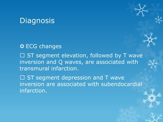
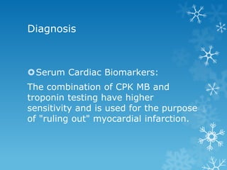
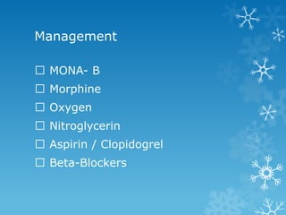
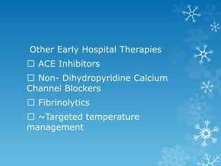
Acute Myocardial Infarction
Acute myocardial infarction (AMI),
commonly known as a heart attack,
is the irreversible necrosis of heart
muscle secondary to prolonged
ischemia
Results from an imbalance in oxygen
supply and demand, caused by plaque
rupture with thrombus formation in a
coronary vessel, resulting in an acute
reduction of blood supply to a portion
of the myocardium
Prevalence of myocardial infarction
by age and sex
Annual number of adults per 1000
having diagnosed heart attack or
fatal coronary heart disease (CHD)
by age and sex
Incidence of heart attack or fatal
coronary heart disease by age, sex,
and race
Risk
 Increasing age and male sex
 Person aged older than 45 years have an
eight times greater risk for AMI.
 Even after menopause, when women's
death rate from heart disease increases,
it's not as great as men's
 Hypertension
 Dyslipidemia
 Diabetes
Risk Factor
 Smoking increases a person's risk for heart disease to
about 4 times greater than non- smokers
 Obesity and physical inactivity People who have excess
body fat — especially at the waist — are more likely to
develop heart disease even if they have no other risk
factors
 Lack of exercise has been linked to 7–12% of cases
 Acute and prolonged intake of high quantities of
alcoholic drinks (3-4 or more) increase the risk of a
heart attack
Pathophysiology
 Coronary Arterial Occlusion.
Rupture of high-risk atheromatous plaque
in the coronary arteries is a primary
causative factor in the development of AMI
When exposed to subendothelial collagen
and necrotic plaque contents, platelets
adhere, become activated, release their
granule contents, and aggregate to form
microthrombi.
Pathophysiology
Vasospasm is stimulated by mediators
released from platelets.
Tissue factor activates the coagulation
pathway, adding to the bulk of the
thrombus.
Within minutes, the thrombus expands to
completely occlude the vessel lumen
Pathophysiology
 In approximately 10% of cases, AMI occurs in the
absence of the typical coronary atherothrombosis.
 Vasospasm
 Emboli
 Others • Intravascular Platelet aggregation • drug
ingestion (e.g., cocaine or ephedrine) • Vegetations
of infective endocarditis, • Intracardiac prosthetic
material • Vasculitis, • Hematologic abnormalities
(e.g., sickle cell disease), • Amyloid deposition, •
Vascular dissection, • Aortic stenosis, • Lowered
systemic blood pressure (e.g., shock)
Pathophysiology
 Myocardial Response.
Coronary arterial obstruction diminishes
blood flow to a region of myocardium
causing ischemia, rapid myocardial
dysfunction, and eventually—with prolonged
vascular compromise — myocyte death.
The anatomic region supplied by that
artery is referred to as the area at risk.
Pathophysiology
Experimental and clinical evidence shows that only
severe ischemia lasting 20 to 30 minutes or longer
leads to irreversible damage (necrosis) of cardiac
myocytes.
This delay in the onset of permanent myocardial
injury provides the rationale for rapid diagnosis in
acute MI—to permit early coronary intervention to
establish reperfusion and salvage as much “at risk”
myocardium as possible
Pathophysiology
The earliest detectable feature of myocyte
necrosis is the disruption of the integrity of the
sarcolemmal membrane, allowing intracellular
macromolecules to leak out of necrotic cells into
the cardiac interstitium and ultimately into the
microvasculature and lymphatics.
Intracellular myocardial proteins into the
circulation forms the basis for blood tests that
can sensitively detect irreversible myocyte
damage, and are important for managing AMI
Pathophysiology
Time to elevation of CKMB, cTnT
and cTnI is 3 to 12 hrs
CK-MB and cTnI peak at 24 hours
CK-MB returns to normal in 48-72
hrs, cTnI in 5-10 days, and cTnT in
5 to 14 days
Main types of acute myocardial infarction,
based on pathology:
Transmural infarction- Transmural infarcts extend
through the whole thickness of the heart muscle and are
usually a result of complete occlusion of the area's blood
supply.
A transmural infarct is sometimes referred to as an “ST
elevation myocardial infarct” (STEMI)
Subendocardial (nontransmural) infarction - involves a
small area in the subendocardial wall of the left ventricle,
ventricular septum, or papillary muscles.
subendocardial infarct as a “non–ST elevation infarct”
(NSTEMI). 19
 A transmural acute myocardial
infarct, predominantly of the
posterolateral left ventricle (arrow).
Note the myocardial hemorrhage at
one edge of the infarct that was
associated with cardiac rupture, and
the anterior scar (arrowhead),
indicative of old infarct.
Sign and symptoms
Chest pain - most common symptom, described as a
sensation of tightness, pressure, or squeezing, not relieved
by rest, position change or nitrate administration, Pain
radiates most often to the left arm, but may also radiate to
the lower jaw, neck, right arm, back, and upper abdomen,
where it may mimic heartburn.
Levine's sign, in which a person localizes the chest pain
by clenching their fists over their sternum.
 SILENT AMI - 20-30% subjects don’t have chest pain,
common in patients with diabetes mellitus,
hypertension, & in elderly patients.
Sign and symptoms
Nausea and Vomiting
Vomiting results as a reflex from severe
pain.
Vasovagal reflexes initiated from area of
ischemia.
Shortness of breath (dyspnea) - the
damage to the heart limits the output of the
left ventricle, causing left ventricular failure
and consequent pulmonary edema.
Sign and symptoms
 Diaphoresis (an excessive form of sweating)
 Light-headedness
 Palpitations
 Loss of consciousness
 inadequate blood flow to the brain and
cardiogenic shock.
 Sudden death - due to the development of
ventricular fibrillation massive surge of
catecholamines from the sympathetic nervous
system
WHO criteria as revised in 2000, a
cardiac troponin rise accompanied
by either typical symptoms,
pathological Q waves, ST elevation
or depression or coronary
intervention are diagnostic of MI.
Diagnosis
 ECG changes
ST segment elevation, followed by T wave
inversion and Q waves, are associated with
transmural infarction.
ST segment depression and T wave
inversion are associated with subendocardial
infarction.
Diagnosis
Serum Cardiac Biomarkers:
The combination of CPK MB and
troponin testing have higher
sensitivity and is used for the purpose
of "ruling out" myocardial infarction.
Management
MONA- B
Morphine
Oxygen
Nitroglycerin
Aspirin / Clopidogrel
Beta-Blockers
Other Early Hospital Therapies
ACE Inhibitors
Non- Dihydropyridine Calcium
Channel Blockers
Fibrinolytics
~Targeted temperature
management
Management
Revascularization procedures
Percutneous Coronary Intervention
/ Angioplasty.
Coronary Artery By –Pass (CABG).
Transmyocardial Laser
Revascularization
Prevention:
 Lifestyle modifications:
 Smoking cessation: Two years after cessation, the risk of
AMI drops by 50%
 Alcohol moderation and prevention of illicit drug use.
 Physical activity and exercise: Exercise 30 minutes
per day 7 days a week. Physical activity can help
control blood cholesterol, diabetes and obesity, as well
as help lower blood pressure.
 Weight Loss - even 10% from current weight, can lower
your heart disease risk
Preventive Measures
 Diet modification Diets rich in soluble fiber,
vegetables, fruits, and whole grains, and low in
saturated fat/trans fat and cholesterol should be
encouraged.
 Lipid management:
 Saturated fat (<7% of total calories),
 Cholesterol and trans fatty acids (<200 mg/day),
 plant stanols/sterols (2 g/day)
 viscous fiber (10 g/day)
 Olive oil, rapeseed oil and related products are to be
used instead of saturated fat
 omega-3 fatty acids (fish)
Hypertension should be managed. Patients with CAD
should have their blood pressure maintained at less than
130/80 mm Hg. This may be achieved using a multimodal
approach, which includes diet modification, lifestyle
changes, exercise, and medications.
Diabetes control should be appropriate - According to the
2007 AHA guideline for management of patients with
STEMI, the goal for HbA1c in diabetic patients should be
less than 7%
Control of comorbid Illnesses:
Patient education:
Patients, their family members, and
the community should be educated
properly, especially on how to detect
and respond to an episode of AMI
Referrence
 Harrison’s Textbook of Medicine
 http://crisbertcualteros.page.tl

Acute Myocardial Infarction

  • 1.
  • 2.
    Acute myocardial infarction(AMI), commonly known as a heart attack, is the irreversible necrosis of heart muscle secondary to prolonged ischemia
  • 3.
    Results from animbalance in oxygen supply and demand, caused by plaque rupture with thrombus formation in a coronary vessel, resulting in an acute reduction of blood supply to a portion of the myocardium
  • 4.
    Prevalence of myocardialinfarction by age and sex Annual number of adults per 1000 having diagnosed heart attack or fatal coronary heart disease (CHD) by age and sex Incidence of heart attack or fatal coronary heart disease by age, sex, and race
  • 5.
    Risk  Increasing ageand male sex  Person aged older than 45 years have an eight times greater risk for AMI.  Even after menopause, when women's death rate from heart disease increases, it's not as great as men's  Hypertension  Dyslipidemia  Diabetes
  • 6.
    Risk Factor  Smokingincreases a person's risk for heart disease to about 4 times greater than non- smokers  Obesity and physical inactivity People who have excess body fat — especially at the waist — are more likely to develop heart disease even if they have no other risk factors  Lack of exercise has been linked to 7–12% of cases  Acute and prolonged intake of high quantities of alcoholic drinks (3-4 or more) increase the risk of a heart attack
  • 7.
    Pathophysiology  Coronary ArterialOcclusion. Rupture of high-risk atheromatous plaque in the coronary arteries is a primary causative factor in the development of AMI When exposed to subendothelial collagen and necrotic plaque contents, platelets adhere, become activated, release their granule contents, and aggregate to form microthrombi.
  • 8.
    Pathophysiology Vasospasm is stimulatedby mediators released from platelets. Tissue factor activates the coagulation pathway, adding to the bulk of the thrombus. Within minutes, the thrombus expands to completely occlude the vessel lumen
  • 9.
    Pathophysiology  In approximately10% of cases, AMI occurs in the absence of the typical coronary atherothrombosis.  Vasospasm  Emboli  Others • Intravascular Platelet aggregation • drug ingestion (e.g., cocaine or ephedrine) • Vegetations of infective endocarditis, • Intracardiac prosthetic material • Vasculitis, • Hematologic abnormalities (e.g., sickle cell disease), • Amyloid deposition, • Vascular dissection, • Aortic stenosis, • Lowered systemic blood pressure (e.g., shock)
  • 10.
    Pathophysiology  Myocardial Response. Coronaryarterial obstruction diminishes blood flow to a region of myocardium causing ischemia, rapid myocardial dysfunction, and eventually—with prolonged vascular compromise — myocyte death. The anatomic region supplied by that artery is referred to as the area at risk.
  • 11.
    Pathophysiology Experimental and clinicalevidence shows that only severe ischemia lasting 20 to 30 minutes or longer leads to irreversible damage (necrosis) of cardiac myocytes. This delay in the onset of permanent myocardial injury provides the rationale for rapid diagnosis in acute MI—to permit early coronary intervention to establish reperfusion and salvage as much “at risk” myocardium as possible
  • 12.
    Pathophysiology The earliest detectablefeature of myocyte necrosis is the disruption of the integrity of the sarcolemmal membrane, allowing intracellular macromolecules to leak out of necrotic cells into the cardiac interstitium and ultimately into the microvasculature and lymphatics. Intracellular myocardial proteins into the circulation forms the basis for blood tests that can sensitively detect irreversible myocyte damage, and are important for managing AMI
  • 13.
    Pathophysiology Time to elevationof CKMB, cTnT and cTnI is 3 to 12 hrs CK-MB and cTnI peak at 24 hours CK-MB returns to normal in 48-72 hrs, cTnI in 5-10 days, and cTnT in 5 to 14 days
  • 14.
    Main types ofacute myocardial infarction, based on pathology: Transmural infarction- Transmural infarcts extend through the whole thickness of the heart muscle and are usually a result of complete occlusion of the area's blood supply. A transmural infarct is sometimes referred to as an “ST elevation myocardial infarct” (STEMI) Subendocardial (nontransmural) infarction - involves a small area in the subendocardial wall of the left ventricle, ventricular septum, or papillary muscles. subendocardial infarct as a “non–ST elevation infarct” (NSTEMI). 19
  • 15.
     A transmuralacute myocardial infarct, predominantly of the posterolateral left ventricle (arrow). Note the myocardial hemorrhage at one edge of the infarct that was associated with cardiac rupture, and the anterior scar (arrowhead), indicative of old infarct.
  • 16.
    Sign and symptoms Chestpain - most common symptom, described as a sensation of tightness, pressure, or squeezing, not relieved by rest, position change or nitrate administration, Pain radiates most often to the left arm, but may also radiate to the lower jaw, neck, right arm, back, and upper abdomen, where it may mimic heartburn. Levine's sign, in which a person localizes the chest pain by clenching their fists over their sternum.  SILENT AMI - 20-30% subjects don’t have chest pain, common in patients with diabetes mellitus, hypertension, & in elderly patients.
  • 17.
    Sign and symptoms Nauseaand Vomiting Vomiting results as a reflex from severe pain. Vasovagal reflexes initiated from area of ischemia. Shortness of breath (dyspnea) - the damage to the heart limits the output of the left ventricle, causing left ventricular failure and consequent pulmonary edema.
  • 18.
    Sign and symptoms Diaphoresis (an excessive form of sweating)  Light-headedness  Palpitations  Loss of consciousness  inadequate blood flow to the brain and cardiogenic shock.  Sudden death - due to the development of ventricular fibrillation massive surge of catecholamines from the sympathetic nervous system
  • 19.
    WHO criteria asrevised in 2000, a cardiac troponin rise accompanied by either typical symptoms, pathological Q waves, ST elevation or depression or coronary intervention are diagnostic of MI.
  • 20.
    Diagnosis  ECG changes STsegment elevation, followed by T wave inversion and Q waves, are associated with transmural infarction. ST segment depression and T wave inversion are associated with subendocardial infarction.
  • 21.
    Diagnosis Serum Cardiac Biomarkers: Thecombination of CPK MB and troponin testing have higher sensitivity and is used for the purpose of "ruling out" myocardial infarction.
  • 22.
  • 23.
    Other Early HospitalTherapies ACE Inhibitors Non- Dihydropyridine Calcium Channel Blockers Fibrinolytics ~Targeted temperature management
  • 24.
    Management Revascularization procedures Percutneous CoronaryIntervention / Angioplasty. Coronary Artery By –Pass (CABG). Transmyocardial Laser Revascularization
  • 25.
    Prevention:  Lifestyle modifications: Smoking cessation: Two years after cessation, the risk of AMI drops by 50%  Alcohol moderation and prevention of illicit drug use.  Physical activity and exercise: Exercise 30 minutes per day 7 days a week. Physical activity can help control blood cholesterol, diabetes and obesity, as well as help lower blood pressure.  Weight Loss - even 10% from current weight, can lower your heart disease risk
  • 26.
    Preventive Measures  Dietmodification Diets rich in soluble fiber, vegetables, fruits, and whole grains, and low in saturated fat/trans fat and cholesterol should be encouraged.  Lipid management:  Saturated fat (<7% of total calories),  Cholesterol and trans fatty acids (<200 mg/day),  plant stanols/sterols (2 g/day)  viscous fiber (10 g/day)  Olive oil, rapeseed oil and related products are to be used instead of saturated fat  omega-3 fatty acids (fish)
  • 27.
    Hypertension should bemanaged. Patients with CAD should have their blood pressure maintained at less than 130/80 mm Hg. This may be achieved using a multimodal approach, which includes diet modification, lifestyle changes, exercise, and medications. Diabetes control should be appropriate - According to the 2007 AHA guideline for management of patients with STEMI, the goal for HbA1c in diabetic patients should be less than 7% Control of comorbid Illnesses:
  • 28.
    Patient education: Patients, theirfamily members, and the community should be educated properly, especially on how to detect and respond to an episode of AMI
  • 29.
  • 30.