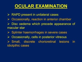
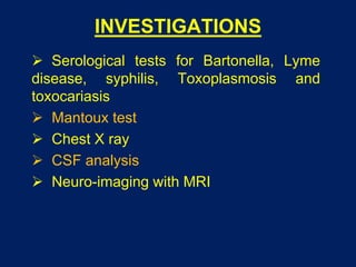
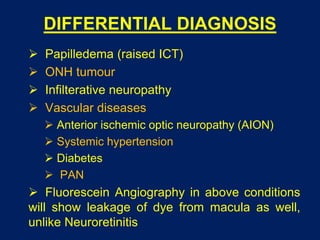
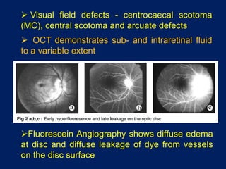
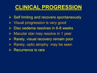
NEURORETINITIS
Dr Saurabh Kushwaha
Resident Ophthalmology
SCOPE
 Background
 Etiology
 Clinical features
 Differential diagnosis
 Investigations
 Clinical Progression
 Treatment
BACKGROUND
 Optic disc edema with
macular star (ODEMS)
 1st described by Leber (1916)
as stellate maculopathy
 Gass (1977) renamed it as
neuroretinitis
 Leber’s idiopathic stellate
neuroretinitis
ETIOLOGY
 Idiopathic - 50%
 Viral infections (MC association)
 Common specific infections:
 Cat scratch disease (Bartonella)
 Syphilis
 Toxoplasmosis
 Lyme disease
 Leptospirosis
 Toxocariasis
 Helminths (Diffuse unilateral subacute neuroretinitis,
DUSN)
CLINICAL FEATURES
 Most commonly affects 3rd and 4th decade
 No sex predilection
 Either eye or both eye can be affected
 Sudden decrease in vision without pain
 Loss of colour vision
 Loss of contrast sensitivity
 Metamorphopsia
EVALUATION
 Complete history for
 Viral infections
 Associations with animals
 Sexually transmitted diseases
 Systemic examination with special emphasis
on neurological examination
OCULAR EXAMINATION
 RAPD present in unilateral cases
 Occasionally, reaction in anterior chamber
 Disc oedema which precede appearance of
macular star
 Splinter haemorrhages in severe cases
 Occasionally, cells in posterior vitreous
 Small, discrete chorioretinal lesions in
idiotpthic cases
Fluorescein Angiography shows diffuse edema
at disc and diffuse leakage of dye from vessels
on the disc surface
 Visual field defects - centrocaecal scotoma
(MC), central scotoma and arcuate defects
 OCT demonstrates sub- and intraretinal fluid
to a variable extent
INVESTIGATIONS
 Serological tests for Bartonella, Lyme
disease, syphilis, Toxoplasmosis and
toxocariasis
 Mantoux test
 Chest X ray
 CSF analysis
 Neuro-imaging with MRI
DIFFERENTIAL DIAGNOSIS
 Papilledema (raised ICT)
 ONH tumour
 Infilterative neuropathy
 Vascular diseases
 Anterior ischemic optic neuropathy (AION)
 Systemic hypertension
 Diabetes
 PAN
 Fluorescein Angiography in above conditions
will show leakage of dye from macula as well,
unlike Neuroretinitis
TREATMENT
 Resolves spontaneously without treatment
 Treatment - speedy recovery and shorter course
of disease
 Specific antibiotic therapy
 Bartonella - Doxycycline
 Syphilis - iv Penicillin
 Toxoplasmosis - pyrimethamine, sulfadiazine,
and corticosteroids
 Leptospirosis - iv Penicillin, Doxycycline
 Recurrent cases - Steroids/immunosuppressant
CLINICAL PROGRESSION
 Self limiting and recovers spontaneously
 Visual progression is very good
 Disc oedema resolves in 6-8 weeks
 Macular star may resolve in 1 year
 Rarely, visual recovery remain poor
 Rarely, optic atrophy may be seen
 Recurrence is rare
SUMMARY
 Resolves spontaneously without treatment
 Idiopathic, unilateral, self limiting, with good
visual prognosis
 Rarely can be recurrent, bilateral, with optic
atrophy and poor visual prognosis
THANK YOU

Neuroretinitis

  • 1.
  • 2.
    SCOPE  Background  Etiology Clinical features  Differential diagnosis  Investigations  Clinical Progression  Treatment
  • 3.
    BACKGROUND  Optic discedema with macular star (ODEMS)  1st described by Leber (1916) as stellate maculopathy  Gass (1977) renamed it as neuroretinitis  Leber’s idiopathic stellate neuroretinitis
  • 4.
    ETIOLOGY  Idiopathic -50%  Viral infections (MC association)  Common specific infections:  Cat scratch disease (Bartonella)  Syphilis  Toxoplasmosis  Lyme disease  Leptospirosis  Toxocariasis  Helminths (Diffuse unilateral subacute neuroretinitis, DUSN)
  • 5.
    CLINICAL FEATURES  Mostcommonly affects 3rd and 4th decade  No sex predilection  Either eye or both eye can be affected  Sudden decrease in vision without pain  Loss of colour vision  Loss of contrast sensitivity  Metamorphopsia
  • 6.
    EVALUATION  Complete historyfor  Viral infections  Associations with animals  Sexually transmitted diseases  Systemic examination with special emphasis on neurological examination
  • 7.
    OCULAR EXAMINATION  RAPDpresent in unilateral cases  Occasionally, reaction in anterior chamber  Disc oedema which precede appearance of macular star  Splinter haemorrhages in severe cases  Occasionally, cells in posterior vitreous  Small, discrete chorioretinal lesions in idiotpthic cases
  • 9.
    Fluorescein Angiography showsdiffuse edema at disc and diffuse leakage of dye from vessels on the disc surface  Visual field defects - centrocaecal scotoma (MC), central scotoma and arcuate defects  OCT demonstrates sub- and intraretinal fluid to a variable extent
  • 10.
    INVESTIGATIONS  Serological testsfor Bartonella, Lyme disease, syphilis, Toxoplasmosis and toxocariasis  Mantoux test  Chest X ray  CSF analysis  Neuro-imaging with MRI
  • 11.
    DIFFERENTIAL DIAGNOSIS  Papilledema(raised ICT)  ONH tumour  Infilterative neuropathy  Vascular diseases  Anterior ischemic optic neuropathy (AION)  Systemic hypertension  Diabetes  PAN  Fluorescein Angiography in above conditions will show leakage of dye from macula as well, unlike Neuroretinitis
  • 12.
    TREATMENT  Resolves spontaneouslywithout treatment  Treatment - speedy recovery and shorter course of disease  Specific antibiotic therapy  Bartonella - Doxycycline  Syphilis - iv Penicillin  Toxoplasmosis - pyrimethamine, sulfadiazine, and corticosteroids  Leptospirosis - iv Penicillin, Doxycycline  Recurrent cases - Steroids/immunosuppressant
  • 13.
    CLINICAL PROGRESSION  Selflimiting and recovers spontaneously  Visual progression is very good  Disc oedema resolves in 6-8 weeks  Macular star may resolve in 1 year  Rarely, visual recovery remain poor  Rarely, optic atrophy may be seen  Recurrence is rare
  • 14.
    SUMMARY  Resolves spontaneouslywithout treatment  Idiopathic, unilateral, self limiting, with good visual prognosis  Rarely can be recurrent, bilateral, with optic atrophy and poor visual prognosis
  • 15.