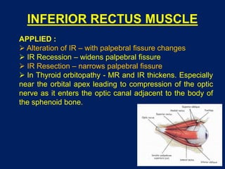
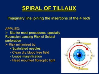
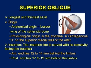
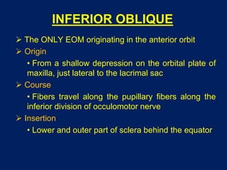
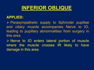
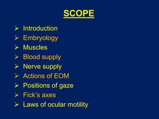
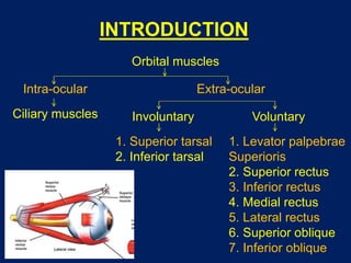
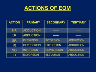
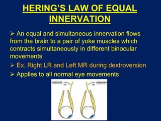
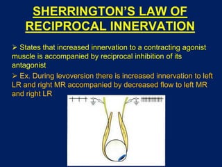
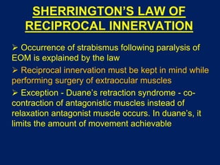
EXTRA OCULAR MUSCLES
Dr Saurabh Kushwaha
Resident Ophthalmology
SCOPE
 Introduction
 Embryology
 Muscles
 Blood supply
 Nerve supply
 Actions of EOM
 Positions of gaze
 Fick’s axes
 Laws of ocular motility
INTRODUCTION
Orbital muscles
Intra-ocular Extra-ocular
Ciliary muscles Involuntary
1. Superior tarsal
2. Inferior tarsal
Voluntary
1. Levator palpebrae
Superioris
2. Superior rectus
3. Inferior rectus
4. Medial rectus
5. Lateral rectus
6. Superior oblique
7. Inferior oblique
EMBRYOLOGY
 Mesodermal origin
 Development begins at 3-4 weeks gestation
 Originate from 03 separate foci of primordial cells
 Muscles receive input from their respective cranial
nerves as early as 1 month of gestation
 Early fascial coverings can be detected around
the extraocular muscles by 3 months of gestation
 The extraocular muscle and their surrounding
tissues are in their final anatomical positions by 6
months of gestation
LEVATOR PALPEBRAE
SUPERIORIS
 Origin
• Inferior surface of lesser wing of sphenoid
 Insertion
• Upper lamina (Voluntary) - Anterior surface of superior
tarsus & skin of upper eyelid
• Middle lamina (Involuntary) - Superior margin of superior
tarsus. (Superior Tarsus Muscle / Muller muscle)
• Lower lamina (Involuntary) - Superior conjunctival fornix
 Nerve Supply
• Voluntary part - Oculomotor Nerve
• Involuntary part - Sympathetic
LEVATOR PALPEBRAE
SUPERIORIS
SUPERIOR RECTUS MUSCLE
 Origin
• Superior part of common annular tendon of
Zinn
 Course
• Passes anterolaterally beneath the levator at
23 degrees with the globeā€˜s AP axis, pierces
Tenon’s capsule
 Insertion
• into sclera by flat tendinous 10 mm broad
insertion, 7.7 mm behind sclero-corneal junction
SUPERIOR RECTUS MUSCLE
APPLIED :
 SR loosely bound to LPS muscle.
• During SR resection- eyelid may be pulled forward,
narrowing palpebral fissure
• In hypotropia, pseudoptosis may be present
 Origin of SR and MR are closely attached to the dural
sheath of the optic nerve
• pain during upward & inward movements of the
globe in RETROBULBAR NEURITIS
 SR - Only elevator in full abduction because IO is
ineffective. Thus when SR is paralysed, abducted eye cant
be elevated
INFERIOR RECTUS MUSCLE
 Origin
• Inferior part of common tendon of Annulus of
Zinn below the optic foramen
 Course
• Passes anterolaterally along the floor of the
orbit, at an angle of 23 degrees
 Insertion
• obliquely in the sclera 6.5 mm behind sclero
corneal junction by a 5.5mm long tendon
INFERIOR RECTUS MUSCLE
APPLIED :
 Alteration of IR – with palpebral fissure changes
 IR Recession – widens palpebral fissure
 IR Resection – narrows palpebral fissure
 In Thyroid orbitopathy - MR and IR thickens. Especially
near the orbital apex leading to compression of the optic
nerve as it enters the optic canal adjacent to the body of
the sphenoid bone.
MEDIAL RECTUS MUSCLE
 Largest ocular muscle, Thicker than the others
 Origin
• widely attached medial and inferior to optic
foramen by common tendon of annulus of Zinn
• From optic nerve sheath
 Course
• Passes along medial wall of the orbit
 Insertion
• into sclera 5.5mm behind sclero-corneal
junction by a tendon 3.7 mm in length
MEDIAL RECTUS MUSCLE
APPLIED:
 In Thyroid orbitopathy, MR and IR thicken; visibility
of muscle insertion through conjunctiva allows swelling
to be detected in Endocrine Exophthalmos
 Pain in Retrobulbar neuritis - Origin close to dural
sheath of Optic Nerve
 Medial rectus inserts closest to the limbus and is
therefore susceptible to injury during anterior segment
surgery
 Inadvertent removal of the MR is a well known
complication of Pterygium excision surgery
LATERAL RECTUS MUSCLE
 Origin
• Annular Tendon of Zinn where it crosses Superior
Orbital Fissure
 Course
• At first adjoins lateral orbital wall separated by fat
• advances anteriorly, it passes medially and pierces
tenon’s Capsule
 Insertion
• On the sclera 6.9 mm behind sclerocorneal
junction by a tendon 8.8 mm long
SPIRAL OF TILLAUX
Imaginary line joining the insertions of the 4 recti
APPLIED:
 Site for most procedures, specially
Recession causing Risk of Scleral
perforation
 Risk minimized by
• Spatulated needles
• Clean dry blood free field
• Loupe magnification
• Head mounted fibreoptic light
SUPERIOR OBLIQUE
 Longest and thinnest EOM
 Origin
• Anatomical origin – Lesser
wing of the sphenoid bone
• Physiological origin is the trochlea, a cartilagenous
ā€œUā€ on the superior medial wall of the orbit
 Insertion: The insertion line is curved with its concavity
facing the trochlea
• Ant. end lies 12 to 14 mm behind the limbus
• Post. end lies 17 to 19 mm behind the limbus
INFERIOR OBLIQUE
 The ONLY EOM originating in the anterior orbit
 Origin
• From a shallow depression on the orbital plate of
maxilla, just lateral to the lacrimal sac
 Course
• Fibers travel along the pupillary fibers along the
inferior division of occulomotor nerve
 Insertion
• Lower and outer part of sclera behind the equator
INFERIOR OBLIQUE
APPLIED:
 Parasympathetic supply to Sphincter pupillae
and ciliary muscle accompanies Nerve to IO,
leading to pupillary abnormalities from surgery in
this area
 Nerve to IO enters lateral portion of muscle
where the muscle crosses IR likely to have
damage in this area
BLOOD SUPPLY
Artery Division Muscle
Ophthalmi
c artery
Medial
muscular
branch
Medial rectus
Inferior rectus
Inferior oblique
Lateral
muscular
branch
Lateral rectus
Superior rectus
Superior oblique
Levator palpebrae
superioris
BLOOD SUPPLY
 The arteries to the four rectus muscles give rise
to anterior ciliary arteries
 The anterior ciliary arteries passes to the
episclera, give branches to the sclera, limbus, and
conjunctiva
 Here they anastomose with the lateral and
medial long ciliary arteries to form
the major arterial circle of the iris
 Veins correspond to the
arteries and empty into the
superior and inferior
ophthalmic veins
BLOOD SUPPLY
APPLIED :
 Accidental risk of severing of Vortex veins during IR and
SR Recession or Resection , IO muscle weakening and
SO muscle tendon exposure
 Blood supply to EOM supplies most of anterior
segment, part of nasal half supplied by long posterior
ciliary artery and thus simultaneous surgery on 3 recti
induces anterior segment ischaemia
 Variations in the number of anterior ciliary arteries
supplied by each muscle become clinically relevant with
regard to the anterior segment blood supply when
disinserting more than two rectus muscle tendons during
muscle surgery
NERVE SUPPLY
Muscle Nerve supply
Superior rectus Superior division of third nerve
Medial rectus Inferior division of third nerve
Inferior rectus Inferior division of third nerve
Inferior oblique Inferior division of third nerve
Superior oblique Trochlear nerve
Lateral rectus Abducent nerve
ACTIONS OF EOM
ACTION PRIMARY SECONDARY TERTIARY
MR ADDUCTION ------ ------
LR ABDUCTION ------ ------
SR ELEVATION INTORSION ADDUCTION
IR DEPRESSION EXTORSION ADDUCTION
SO INTORSION DEPRESSION ABDUCTION
IO EXTORSION ELEVATION ABDUCTION
POSITIONS OF GAZE
 Primary position
• fixating straight at a distant object with head erect
• visual axis of the two eyes are parallel
 Secondary positions
• straight up (supraversion)
• straight down (infraversion)
• right gaze (dextroversion)
• left gaze (levoversion)
 Tertiary positions
• up and right (dextroelevation)
• up and left (levoelevation)
• down and right (dextrodepression)
• down and left (levodepression)
CARDINAL POSITIONS OF GAZE
 Refer to positions of the globe that minimize the angle
between the axis of the EOM being evaluated and the visual
axis
 Minimizing this angle minimizes the secondary and
tertiary actions of the muscle on the globe
FICK’S AXES
 X (horizontal) axis
• Lies horizontally when head is upright
• Elevation / Depression
 Y (antero-posterior) axis
• Torsional movements
• Extorsion / Intorsion
 Z (vertical axis)
• Lies vertically
• Adduction / Abduction
LAWS OF OCULAR MOTILITY
 Agonists
• Any particular EOM producing specific ocular
movement
• Ex. Right LR for right eye abduction
 Synergists
• Muscles of the same eye that move the eye
in the same direction
• Ex. Right SR and right IO for right eye
elevation
 Antagonists
• A pair of muscles in the same eye that move the
eye in opposite directions
• Ex. right LR and right MR
 Yoke muscles (contralateral synergists)
• Pair of muscles, one in each eye, that produce
conjugate ocular movements
• Ex. right LR and left MR in dextroversion
LISTING’S LAW
 All achieved eye orientations can be reached by
starting from one specific "primaryā€œ reference
orientation and then rotating about an axis that lies
within the plane orthogonal to the primary
orientation's gaze direction (line of sight / visual
axis)
 This plane is called Listing's plane
DONDER’S LAW
 Donder stated that each position of line of sight
belongs to the definite orientation of vertical and
horizontal retinal meridian relative to the coordinate of
the space.
 Orientation of retinal meridian is always same
irrespective of the path the eye has taken to reach that
position and depends upon the amount of elevation or
depression and lateral rotation of the globe, after
returning to the initial position the retinal meridian is
oriented exactly as it was before the movement was
initiated
HERING’S LAW OF EQUAL
INNERVATION
 An equal and simultaneous innervation flows
from the brain to a pair of yoke muscles which
contracts simultaneously in different binocular
movements
 Ex. Right LR and Left MR during dextroversion
 Applies to all normal eye movements
HERING’S LAW OF EQUAL
INNERVATION
APPLIED:
 Secondary Deviation in patient with paralytic
squint is more than the primary deviation
 Excess innervation is required to the paralysed
muscle to fixate
 Concomitant excess supply leads to more
secondary deviation
SHERRINGTON’S LAW OF
RECIPROCAL INNERVATION
 States that increased innervation to a contracting agonist
muscle is accompanied by reciprocal inhibition of its
antagonist
 Ex. During levoversion there is increased innervation to left
LR and right MR accompanied by decreased flow to left MR
and right LR
SHERRINGTON’S LAW OF
RECIPROCAL INNERVATION
 Occurrence of strabismus following paralysis of
EOM is explained by the law
 Reciprocal innervation must be kept in mind while
performing surgery of extraocular muscles
 Exception - Duane’s retraction syndrome - co-
contraction of antagonistic muscles instead of
relaxation antagonist muscle occurs. In duane’s, it
limits the amount of movement achievable
THANK YOU

Extra ocular muscles

  • 1.
    EXTRA OCULAR MUSCLES DrSaurabh Kushwaha Resident Ophthalmology
  • 2.
    SCOPE  Introduction  Embryology Muscles  Blood supply  Nerve supply  Actions of EOM  Positions of gaze  Fick’s axes  Laws of ocular motility
  • 3.
    INTRODUCTION Orbital muscles Intra-ocular Extra-ocular Ciliarymuscles Involuntary 1. Superior tarsal 2. Inferior tarsal Voluntary 1. Levator palpebrae Superioris 2. Superior rectus 3. Inferior rectus 4. Medial rectus 5. Lateral rectus 6. Superior oblique 7. Inferior oblique
  • 4.
    EMBRYOLOGY  Mesodermal origin Development begins at 3-4 weeks gestation  Originate from 03 separate foci of primordial cells  Muscles receive input from their respective cranial nerves as early as 1 month of gestation  Early fascial coverings can be detected around the extraocular muscles by 3 months of gestation  The extraocular muscle and their surrounding tissues are in their final anatomical positions by 6 months of gestation
  • 5.
    LEVATOR PALPEBRAE SUPERIORIS  Origin •Inferior surface of lesser wing of sphenoid  Insertion • Upper lamina (Voluntary) - Anterior surface of superior tarsus & skin of upper eyelid • Middle lamina (Involuntary) - Superior margin of superior tarsus. (Superior Tarsus Muscle / Muller muscle) • Lower lamina (Involuntary) - Superior conjunctival fornix  Nerve Supply • Voluntary part - Oculomotor Nerve • Involuntary part - Sympathetic
  • 6.
  • 7.
    SUPERIOR RECTUS MUSCLE Origin • Superior part of common annular tendon of Zinn  Course • Passes anterolaterally beneath the levator at 23 degrees with the globeā€˜s AP axis, pierces Tenon’s capsule  Insertion • into sclera by flat tendinous 10 mm broad insertion, 7.7 mm behind sclero-corneal junction
  • 8.
    SUPERIOR RECTUS MUSCLE APPLIED:  SR loosely bound to LPS muscle. • During SR resection- eyelid may be pulled forward, narrowing palpebral fissure • In hypotropia, pseudoptosis may be present  Origin of SR and MR are closely attached to the dural sheath of the optic nerve • pain during upward & inward movements of the globe in RETROBULBAR NEURITIS  SR - Only elevator in full abduction because IO is ineffective. Thus when SR is paralysed, abducted eye cant be elevated
  • 9.
    INFERIOR RECTUS MUSCLE Origin • Inferior part of common tendon of Annulus of Zinn below the optic foramen  Course • Passes anterolaterally along the floor of the orbit, at an angle of 23 degrees  Insertion • obliquely in the sclera 6.5 mm behind sclero corneal junction by a 5.5mm long tendon
  • 10.
    INFERIOR RECTUS MUSCLE APPLIED:  Alteration of IR – with palpebral fissure changes  IR Recession – widens palpebral fissure  IR Resection – narrows palpebral fissure  In Thyroid orbitopathy - MR and IR thickens. Especially near the orbital apex leading to compression of the optic nerve as it enters the optic canal adjacent to the body of the sphenoid bone.
  • 11.
    MEDIAL RECTUS MUSCLE Largest ocular muscle, Thicker than the others  Origin • widely attached medial and inferior to optic foramen by common tendon of annulus of Zinn • From optic nerve sheath  Course • Passes along medial wall of the orbit  Insertion • into sclera 5.5mm behind sclero-corneal junction by a tendon 3.7 mm in length
  • 12.
    MEDIAL RECTUS MUSCLE APPLIED: In Thyroid orbitopathy, MR and IR thicken; visibility of muscle insertion through conjunctiva allows swelling to be detected in Endocrine Exophthalmos  Pain in Retrobulbar neuritis - Origin close to dural sheath of Optic Nerve  Medial rectus inserts closest to the limbus and is therefore susceptible to injury during anterior segment surgery  Inadvertent removal of the MR is a well known complication of Pterygium excision surgery
  • 13.
    LATERAL RECTUS MUSCLE Origin • Annular Tendon of Zinn where it crosses Superior Orbital Fissure  Course • At first adjoins lateral orbital wall separated by fat • advances anteriorly, it passes medially and pierces tenon’s Capsule  Insertion • On the sclera 6.9 mm behind sclerocorneal junction by a tendon 8.8 mm long
  • 14.
    SPIRAL OF TILLAUX Imaginaryline joining the insertions of the 4 recti APPLIED:  Site for most procedures, specially Recession causing Risk of Scleral perforation  Risk minimized by • Spatulated needles • Clean dry blood free field • Loupe magnification • Head mounted fibreoptic light
  • 15.
    SUPERIOR OBLIQUE  Longestand thinnest EOM  Origin • Anatomical origin – Lesser wing of the sphenoid bone • Physiological origin is the trochlea, a cartilagenous ā€œUā€ on the superior medial wall of the orbit  Insertion: The insertion line is curved with its concavity facing the trochlea • Ant. end lies 12 to 14 mm behind the limbus • Post. end lies 17 to 19 mm behind the limbus
  • 16.
    INFERIOR OBLIQUE  TheONLY EOM originating in the anterior orbit  Origin • From a shallow depression on the orbital plate of maxilla, just lateral to the lacrimal sac  Course • Fibers travel along the pupillary fibers along the inferior division of occulomotor nerve  Insertion • Lower and outer part of sclera behind the equator
  • 17.
    INFERIOR OBLIQUE APPLIED:  Parasympatheticsupply to Sphincter pupillae and ciliary muscle accompanies Nerve to IO, leading to pupillary abnormalities from surgery in this area  Nerve to IO enters lateral portion of muscle where the muscle crosses IR likely to have damage in this area
  • 18.
    BLOOD SUPPLY Artery DivisionMuscle Ophthalmi c artery Medial muscular branch Medial rectus Inferior rectus Inferior oblique Lateral muscular branch Lateral rectus Superior rectus Superior oblique Levator palpebrae superioris
  • 19.
    BLOOD SUPPLY  Thearteries to the four rectus muscles give rise to anterior ciliary arteries  The anterior ciliary arteries passes to the episclera, give branches to the sclera, limbus, and conjunctiva  Here they anastomose with the lateral and medial long ciliary arteries to form the major arterial circle of the iris  Veins correspond to the arteries and empty into the superior and inferior ophthalmic veins
  • 20.
    BLOOD SUPPLY APPLIED : Accidental risk of severing of Vortex veins during IR and SR Recession or Resection , IO muscle weakening and SO muscle tendon exposure  Blood supply to EOM supplies most of anterior segment, part of nasal half supplied by long posterior ciliary artery and thus simultaneous surgery on 3 recti induces anterior segment ischaemia  Variations in the number of anterior ciliary arteries supplied by each muscle become clinically relevant with regard to the anterior segment blood supply when disinserting more than two rectus muscle tendons during muscle surgery
  • 21.
    NERVE SUPPLY Muscle Nervesupply Superior rectus Superior division of third nerve Medial rectus Inferior division of third nerve Inferior rectus Inferior division of third nerve Inferior oblique Inferior division of third nerve Superior oblique Trochlear nerve Lateral rectus Abducent nerve
  • 22.
    ACTIONS OF EOM ACTIONPRIMARY SECONDARY TERTIARY MR ADDUCTION ------ ------ LR ABDUCTION ------ ------ SR ELEVATION INTORSION ADDUCTION IR DEPRESSION EXTORSION ADDUCTION SO INTORSION DEPRESSION ABDUCTION IO EXTORSION ELEVATION ABDUCTION
  • 23.
    POSITIONS OF GAZE Primary position • fixating straight at a distant object with head erect • visual axis of the two eyes are parallel  Secondary positions • straight up (supraversion) • straight down (infraversion) • right gaze (dextroversion) • left gaze (levoversion)
  • 24.
     Tertiary positions •up and right (dextroelevation) • up and left (levoelevation) • down and right (dextrodepression) • down and left (levodepression)
  • 25.
    CARDINAL POSITIONS OFGAZE  Refer to positions of the globe that minimize the angle between the axis of the EOM being evaluated and the visual axis  Minimizing this angle minimizes the secondary and tertiary actions of the muscle on the globe
  • 26.
    FICK’S AXES  X(horizontal) axis • Lies horizontally when head is upright • Elevation / Depression  Y (antero-posterior) axis • Torsional movements • Extorsion / Intorsion  Z (vertical axis) • Lies vertically • Adduction / Abduction
  • 27.
    LAWS OF OCULARMOTILITY  Agonists • Any particular EOM producing specific ocular movement • Ex. Right LR for right eye abduction  Synergists • Muscles of the same eye that move the eye in the same direction • Ex. Right SR and right IO for right eye elevation
  • 28.
     Antagonists • Apair of muscles in the same eye that move the eye in opposite directions • Ex. right LR and right MR  Yoke muscles (contralateral synergists) • Pair of muscles, one in each eye, that produce conjugate ocular movements • Ex. right LR and left MR in dextroversion
  • 29.
    LISTING’S LAW  Allachieved eye orientations can be reached by starting from one specific "primaryā€œ reference orientation and then rotating about an axis that lies within the plane orthogonal to the primary orientation's gaze direction (line of sight / visual axis)  This plane is called Listing's plane
  • 30.
    DONDER’S LAW  Donderstated that each position of line of sight belongs to the definite orientation of vertical and horizontal retinal meridian relative to the coordinate of the space.  Orientation of retinal meridian is always same irrespective of the path the eye has taken to reach that position and depends upon the amount of elevation or depression and lateral rotation of the globe, after returning to the initial position the retinal meridian is oriented exactly as it was before the movement was initiated
  • 31.
    HERING’S LAW OFEQUAL INNERVATION  An equal and simultaneous innervation flows from the brain to a pair of yoke muscles which contracts simultaneously in different binocular movements  Ex. Right LR and Left MR during dextroversion  Applies to all normal eye movements
  • 32.
    HERING’S LAW OFEQUAL INNERVATION APPLIED:  Secondary Deviation in patient with paralytic squint is more than the primary deviation  Excess innervation is required to the paralysed muscle to fixate  Concomitant excess supply leads to more secondary deviation
  • 33.
    SHERRINGTON’S LAW OF RECIPROCALINNERVATION  States that increased innervation to a contracting agonist muscle is accompanied by reciprocal inhibition of its antagonist  Ex. During levoversion there is increased innervation to left LR and right MR accompanied by decreased flow to left MR and right LR
  • 34.
    SHERRINGTON’S LAW OF RECIPROCALINNERVATION  Occurrence of strabismus following paralysis of EOM is explained by the law  Reciprocal innervation must be kept in mind while performing surgery of extraocular muscles  Exception - Duane’s retraction syndrome - co- contraction of antagonistic muscles instead of relaxation antagonist muscle occurs. In duane’s, it limits the amount of movement achievable
  • 35.