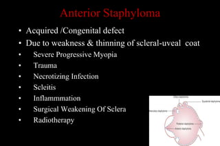
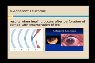
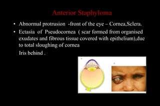
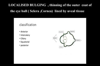
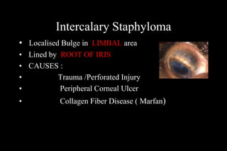
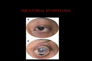
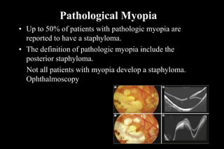
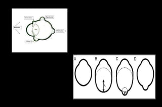
STAPHYLOMA VIRAL KERATITIS
DR M SAQUIB
MBBS,MS , FSCEH DELHI,FHVDESAI PUNE,
EX REGISTRARA JNMCH,AMU
CONSULTANT OPHTHALMOLOGIST
HOD D/O OPHTHALMOLOGY
G.S .MEDICAL COLLEGE
Founder sec: MEDICS India , www.medicsngo.org
Anterior Staphyloma
• Acquired /Congenital defect
• Due to weakness & thinning of scleral-uveal coat
• Severe Progressive Myopia
• Trauma
• Necrotizing Infection
• Scleitis
• Inflammmation
• Surgical Weakening Of Sclera
• Radiotherapy
Ant Staphyloma ( dd Adherent Leucoma)
Pseudocornea ( scar formed from organised
exudates and fibrous tissue covered with
epithelium)ughing of cornea
Anterior Staphyloma
• Abnormal protrusion -front of the eye – Cornea,Sclera.
• Ectasia of Pseudocornea ( scar formed from organised
exudates and fibrous tissue covered with epithelium),due
to total sloughing of cornea
Iris behind .
LOCALISED BULGING , thinning of the outer, coat of
the eye ball ( Sclera ,Cornea) lined by uveal tissue ..f
the eye
Intercalary Staphyloma
• Localised Bulge in LIMBAL area
• Lined by ROOT OF IRIS
• CAUSES :
• Trauma /Perforated Injury
• Peripheral Corneal Ulcer
• Collagen Fiber Disease ( Marfan)
• Sequelae:
Secondary Angle Closure Glaucoma
Corneal Astigmatism ---VA
• Treatment : Localised Staphylectomy
Oral Steroid
Ciliary Staphyloma
• Bulge of weak Sclera Lined by CILIARY BODY
• 2- 3 mm away from Limbus
• CAUSES : Perforation
Scleritis
Absolute Glaucoma
Equatorial Staphyloma
• Bulge of Sclera in Equatorial region
• Lined by Choroid
• Common where vortex vein perforate sclera (14mm behind limbus)
• CAUSES: Scleritis
• Scleral degeneration
• Pathological Myopia
• Chronic uncontrolled Glaucoma
EQUATORIAL STAPHYLOMA
Posterior Staphyloma
• Circumscribed outpouching of the posterior
fundus .Bulge of weak Sclera lined by Choroid
behind the Equator .
Cause :
Pathological Myopia
Posterior Scleritis
Perforating Injury
Infection
• Congenital Weak Bruch’s membrane, Retinitis pigmentosa,
Aalport’s syndrome,Pseudoxanthoma elasticum, and Tilted-disc
syndrom
Pathological Myopia
• Up to 50% of patients with pathologic myopia are
reported to have a staphyloma.
• The definition of pathologic myopia include the
posterior staphyloma.
Not all patients with myopia develop a staphyloma.
Ophthalmoscopy
• Staphylomas are associated with other macular
Complations Related To Myopia,
• Choroidal neovascularization,
• Retinoschisis,
• Glaucomatous optic neuropathy.
• In the Non-Myopic-to trauma or infection,
and rarely, surgery.
BLUE SCLERA
• A bluish coloration of the whites of the eyes.
• The blue color is caused by thinness and transparency of
the collagen fibers of the sclera, allowing the veins in the
underlying tissue to show through.
• Conditions,
• particularly connective tissue disorders. These include
Osteogenesis imperfecta, Marfan's syndrome, Ehlers-
Danlos syndrome, Pseudoxanthoma elasticum, and
Willems De Vries syndrome.
BLUE -HOPES
• Buphthalmos
• High Myopia
• OI
• Pseudoxanthoma Elasticum
• Ehlers Danlos Synd
• Scleritis
Spheroidal degeneration
Treatment
Central spread, coalescence and
opacification
Advanced lesions become
nodular and elevated
• Rare, typically affects outdoor workers
• Starts with peripheral, interpalpebral, small amber-coloured
granules in superficial stroma
• Debridement or superficial keratotomy if mild
• Keratoplasty if severe
Progression
Salzmann nodular degeneration
•
Uncommon
, unilateral
or bilateral
•Raised
hyaline
plaque
•Between
Epithelium
and
Bowmans .
• Secondary to
chronic
keratitis,
Phlyctenular
keratitis,
Rosacea
Trachoma
• Discrete superficial stromal opacities
and nodules
• Base of nodule may be surrounded by
iron deposits
Treatment - similar to spheroid degeneration

Staphyloma

  • 1.
    STAPHYLOMA VIRAL KERATITIS DRM SAQUIB MBBS,MS , FSCEH DELHI,FHVDESAI PUNE, EX REGISTRARA JNMCH,AMU CONSULTANT OPHTHALMOLOGIST HOD D/O OPHTHALMOLOGY G.S .MEDICAL COLLEGE Founder sec: MEDICS India , www.medicsngo.org
  • 2.
    Anterior Staphyloma • Acquired/Congenital defect • Due to weakness & thinning of scleral-uveal coat • Severe Progressive Myopia • Trauma • Necrotizing Infection • Scleitis • Inflammmation • Surgical Weakening Of Sclera • Radiotherapy
  • 3.
    Ant Staphyloma (dd Adherent Leucoma) Pseudocornea ( scar formed from organised exudates and fibrous tissue covered with epithelium)ughing of cornea
  • 5.
    Anterior Staphyloma • Abnormalprotrusion -front of the eye – Cornea,Sclera. • Ectasia of Pseudocornea ( scar formed from organised exudates and fibrous tissue covered with epithelium),due to total sloughing of cornea Iris behind .
  • 6.
    LOCALISED BULGING ,thinning of the outer, coat of the eye ball ( Sclera ,Cornea) lined by uveal tissue ..f the eye
  • 7.
    Intercalary Staphyloma • LocalisedBulge in LIMBAL area • Lined by ROOT OF IRIS • CAUSES : • Trauma /Perforated Injury • Peripheral Corneal Ulcer • Collagen Fiber Disease ( Marfan)
  • 8.
    • Sequelae: Secondary AngleClosure Glaucoma Corneal Astigmatism ---VA • Treatment : Localised Staphylectomy Oral Steroid
  • 9.
    Ciliary Staphyloma • Bulgeof weak Sclera Lined by CILIARY BODY • 2- 3 mm away from Limbus • CAUSES : Perforation Scleritis Absolute Glaucoma
  • 10.
    Equatorial Staphyloma • Bulgeof Sclera in Equatorial region • Lined by Choroid • Common where vortex vein perforate sclera (14mm behind limbus) • CAUSES: Scleritis • Scleral degeneration • Pathological Myopia • Chronic uncontrolled Glaucoma
  • 11.
  • 12.
    Posterior Staphyloma • Circumscribedoutpouching of the posterior fundus .Bulge of weak Sclera lined by Choroid behind the Equator . Cause : Pathological Myopia Posterior Scleritis Perforating Injury Infection • Congenital Weak Bruch’s membrane, Retinitis pigmentosa, Aalport’s syndrome,Pseudoxanthoma elasticum, and Tilted-disc syndrom
  • 13.
    Pathological Myopia • Upto 50% of patients with pathologic myopia are reported to have a staphyloma. • The definition of pathologic myopia include the posterior staphyloma. Not all patients with myopia develop a staphyloma. Ophthalmoscopy
  • 14.
    • Staphylomas areassociated with other macular Complations Related To Myopia, • Choroidal neovascularization, • Retinoschisis, • Glaucomatous optic neuropathy. • In the Non-Myopic-to trauma or infection, and rarely, surgery.
  • 16.
    BLUE SCLERA • Abluish coloration of the whites of the eyes. • The blue color is caused by thinness and transparency of the collagen fibers of the sclera, allowing the veins in the underlying tissue to show through. • Conditions, • particularly connective tissue disorders. These include Osteogenesis imperfecta, Marfan's syndrome, Ehlers- Danlos syndrome, Pseudoxanthoma elasticum, and Willems De Vries syndrome.
  • 17.
    BLUE -HOPES • Buphthalmos •High Myopia • OI • Pseudoxanthoma Elasticum • Ehlers Danlos Synd • Scleritis
  • 29.
    Spheroidal degeneration Treatment Central spread,coalescence and opacification Advanced lesions become nodular and elevated • Rare, typically affects outdoor workers • Starts with peripheral, interpalpebral, small amber-coloured granules in superficial stroma • Debridement or superficial keratotomy if mild • Keratoplasty if severe Progression
  • 30.
    Salzmann nodular degeneration • Uncommon ,unilateral or bilateral •Raised hyaline plaque •Between Epithelium and Bowmans . • Secondary to chronic keratitis, Phlyctenular keratitis, Rosacea Trachoma • Discrete superficial stromal opacities and nodules • Base of nodule may be surrounded by iron deposits Treatment - similar to spheroid degeneration