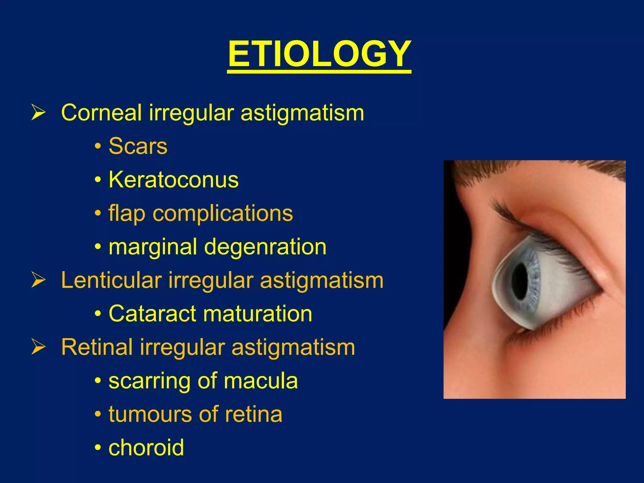
ERRORS OF REFRACTION
Dr Saurabh Kushwaha
Resident Ophthalmology
SCOPE
 Emmetropia
 Ametropia
 Hypermetropia
 Myopia
 Astigmatism
EMMETROPIA
 State of refraction where parallel rays of
light coming from infinity are focused at the
sensitive layer of retina with accommodation
being at rest
NORMAL VARIATION WITH AGE
 At birth
• Eyeball is relatively short
• +2 to +3D hyperopia which gradually reduces
 By the age of 5 -7 years
• Eye is emmetropic till the age of 50 years
• In 50% of the population emmetropia is not
reached and some degree of hypermetropia
persists
• On the other hand, the mark maybe overshot,
and the eye may become myopic
 After 50 years of age
• Tendency to develop hypermetropia again
• It due to 2 factors, both associated with lens
• Outer cortical fibers have lesser
curvature, decreasing the converging
power
• Refractive index of cortex increases and
lens becomes homogenous decreasing
the converging power
 After 65 years of age, all of the hypermetropia
becomes absolute due to loss of accommodation
AMETROPIA
 State of refraction where parallel rays of light
coming from infinity are focused either in front or
behind the sensitive layer of retina in one or
both meridian
 Components of ametropia
• Corneal power
• Anterior chamber depth
• Crystalline lens power
• Axial length
PREVALENCE OF AMETROPIA
 Stenstrom’s study from Uppsala, Sweden
• Low myopia (≤ 2D) : 29%
• Moderate myopia (2-6D) : 7%
• High myopia (>6D) : 2.5%
• Emmetropia & hypermetropia up to 2D: 61%
• High hypermetropia : 0.5%
HYPERMETROPIA
 State of refraction where parallel rays of
light coming from infinity are focused behind
the retina with accommodation being at rest
CLASSIFICATION
 Aetiological types
 Clinical types
 Classification by extent of error
AETIOLOGICAL TYPES
 Axial hypermetropia
 Curvatural hypermetropia
 Index hypermetropia
 Positional hypermetropia
 Absence of crystalline lens
 Consecutive hypermetropia
CLINICAL TYPES
 Simple hypermetropia
• Axial hypermetropia
• Curvatural hypermetropia
 Pathological hypermetropia
• Congenital hypermetropia
• Acquired hypermetropia
 Functional hypermetropia
CLASSIFICATION ON EXTENT
OF ERROR
 Low - refractive error of +2D or less
 Moderate - refractive error of +2.25 to +5.0D
 High - refractive error of +5.25D or more
COMPONENTS
 Total hypermetropia
 Latent hypermetropia
 Manifest hypermetropia
• Facultative hypermetropia
• Absolute hypermetropia
Total hypermetropia = Latent + Manifest (Facultative + Absolute)
SYMPTOMS
 Asymptomatic (<1D) in young patients is corrected
by mild accommodative effort
 Aesthenopic symptoms (1-2D), patients develops
aesthenopic symptoms due to sustained
accommodative efforts
 Defective vision (2-4D) with aesthenopic symptoms
 Defective vision (>4D ) only
SIGNS
 Reduced visual acuity
 Size of the eyeball may be normal or may
appear small as a whole. A scan may reveal short
anteroposterior length of the eyeball
 Cornea may be slightly smaller than normal,
may be cornea plana
 Anterior chamber is comparatively shallow since
the eyeball is small and the size of lens varies very
little and angle is narrow (predisposition to narrow
angle glaucoma)
FUNDUS EXAMINATION
 Optic disc may appear small and hyperaemic
with ill-defined margins and may mimic papillitis
 The vascular reflex may be accentuated and
the vessels may show undue tortuosity and
abnormal branching
 Foveal reflex may be situated at greater
distance from disc margin.
 The retina as a whole may shine due to
greater brilliance of light reflection (shot silk
appearance)
COMPLICATIONS
 Recurrent styes, blepharitis or chalazion
 Accommodative convergent squint
 Amblyopia
 Anisometropic
 Strabismus
 Uncorrected bilateral high hypermetropia
 Predisposition to develop primary narrow
angle glaucoma
MANAGEMENT
 Optical treatment
 Surgical treatment
 Visual hygiene
OPTICAL TREATMENT
 Basic principle is to prescribe convex lenses, so
that the light rays are brought to focus on the retina
 Total amount of hypermetropia should always be
discovered under complete cycloplegia
 Total manifest refractive error when small (≤1D),
correction is given only if the patient is
symptomatic
 Spherical correction given should be comfortably
acceptable to the patient.
CORRECTION IN CHILDREN
 Children < 4 years usually accept full cycloplegic measurement
 Once child reaches the school age, consider reducing the plus
power by about 1/3, but the child should not accommodate more
than 2.5D continually for distance
 Child may not accept the power prescribed. So, always first
under correct that the child accepts comfortably. Gradually
increase the correction at 6 months interval.
 If there is associated exophoria, hypermetropia is corrected by
1-2D
 In presence of accommodative convergent squint, full correction
should be given in the first sitting
 It is important to remember that hypermetropia may diminish
with the growth of the child, so refraction should be carried out
every 6 months
CORRECTION IN ADULTS
 Manifest hypermetropia is corrected
 Absolute + Facultative
 Maximum power with clear vision should be
prescribed which the patient is comfortable with
SURGICAL CORRECTION
 Conductive keratoplasty/corneal refractive
therapy
 Laser thermal keratoplasty
 Photorefractive Keratectomy
 Hyperopic LASIK
 Phakic IOLs
 Refractive lens exchange
VISUAL HYGIENE
 While reading or doing intensive near work,
take a break about every 30 mins
 Maintain proper reading distance
 Sufficient illumination
 Limit on time spent watching TV, videogames
 Sit 5 – 6 feet away from TV
MYOPIA
 State of refraction where parallel rays of light
entering the eye are focused in front of retina
with accommodation being at rest
CLASSIFICATION
 Aetiological types
 Clinical types
AETIOLOGICAL TYPES
 Axial myopia
 Curvatural myopia
 Index myopia
 Positional myopia
 Myopia due to excess accommodation
CLINICAL TYPES
 Congenital myopia
 Simple or developmental myopia
 Degenerative or pathological myopia
 Acquired myopia
CONGENITAL MYOPIA
 Common in premature babies or with birth
defects
 Stationary (8-10D)
 Associated with
• Increase in axial length
• Esotropia
• Other congenital anomalies of eye
 Early and full correction under cycloplegia
 Poor prognosis in unilateral cases with
severe myopia and anisometropia
SIMPLE MYOPIA
 Physiological/ school myopia
 Commonest type
 Results due to normal biological variations in
development of eye
 Age of onset is 7-10 yrs
 Moderate severity of <5D, never exceeds 8D
DEGENERATIVE MYOPIA
 Progressive in nature
 Related to heredity, general growth process
 Heredity linked growth of retina
 Factors affecting general growth process
 Age of onset is early adult life
 Severe >6D
SYMPTOMS
 Distant blurred vision
 Half shutting of eyes
 Asthenopic symptoms
 Muscae volitantes
 Night blindness
 Divergent squint
SIGNS
 Prominent eyeballs
 Large cornea
 Anterior chamber is deep
 Large & sluggishly reacting pupil
FUNDUS EXAMINATION
 Optic disc : appears large and pale & at its temporal edge,
myopic crescent.
 Sub retinal neovascularization
 Sub retinal hemorrhage
 Posterior staphyloma
Foster – Fuchs's spots
COMPLICATIONS
 Macular hemorrhage
 Retinal tears, detachment
 Vitreous hemorrhage
 Choroidal hemorrhage
 Complicated cataract
 Nuclear sclerosis
 Primary open angle glaucoma
MANAGEMENT
 Optical treatment
 Surgical treatment
OPTICAL TREATMENT
 Basic principle is to prescribe concave lenses, so
that the light rays are brought to focus on the retina
 Minimum acceptance providing maximum vision
 HIGH MYOPIA - undercorrection is done to avoid
• near vision problem
• minification of images
 Contact lenses are better
 LOW MYOPIA(<6D):Young children, glasses required only if
• Isometropia
<2years ≥ -4.0D
2-3years ≥ -3.0D
• Anisometropia:
≥ -2.5D
• Give full correction under cycloplegia
• Avoid overcorrection
 LOW MYOPIA(<6D): Adults
• <30years-full correction
• >30years-less than full correction with which patient is
comfortable for near vision
SURGICAL CORRECTION
 Radial keratotomy
 Lamellar corneal refractive procedures
 Laser based procedures
• PRK, LASIK, LASEK, C-LASIK, E-LASIK
 Miscellaneous corneal refractive procedures
• Orthokeratology
• Intracorneal contact leses
• Intra stromal corneal ring segments
• Gel injectable adjustable keratoplasty
 Intraocular refractive procedures
• Phakic refractive lenses
• Refractive lens exchange
ASTIGMATISM
 State of refraction where parallel rays of
light from a point source fail to meet in a focal
point, but form focal lines, resulting in a blurred
and imperfect image
 Types
•Regular astigmatism
•Irregular astigmatism
REGULAR ASTIGMATISM
 Refractive power changes uniformly from
one meridian to another principal meridian
 Depending upon axis and angle between
the two principal meridian
• With-the-rule astigmatism
• Against-the-rule astigmatism
• Oblique astigmatism
• Bi-oblique astigmatism
TYPES
 Simple astigmatism
• Simple hyperopic astigmatism
• Simple myopic astigmatism
 Compound astigmatism
• Compound hyperopic astigmatism
• Compound myopic astigmatism
 Mixed astigmatism
ETIOLOGY
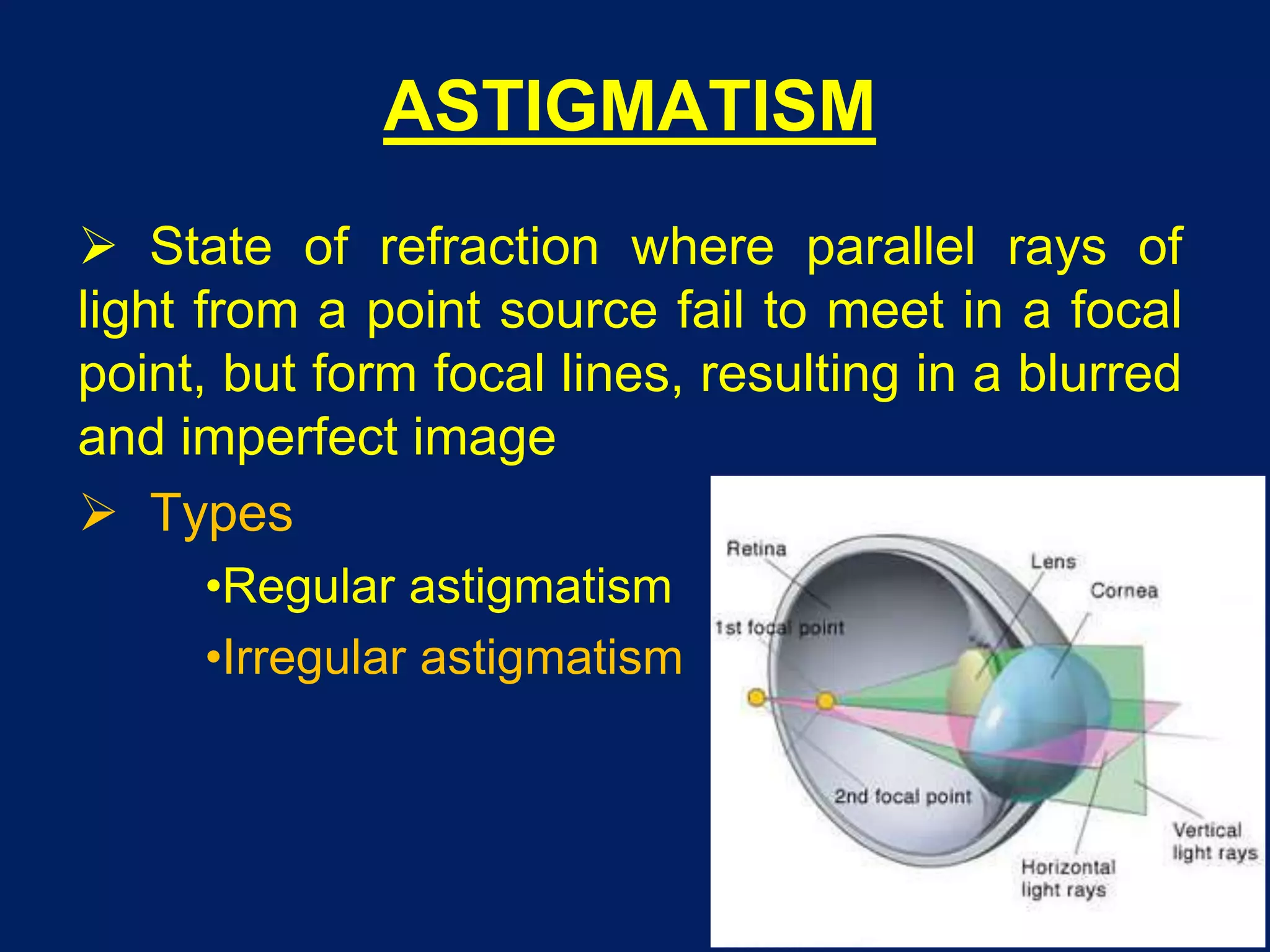
 Corneal astigmatism - curvatural [common]
 Lenticular is rare. It may be:
• Curvatural - lenticonus
• Positional - tilting or oblique placement
of lens, subluxation
 Retinal - oblique placement of macula [rare]
SYMPTOMS
 Blurring of vision
 Asthenopic symptoms
 Tilting of head
 Squint
INVESTIGATIONS
 Retinoscopy
 Keratometry
 Computerized corneal tomography
 Astigmatic fan test
 Jackson cross cylinder
MANAGEMENT
 Optical treatment
• Spectacles
• Contact lenses
 Surgical treatment
 Small astigmatism- treatment is required only
• In presence of asthenopic symptoms
• Decreased vision
 High astigmatism- full correction
• Better to avoid new astigmatic correction in adults
because of intolerable distraction
• Bi-oblique, mixed, high astigmatism are better
treated by contact lenses
IRREGULAR ASTIGMATISM
 Irregular change of refractive power in
different meridia
ETIOLOGY
 Corneal irregular astigmatism
• Scars
• Keratoconus
• flap complications
• marginal degenration
 Lenticular irregular astigmatism
• Cataract maturation
 Retinal irregular astigmatism
• scarring of macula
• tumours of retina
• choroid
SYMPTOMS
 Defective vision
 Distortion of objects
 Polyopia
INVESTIGATIONS
 Placido's disc test reveals distorted circles
 Computerized corneal topography
MANAGEMENT
 Optical treatment
• Contact lenses
• Scleral lenses
• Piggyback lens
 Surgical treatment
• Penetrating keratoplasty
THANK YOU

Errors of refraction

  • 1.
    ERRORS OF REFRACTION DrSaurabh Kushwaha Resident Ophthalmology
  • 2.
    SCOPE  Emmetropia  Ametropia Hypermetropia  Myopia  Astigmatism
  • 3.
    EMMETROPIA  State ofrefraction where parallel rays of light coming from infinity are focused at the sensitive layer of retina with accommodation being at rest
  • 4.
    NORMAL VARIATION WITHAGE  At birth • Eyeball is relatively short • +2 to +3D hyperopia which gradually reduces  By the age of 5 -7 years • Eye is emmetropic till the age of 50 years • In 50% of the population emmetropia is not reached and some degree of hypermetropia persists • On the other hand, the mark maybe overshot, and the eye may become myopic
  • 5.
     After 50years of age • Tendency to develop hypermetropia again • It due to 2 factors, both associated with lens • Outer cortical fibers have lesser curvature, decreasing the converging power • Refractive index of cortex increases and lens becomes homogenous decreasing the converging power  After 65 years of age, all of the hypermetropia becomes absolute due to loss of accommodation
  • 6.
    AMETROPIA  State ofrefraction where parallel rays of light coming from infinity are focused either in front or behind the sensitive layer of retina in one or both meridian  Components of ametropia • Corneal power • Anterior chamber depth • Crystalline lens power • Axial length
  • 7.
    PREVALENCE OF AMETROPIA Stenstrom’s study from Uppsala, Sweden • Low myopia (≤ 2D) : 29% • Moderate myopia (2-6D) : 7% • High myopia (>6D) : 2.5% • Emmetropia & hypermetropia up to 2D: 61% • High hypermetropia : 0.5%
  • 8.
    HYPERMETROPIA  State ofrefraction where parallel rays of light coming from infinity are focused behind the retina with accommodation being at rest
  • 9.
    CLASSIFICATION  Aetiological types Clinical types  Classification by extent of error
  • 10.
    AETIOLOGICAL TYPES  Axialhypermetropia  Curvatural hypermetropia  Index hypermetropia  Positional hypermetropia  Absence of crystalline lens  Consecutive hypermetropia
  • 11.
    CLINICAL TYPES  Simplehypermetropia • Axial hypermetropia • Curvatural hypermetropia  Pathological hypermetropia • Congenital hypermetropia • Acquired hypermetropia  Functional hypermetropia
  • 12.
    CLASSIFICATION ON EXTENT OFERROR  Low - refractive error of +2D or less  Moderate - refractive error of +2.25 to +5.0D  High - refractive error of +5.25D or more
  • 13.
    COMPONENTS  Total hypermetropia Latent hypermetropia  Manifest hypermetropia • Facultative hypermetropia • Absolute hypermetropia Total hypermetropia = Latent + Manifest (Facultative + Absolute)
  • 14.
    SYMPTOMS  Asymptomatic (<1D)in young patients is corrected by mild accommodative effort  Aesthenopic symptoms (1-2D), patients develops aesthenopic symptoms due to sustained accommodative efforts  Defective vision (2-4D) with aesthenopic symptoms  Defective vision (>4D ) only
  • 15.
    SIGNS  Reduced visualacuity  Size of the eyeball may be normal or may appear small as a whole. A scan may reveal short anteroposterior length of the eyeball  Cornea may be slightly smaller than normal, may be cornea plana  Anterior chamber is comparatively shallow since the eyeball is small and the size of lens varies very little and angle is narrow (predisposition to narrow angle glaucoma)
  • 16.
    FUNDUS EXAMINATION  Opticdisc may appear small and hyperaemic with ill-defined margins and may mimic papillitis  The vascular reflex may be accentuated and the vessels may show undue tortuosity and abnormal branching  Foveal reflex may be situated at greater distance from disc margin.  The retina as a whole may shine due to greater brilliance of light reflection (shot silk appearance)
  • 17.
    COMPLICATIONS  Recurrent styes,blepharitis or chalazion  Accommodative convergent squint  Amblyopia  Anisometropic  Strabismus  Uncorrected bilateral high hypermetropia  Predisposition to develop primary narrow angle glaucoma
  • 18.
    MANAGEMENT  Optical treatment Surgical treatment  Visual hygiene
  • 19.
    OPTICAL TREATMENT  Basicprinciple is to prescribe convex lenses, so that the light rays are brought to focus on the retina  Total amount of hypermetropia should always be discovered under complete cycloplegia  Total manifest refractive error when small (≤1D), correction is given only if the patient is symptomatic  Spherical correction given should be comfortably acceptable to the patient.
  • 20.
    CORRECTION IN CHILDREN Children < 4 years usually accept full cycloplegic measurement  Once child reaches the school age, consider reducing the plus power by about 1/3, but the child should not accommodate more than 2.5D continually for distance  Child may not accept the power prescribed. So, always first under correct that the child accepts comfortably. Gradually increase the correction at 6 months interval.  If there is associated exophoria, hypermetropia is corrected by 1-2D  In presence of accommodative convergent squint, full correction should be given in the first sitting  It is important to remember that hypermetropia may diminish with the growth of the child, so refraction should be carried out every 6 months
  • 21.
    CORRECTION IN ADULTS Manifest hypermetropia is corrected  Absolute + Facultative  Maximum power with clear vision should be prescribed which the patient is comfortable with
  • 22.
    SURGICAL CORRECTION  Conductivekeratoplasty/corneal refractive therapy  Laser thermal keratoplasty  Photorefractive Keratectomy  Hyperopic LASIK  Phakic IOLs  Refractive lens exchange
  • 23.
    VISUAL HYGIENE  Whilereading or doing intensive near work, take a break about every 30 mins  Maintain proper reading distance  Sufficient illumination  Limit on time spent watching TV, videogames  Sit 5 – 6 feet away from TV
  • 24.
    MYOPIA  State ofrefraction where parallel rays of light entering the eye are focused in front of retina with accommodation being at rest
  • 25.
  • 26.
    AETIOLOGICAL TYPES  Axialmyopia  Curvatural myopia  Index myopia  Positional myopia  Myopia due to excess accommodation
  • 27.
    CLINICAL TYPES  Congenitalmyopia  Simple or developmental myopia  Degenerative or pathological myopia  Acquired myopia
  • 28.
    CONGENITAL MYOPIA  Commonin premature babies or with birth defects  Stationary (8-10D)  Associated with • Increase in axial length • Esotropia • Other congenital anomalies of eye  Early and full correction under cycloplegia  Poor prognosis in unilateral cases with severe myopia and anisometropia
  • 29.
    SIMPLE MYOPIA  Physiological/school myopia  Commonest type  Results due to normal biological variations in development of eye  Age of onset is 7-10 yrs  Moderate severity of <5D, never exceeds 8D
  • 30.
    DEGENERATIVE MYOPIA  Progressivein nature  Related to heredity, general growth process  Heredity linked growth of retina  Factors affecting general growth process  Age of onset is early adult life  Severe >6D
  • 31.
    SYMPTOMS  Distant blurredvision  Half shutting of eyes  Asthenopic symptoms  Muscae volitantes  Night blindness  Divergent squint
  • 32.
    SIGNS  Prominent eyeballs Large cornea  Anterior chamber is deep  Large & sluggishly reacting pupil
  • 33.
    FUNDUS EXAMINATION  Opticdisc : appears large and pale & at its temporal edge, myopic crescent.
  • 34.
     Sub retinalneovascularization  Sub retinal hemorrhage
  • 35.
  • 36.
    COMPLICATIONS  Macular hemorrhage Retinal tears, detachment  Vitreous hemorrhage  Choroidal hemorrhage  Complicated cataract  Nuclear sclerosis  Primary open angle glaucoma
  • 37.
  • 38.
    OPTICAL TREATMENT  Basicprinciple is to prescribe concave lenses, so that the light rays are brought to focus on the retina  Minimum acceptance providing maximum vision  HIGH MYOPIA - undercorrection is done to avoid • near vision problem • minification of images  Contact lenses are better
  • 39.
     LOW MYOPIA(<6D):Youngchildren, glasses required only if • Isometropia <2years ≥ -4.0D 2-3years ≥ -3.0D • Anisometropia: ≥ -2.5D • Give full correction under cycloplegia • Avoid overcorrection  LOW MYOPIA(<6D): Adults • <30years-full correction • >30years-less than full correction with which patient is comfortable for near vision
  • 40.
    SURGICAL CORRECTION  Radialkeratotomy  Lamellar corneal refractive procedures  Laser based procedures • PRK, LASIK, LASEK, C-LASIK, E-LASIK  Miscellaneous corneal refractive procedures • Orthokeratology • Intracorneal contact leses • Intra stromal corneal ring segments • Gel injectable adjustable keratoplasty  Intraocular refractive procedures • Phakic refractive lenses • Refractive lens exchange
  • 41.
    ASTIGMATISM  State ofrefraction where parallel rays of light from a point source fail to meet in a focal point, but form focal lines, resulting in a blurred and imperfect image  Types •Regular astigmatism •Irregular astigmatism
  • 42.
    REGULAR ASTIGMATISM  Refractivepower changes uniformly from one meridian to another principal meridian  Depending upon axis and angle between the two principal meridian • With-the-rule astigmatism • Against-the-rule astigmatism • Oblique astigmatism • Bi-oblique astigmatism
  • 43.
    TYPES  Simple astigmatism •Simple hyperopic astigmatism • Simple myopic astigmatism  Compound astigmatism • Compound hyperopic astigmatism • Compound myopic astigmatism  Mixed astigmatism
  • 44.
    ETIOLOGY  Corneal astigmatism- curvatural [common]  Lenticular is rare. It may be: • Curvatural - lenticonus • Positional - tilting or oblique placement of lens, subluxation  Retinal - oblique placement of macula [rare]
  • 45.
    SYMPTOMS  Blurring ofvision  Asthenopic symptoms  Tilting of head  Squint
  • 46.
    INVESTIGATIONS  Retinoscopy  Keratometry Computerized corneal tomography  Astigmatic fan test  Jackson cross cylinder
  • 47.
    MANAGEMENT  Optical treatment •Spectacles • Contact lenses  Surgical treatment  Small astigmatism- treatment is required only • In presence of asthenopic symptoms • Decreased vision  High astigmatism- full correction • Better to avoid new astigmatic correction in adults because of intolerable distraction • Bi-oblique, mixed, high astigmatism are better treated by contact lenses
  • 48.
    IRREGULAR ASTIGMATISM  Irregularchange of refractive power in different meridia
  • 49.
    ETIOLOGY  Corneal irregularastigmatism • Scars • Keratoconus • flap complications • marginal degenration  Lenticular irregular astigmatism • Cataract maturation  Retinal irregular astigmatism • scarring of macula • tumours of retina • choroid
  • 50.
    SYMPTOMS  Defective vision Distortion of objects  Polyopia
  • 51.
    INVESTIGATIONS  Placido's disctest reveals distorted circles  Computerized corneal topography
  • 52.
    MANAGEMENT  Optical treatment •Contact lenses • Scleral lenses • Piggyback lens  Surgical treatment • Penetrating keratoplasty
  • 53.