Downloaded 205 times
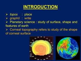
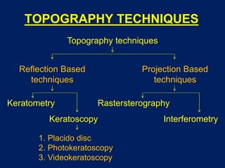
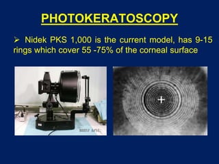
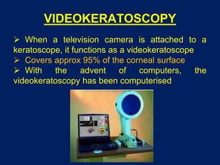
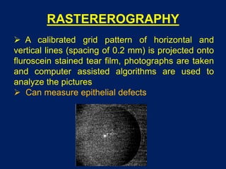
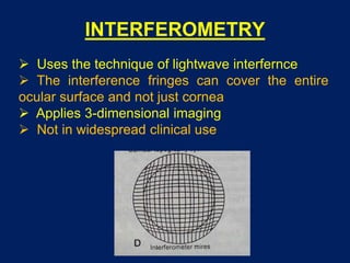
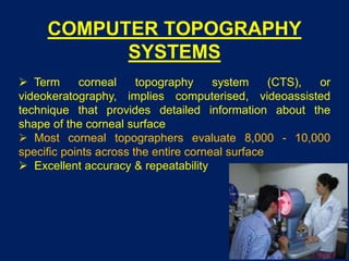

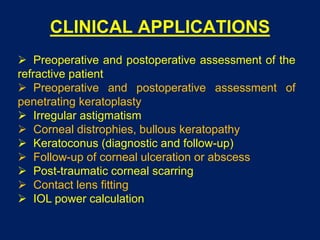
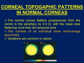
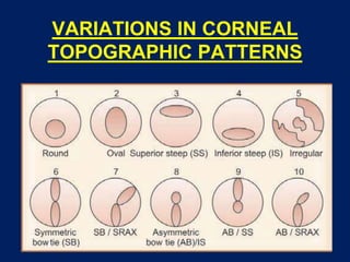


This document provides an overview of corneal topography. It begins by defining corneal topography as the study of the shape of the corneal surface. It then describes several techniques for evaluating corneal topography including keratometry, keratoscopy using Placido discs and photokeratoscopy, rasterstereography, and interferometry. Computerized topography systems that provide detailed maps of the corneal surface are also discussed. The document outlines clinical applications of corneal topography and variations in topographic patterns seen in normal and diseased corneas.


![Hypothalamus short ppt by Dr. Neha [PT].pptx](https://cdn.slidesharecdn.com/ss_thumbnails/hypothalamusbydr-260124145759-b9f94a93-thumbnail.jpg?width=640&height=640&fit=bounds)

