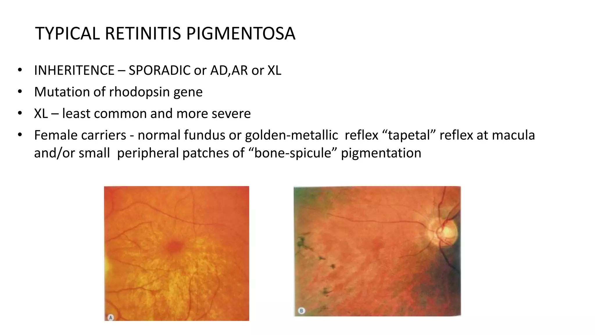
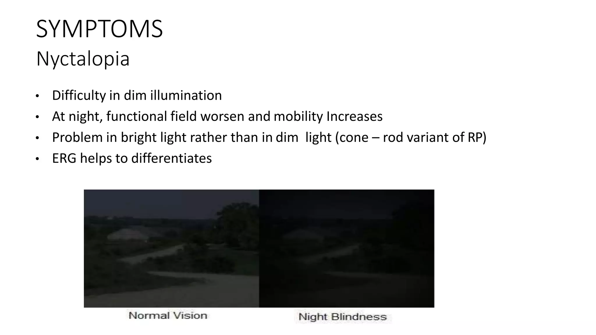
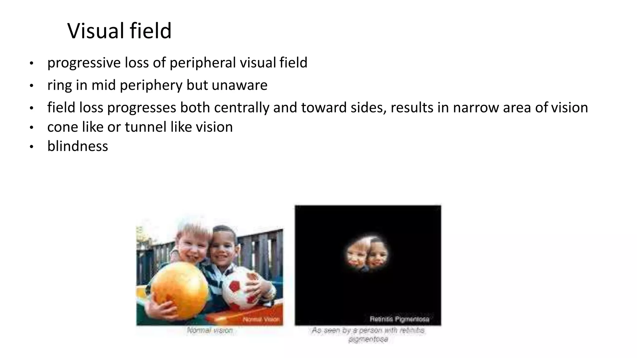
Retinitis pigmentosa is a group of inherited retinal diseases characterized by progressive vision loss. It has a prevalence of 1 in 3500 people. The document discusses the various types of retinitis pigmentosa based on inheritance patterns and symptoms. It also summarizes the clinical features, investigations, management, and associated syndromes of retinitis pigmentosa.
It describes about the procedure of Hess charting. it serves as a great tool to understand the concepts involved. Suitable for optometry course. This is not a routine procedure but an important procedure which is used in diagnosis.
Most retinal surgeons are trained to create formal retinal drawings of the fundus.
Retinal drawings are useful to document pathology, although more and more people now prefer fundus photographs.
Can be used for serial follow up of patients to document changes in the pathology.
This presentation is about a group of eye disorder, Retinitis Pigmentosa. RP is a non-treatable genetic disorder which cause due to degradation of photoreceptor cells in retina. Unfortunately we do not have a proper treatment for RP, but in this presentation I suggest some of options that can help a individual having RP.
Retinitis pigmentosa is rare heritable diseases which affect photoreceptor and make the patient unable to see normally in dimlight then loss of vision.
Similar to Retinitis pigmentosa and allied disorders (20)
Anti ulcer drugs and their Advance pharmacology ||
Anti-ulcer drugs are medications used to prevent and treat ulcers in the stomach and upper part of the small intestine (duodenal ulcers). These ulcers are often caused by an imbalance between stomach acid and the mucosal lining, which protects the stomach lining.
||Scope: Overview of various classes of anti-ulcer drugs, their mechanisms of action, indications, side effects, and clinical considerations.
Explore natural remedies for syphilis treatment in Singapore. Discover alternative therapies, herbal remedies, and lifestyle changes that may complement conventional treatments. Learn about holistic approaches to managing syphilis symptoms and supporting overall health.
- Video recording of this lecture in English language: https://youtu.be/lK81BzxMqdo
- Video recording of this lecture in Arabic language: https://youtu.be/Ve4P0COk9OI
- Link to download the book free: https://nephrotube.blogspot.com/p/nephrotube-nephrology-books.html
- Link to NephroTube website: www.NephroTube.com
- Link to NephroTube social media accounts: https://nephrotube.blogspot.com/p/join-nephrotube-on-social-media.html
Prix Galien International 2024 Forum ProgramLevi Shapiro
June 20, 2024, Prix Galien International and Jerusalem Ethics Forum in ROME. Detailed agenda including panels:
- ADVANCES IN CARDIOLOGY: A NEW PARADIGM IS COMING
- WOMEN’S HEALTH: FERTILITY PRESERVATION
- WHAT’S NEW IN THE TREATMENT OF INFECTIOUS,
ONCOLOGICAL AND INFLAMMATORY SKIN DISEASES?
- ARTIFICIAL INTELLIGENCE AND ETHICS
- GENE THERAPY
- BEYOND BORDERS: GLOBAL INITIATIVES FOR DEMOCRATIZING LIFE SCIENCE TECHNOLOGIES AND PROMOTING ACCESS TO HEALTHCARE
- ETHICAL CHALLENGES IN LIFE SCIENCES
- Prix Galien International Awards Ceremony
Title: Sense of Taste
Presenter: Dr. Faiza, Assistant Professor of Physiology
Qualifications:
MBBS (Best Graduate, AIMC Lahore)
FCPS Physiology
ICMT, CHPE, DHPE (STMU)
MPH (GC University, Faisalabad)
MBA (Virtual University of Pakistan)
Learning Objectives:
Describe the structure and function of taste buds.
Describe the relationship between the taste threshold and taste index of common substances.
Explain the chemical basis and signal transduction of taste perception for each type of primary taste sensation.
Recognize different abnormalities of taste perception and their causes.
Key Topics:
Significance of Taste Sensation:
Differentiation between pleasant and harmful food
Influence on behavior
Selection of food based on metabolic needs
Receptors of Taste:
Taste buds on the tongue
Influence of sense of smell, texture of food, and pain stimulation (e.g., by pepper)
Primary and Secondary Taste Sensations:
Primary taste sensations: Sweet, Sour, Salty, Bitter, Umami
Chemical basis and signal transduction mechanisms for each taste
Taste Threshold and Index:
Taste threshold values for Sweet (sucrose), Salty (NaCl), Sour (HCl), and Bitter (Quinine)
Taste index relationship: Inversely proportional to taste threshold
Taste Blindness:
Inability to taste certain substances, particularly thiourea compounds
Example: Phenylthiocarbamide
Structure and Function of Taste Buds:
Composition: Epithelial cells, Sustentacular/Supporting cells, Taste cells, Basal cells
Features: Taste pores, Taste hairs/microvilli, and Taste nerve fibers
Location of Taste Buds:
Found in papillae of the tongue (Fungiform, Circumvallate, Foliate)
Also present on the palate, tonsillar pillars, epiglottis, and proximal esophagus
Mechanism of Taste Stimulation:
Interaction of taste substances with receptors on microvilli
Signal transduction pathways for Umami, Sweet, Bitter, Sour, and Salty tastes
Taste Sensitivity and Adaptation:
Decrease in sensitivity with age
Rapid adaptation of taste sensation
Role of Saliva in Taste:
Dissolution of tastants to reach receptors
Washing away the stimulus
Taste Preferences and Aversions:
Mechanisms behind taste preference and aversion
Influence of receptors and neural pathways
Impact of Sensory Nerve Damage:
Degeneration of taste buds if the sensory nerve fiber is cut
Abnormalities of Taste Detection:
Conditions: Ageusia, Hypogeusia, Dysgeusia (parageusia)
Causes: Nerve damage, neurological disorders, infections, poor oral hygiene, adverse drug effects, deficiencies, aging, tobacco use, altered neurotransmitter levels
Neurotransmitters and Taste Threshold:
Effects of serotonin (5-HT) and norepinephrine (NE) on taste sensitivity
Supertasters:
25% of the population with heightened sensitivity to taste, especially bitterness
Increased number of fungiform papillae
Report Back from SGO 2024: What’s the Latest in Cervical Cancer?bkling
Are you curious about what’s new in cervical cancer research or unsure what the findings mean? Join Dr. Emily Ko, a gynecologic oncologist at Penn Medicine, to learn about the latest updates from the Society of Gynecologic Oncology (SGO) 2024 Annual Meeting on Women’s Cancer. Dr. Ko will discuss what the research presented at the conference means for you and answer your questions about the new developments.
Factory Supply Best Quality Pmk Oil CAS 28578–16–7 PMK Powder in Stockrebeccabio
Factory Supply Best Quality Pmk Oil CAS 28578–16–7 PMK Powder in Stock
Telegram: bmksupplier
signal: +85264872720
threema: TUD4A6YC
You can contact me on Telegram or Threema
Communicate promptly and reply
Free of customs clearance, Double Clearance 100% pass delivery to USA, Canada, Spain, Germany, Netherland, Poland, Italy, Sweden, UK, Czech Republic, Australia, Mexico, Russia, Ukraine, Kazakhstan.Door to door service
Hot Selling Organic intermediates
These lecture slides, by Dr Sidra Arshad, offer a quick overview of physiological basis of a normal electrocardiogram.
Learning objectives:
1. Define an electrocardiogram (ECG) and electrocardiography
2. Describe how dipoles generated by the heart produce the waveforms of the ECG
3. Describe the components of a normal electrocardiogram of a typical bipolar leads (limb II)
4. Differentiate between intervals and segments
5. Enlist some common indications for obtaining an ECG
Study Resources:
1. Chapter 11, Guyton and Hall Textbook of Medical Physiology, 14th edition
2. Chapter 9, Human Physiology - From Cells to Systems, Lauralee Sherwood, 9th edition
3. Chapter 29, Ganong’s Review of Medical Physiology, 26th edition
4. Electrocardiogram, StatPearls - https://www.ncbi.nlm.nih.gov/books/NBK549803/
5. ECG in Medical Practice by ABM Abdullah, 4th edition
6. ECG Basics, http://www.nataliescasebook.com/tag/e-c-g-basics
Ethanol (CH3CH2OH), or beverage alcohol, is a two-carbon alcohol
that is rapidly distributed in the body and brain. Ethanol alters many
neurochemical systems and has rewarding and addictive properties. It
is the oldest recreational drug and likely contributes to more morbidity,
mortality, and public health costs than all illicit drugs combined. The
5th edition of the Diagnostic and Statistical Manual of Mental Disorders
(DSM-5) integrates alcohol abuse and alcohol dependence into a single
disorder called alcohol use disorder (AUD), with mild, moderate,
and severe subclassifications (American Psychiatric Association, 2013).
In the DSM-5, all types of substance abuse and dependence have been
combined into a single substance use disorder (SUD) on a continuum
from mild to severe. A diagnosis of AUD requires that at least two of
the 11 DSM-5 behaviors be present within a 12-month period (mild
AUD: 2–3 criteria; moderate AUD: 4–5 criteria; severe AUD: 6–11 criteria).
The four main behavioral effects of AUD are impaired control over
drinking, negative social consequences, risky use, and altered physiological
effects (tolerance, withdrawal). This chapter presents an overview
of the prevalence and harmful consequences of AUD in the U.S.,
the systemic nature of the disease, neurocircuitry and stages of AUD,
comorbidities, fetal alcohol spectrum disorders, genetic risk factors, and
pharmacotherapies for AUD.
The prostate is an exocrine gland of the male mammalian reproductive system
It is a walnut-sized gland that forms part of the male reproductive system and is located in front of the rectum and just below the urinary bladder
Function is to store and secrete a clear, slightly alkaline fluid that constitutes 10-30% of the volume of the seminal fluid that along with the spermatozoa, constitutes semen
A healthy human prostate measures (4cm-vertical, by 3cm-horizontal, 2cm ant-post ).
It surrounds the urethra just below the urinary bladder. It has anterior, median, posterior and two lateral lobes
It’s work is regulated by androgens which are responsible for male sex characteristics
Generalised disease of the prostate due to hormonal derangement which leads to non malignant enlargement of the gland (increase in the number of epithelial cells and stromal tissue)to cause compression of the urethra leading to symptoms (LUTS
Pulmonary Thromboembolism - etilogy, types, medical- Surgical and nursing man...VarunMahajani
Disruption of blood supply to lung alveoli due to blockage of one or more pulmonary blood vessels is called as Pulmonary thromboembolism. In this presentation we will discuss its causes, types and its management in depth.
4. Also known as
“tapetoretinal degeneration” (by Leber in 1916)
“primary pigmentary retinal degeneration,”
“pigmentary retinopathy,”
“Rod cone dystrophy.”
5. PREVALENCE
• Prevalence 1 in 3500 to 1 in 4500
• South India – common retinal cause of blindness
• Andhra Pradesh – prevalence of 0.8% in a population
7. • INHERITENCE – SPORADIC or AD,AR or XL
• Mutation of rhodopsin gene
• XL – least common and more severe
• Female carriers - normal fundus or golden-metallic reflex “tapetal” reflex at macula
and/or small peripheral patches of “bone-spicule” pigmentation
TYPICAL RETINITIS PIGMENTOSA
8. Modes of inheritence
• XLR is the least common but most sever e form, and may
result in complete blindness by the third or fourth decades
• AR disease can also be severe
• Sporadic cases may have a more favourable prognosis, with
retention of central vision until the sixth decade or later
• AD disease generally has the best prognosis
9. SYMPTOMS
Nyctalopia
• Difficulty in dim illumination
• At night, functional field worsen and mobility Increases
• Problem in bright light rather than in dim light (cone – rod variant of RP)
• ERG helps to differentiates
10. Visual field
• progressive loss of peripheral visual field
• ring in mid periphery but unaware
• field loss progresses both centrally and toward sides, results in narrow area of vision
• cone like or tunnel like vision
• blindness
11. • In general, there is a strong tendency for the visual field loss to be symmetric
between the two eyes.
• A notable exception to this is the phenotype expressed in female carriers of X-
linked RP.
• In this situation, the distribution of mutant photoreceptors in the retina is
determined by lyonization (X-chromosomal inactivation).
• This is a random event determining which genes of the two X chromosomes (the
normal or mutant copy) are expressed in a particular cell.
• This leads not only to unusual, irregular patterns of visual field loss in individual
eyes but also to quite striking differences in field loss between the two eyes.
12. LIGHT AND GLARE PROBLEMS
• Photopsia or photophobia
• Photopsias are described as tiny, blinking or shimmering lights or as a coarse, sparkling
graininess to vision
• As the scotomas become denser over the years, the photopsia decrease and finally
disappear
• The cellular or tissue correlates that underlie photopsia in RP are unknown but may
include photoreceptor dysfunction, neural sprouting, aberrant synapse formation, and
secondary remodeling of the retina, all of which occur as sequelae of photoreceptor
degeneration.
13. Central vision loss
• Cystoid macular edema (CME)
• diffuse retinal vascular leakage
• macular preretinal fibrosis
• retinal pigment epithelial (RPE) defects in the macula or fovea
• frequent fluctuations in vision “good days and bad days”
14. Fundus Appearance
• attenuated retinal vessels,
• mottling and granularity of the RPE,
• bone spicule intraretinal pigmentation,
• optic nerve head pallor
15. • Patients who have very early RP without fundus pigmentary abnormalities are
often diagnosed as having RP sine pigmento or pauci pigmentary RP.
• This is no longer considered a specific subtype of RP but a stage through which
many, if not most, patients with RP pass.
• The sine pigmento stage may exist for decades before typical RP signs appear.
16. • Intraretinal, bone spicule pigment formations represent migration of pigment from disintegration
of RPE cells with accumulation in the interstitial spaces surrounding retinal vessels.
• This process occurs most prominently at the junctions of vessels producing perivascular pigmentary
cuffing and spicule-shaped deposits.
• The loss of pigment within the pigment epithelial cells often produces an overall gray, desaturated
appearance to the retina with greater visibility of underlying choroidal vessels through the more
transparent pigment epithelium.
• Eventually, the normal salmon-pink color of the entire mid- and far peripheral fundus is replaced by
dense bone spicule pigmentary formations
17. GRADUAL INCREASE IN DENSITY OF THE
PIGMENTARY CHANGES WITH ANTERIOR AND
POSTERIOR SPREAD
19. Ganglion cell degeneration
Increase in optic atrophy
Wax like yellowish appearance
Consecutive atrophy with macular atrophy
Epiretinal
Membrane
20. The optic disc may be normal in early RP, show a waxy fullness with hyperemia,
or appear waxy and pale A “golden ring” or yellowish-white halo
Golden ring
Waxy pallor
Peripappilary drusen-common
in Ushers syndrome
21. Vitreous abnormality
• presence of fine, dust-like pigmented cells released from degeneration of the
RPE.
• complete posterior detachment of the vitreous
• “cotton-ball” opacities
• interwoven filaments in the retrocortical space
• spindle shaped vitreous condensations
22. Severe and generalized pigmentary changes throughout the macula and periphery as
well as inferior exudate and a serous retinal detachment .
The patient received laser photocoagulation leading to resolution of the subretinal
fluid.
23. • Posterior subcapsular cataract
• Open angle glaucoma
• Myopia common
• Keratoconus uncommon
• Vitreous changes – post. Vitreous Detachment
• Optic disc drusen very frequent
ANTERIOR SEGMENT COMPLICATION
24. INVESTIGATIONS
HISTORY – Rate of progression, functional difficulties, systemic associations,
family history, allergic , psychosocial and educational history
VISUAL ACUITY – Helps to know the extent of the disease
Early stage – partial or complete annular or ring
scotoma seen corresponding to degenerated zone
of retina.
Late stage – field becomes smaller, reduces to restricted area
around the fixation point
Long duration Central Vision remains a.k.a “Tubular Vision”
25. • COLOUR VISION –
Good Central Vision – Normal colour vision
Involvement of CONES – colour vision impaired
• AMSLER GRID – for any distortion/scotoma in central 10 degree of visual field
• Flash light evaluation – squint, ptosis(kearns-sayre syndrome), nystagmus, pupillary
reactions.
• S.L.E – Conjunctival or Corneal deposits ( e.g in cystinosis) with Lens Status
• Anterior Chamber Depth and Gonioscopy I.O.P
• I.D.O and 90D/78D examination
• Systemic evaluation
26. ERG IN RETINITIS PIGMENTOSA
• In Diagnosis of RP.
• Distinguish some types of RP.
• Screening of Relatives of patient of RP.
• Follow up of disease .
• D D from other similar dystrophies.
27. DIAGNOSIS
AMPLITUDE
• Usual ERG findings (Wide Field ERG) in RP include general reduction in the amplitude(a
& b waves) of ERG responses .
• Reduction of scotopic rod response is the first ERG sign.
• Maximum response which reflects rod and cone activity may be affected after scotopic
responses become abnormal.
• Visual electrophysiologists usually concentrate on scotopic and mesopic results for early
detection of RP.
28. 2) Implicit Time:
• RP patients often show prolonged b-wave implicit times.
• However, in the early stages of the disease, ERG may show normal amplitude and
implicit time values.
29. MERG IN DIAGNOSIS OF RP
MERG from a
patient with RP. No
activity is recorded
in the periphery.
Responses are
obtained centrally,
but are abnormal in
both amplitude and
implicit time
30. DISTINGUISH SOME TYPES OF RP
Rod responses to dim blue light under dark-adapted conditions are reduced in all
genetic types and, when detectable, are delayed in b- wave implicit times.
In the dominant with reduced penetrance, X-linked, and autosomal recessive forms of
RP,cone b-wave implicit times, are so delayed
The subnormal responses with delayed b-wave implicit times seen in the widespread
progressive forms of RP contrast with the subnormal responses with normal b-wave
implicit times seen in self- limited sector RP
31. Electroretinographic
responses for a normal
subject and four patients with
retinitis pigmentosa (ages 13,
14, 14, and 9).
Responses were obtained after
45 minutes of dark adaptation
to single flashes of blue light
(left column) and white light
(middle column). Responses
(right column) were obtained
to 30- cycles-per-second (or
30-Hz) white flickering light
32. SCREENING OF RELATIVES OF PATIENT OF RP
Full-field ERGs can be used not only to detect which patients are affected with the early
stages of RP but also to determine which relatives are normal. In families with RP,
patients age 6 and older with normal full-field ERGs with normal cone and rod
amplitudes and normal cone and rod b- wave implicit times have not been observed to
develop RP at a later time.
33. MANAGEMENT
• REFRACTIVE ERROR
Refractive correction
HIGH HYPERMETROPIA -leber’s congenital amaurosis
RP and keratoconus could coexist.
• CATARACT –
Develops in early stages
VA decrease and glare increase
34. MACULAR EDEMA and EPIRETINAL MEMBRANES
• Affect of Central Vision by C.M.E
• best detected by O.C.T and F.F.A
• Thin Epiretinal Membrane in front of
Macula causes Central Vision Loss
• Vitreous Surgery to peel the membrane
also removes Vitreous opacities
• Systemic Acetazolamide
35. GENETIC STUDY –
• Modes - AD,AR,XL
• Closed relative marriage increase risk
• Pedigree tree drawn for all patients
• Genetic counseling
VISUAL REHABLITATION IN END STAGE
38. Usher syndrome
• About 15% to 20% of affected individuals with retinitis pigmentosa
have associated hearing loss
three major types
Type I (75%) - profound congenital sensorineural deafness and severe RP
with an extinguished ERG in the first decade plus unintelligible speech &
vestibular ataxia
Type III (2%), with progressive hearing loss, vestibular dysfunction and
relatively late-onset pigmentary retinopathy
39. Kearns–Sayre syndrome
• Part of chronic progressive external
ophthalmoplegia
• Mitochondrial inheritance
• Abnormalities include
• Ptosis
• diffuse disturbance of the RPE
• ERGs that are usually reduced in amplitude
• respiratory distress
• heart block which may require a pacemaker
40. Bassen–Kornzweig syndrome
• Abetalipoproteinaemia
• Malabsorption of fat and fat-soluble vitamin
• Failure to thrive in infancy, acanthocytosis
• Spinocerebelar ataxia
• The fundus exhibits scattered white dots followed by RP-like
changes developing towards the end of the first decade; there
may also be ptosis, ophthalmoplegia, strabismus and
nystagmus
41. Refsum disease
• The patient accumulates exogenous phytanic acid
• Findings include a peripheral neuropathy, ataxia, an increase in CSF protein with a
normal cell count, retinitis pigmentosa ,ichtyosis
• All have elevated serum phytanic acid
• A defect exists in the conversion of phytanic acid to alpha- hydroxy phytanic acid
which results in its accumulation
• Treatment consists of restricting not only animal fats and milk products (i.e., foods that
contain phytanic acid) but also green leafy vegetables containing phytol
42.
43. Bardet–Biedl syndrome
• Includes RP, mental retardation, polydactylism, apple-
shaped obesity, and hypogonadism.
• Almost 80% have severe changes by the age of 20
years
44. Retinitis pigmentosa sine pigmento
• Sine pimento = Without pigment
• Absence or paucity of pigment accumulation
• May subsequently appear with time
• Functional manifestations are similar to typical RP
45. Retinitis punctata albescens
• Albescens = whitish
• Scattered whitish-yellow spots, most numerous at the
equator, usually sparing the macula, and associated with
arteriolar attenuation
• Nyctalopia and progressive field loss occur
46. Sector retinitis pigmentosa
• Sectoral RP
• AD
• generally involvement of inferior quadrants
• Progression is slow (many cases are apparently stationary)
• Unilateral RP can also occur
47. Leber congenital amaurosis
• Severe rod-cone dystrophy
• The commonest genetically defined cause of visual impairment in
children
• ERG is usually non-recordable even in early cases
• Systemic associations include
• mental handicap, deafness, epilepsy, central nervous system and
renal anomalies, skeletal malformations and endocrine dysfunction
48. • Presentation
• Blindness at birth or early infancy
• associated with roving eye movements
• Photoaversion
• Cataract
• Hypermetropia
• Nystagmus
49. • Signs are variable but may include:
• Absent or diminished pupillary light reflex
• The fundi may be normal in early life apart from mild arteriolar narrowing
• Initially mild peripheral pigmentary retinopathy, salt and pepper changes, and less
frequently yellow flecks
• Severe macular pigmentation
• Pigmentary retinopathy, optic atrophy and severe arteriolar narrowing in later
childhood
• Oculodigital syndrome: constant rubbing of the eyes may cause orbital fat atrophy
with enophthalmos, and subsequent keratoconus or keratoglobus
50.
51. Pigmented paravenous chorioretinal atrophy
• Usually asymptomatic and non-progressive
• ERG is normal
• Paravenous bone-spicule pigmentation
together with sharply outlined zones of
chorioretinal atrophy that follow the course of
the major retinal veins
• Changes may also encircle the optic disc
• The optic disc and vascular calibre are
usually normal