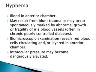
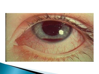
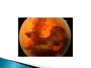
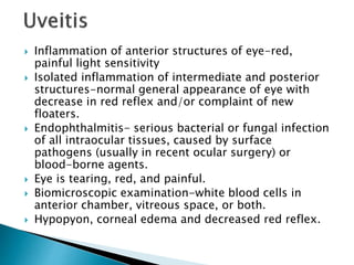
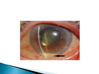
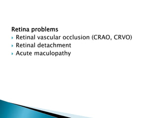
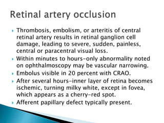
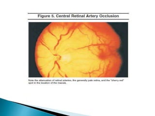
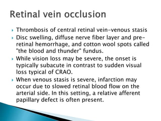
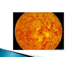
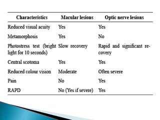
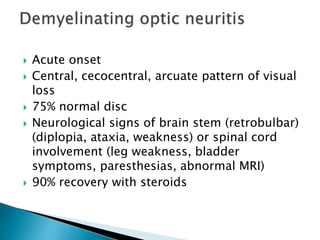
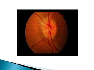
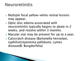
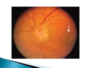
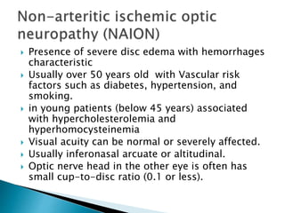
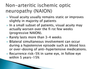
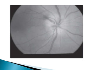
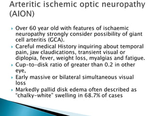
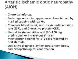
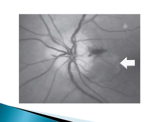
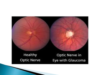
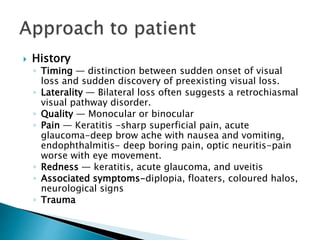
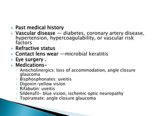
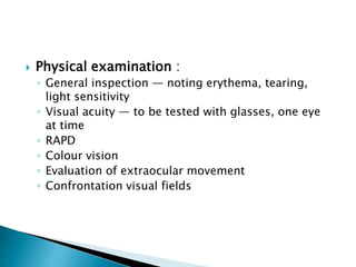
Dr. Parag Moon
Senior resident
Dept. of Neurology
GMC, Kota.
 Transient visual loss (<24hr)
 Persistent visual loss(>24 hr)
◦ Sudden, painless
◦ Gradual, painless
◦ Painful
 Monocular: anterior to chiasm
 Binocular: posterior to chiasm
1. Embolic cerebrovascular disease
2. Ocular (intermittent angle closure
glaucoma, hyphema, impending central
retinal vein occlusion, optic disc edema)
3. Vasculitis (GCA)
4. Migraine/vasospasm
5. Other (Uhthoff phenomenon, idiopathyic,
nonorganic)
Pathologically can be broadly divided into
three major categories:
 Media problems
 Retinal problems
 Neural visual pathway problems
 Keratitis
• Infectious or non-infectious
 Corneal edema
• Acute glaucoma (primarily)
 Hyphema
• Spontaneous or traumatic
 Alterations in crystalline lens
• Thickening, clouding or dislocation
 Vitreous hemorrhage
• Spontaneous or traumatic Uveitis
 Inflammation of cornea due to trauma, abrasive
exposure, allergy or infection.
 Marked by cloudiness, irregularity or loss of
epithelial or sub-epithelial corneal tissues.
 Typically eye is tearing, red, painful or irritated.
 Loss of epithelial cells demonstrated by corneal
uptake of fluorescein dye, creating a focal or
diffuse green glow under a cobalt blue light.
 Deeper corneal disease may be visible as a focal or
diffuse white opacity, or by dulling of usually
distinct reflection of light off of cornea (corneal
light reflex).
 Loss of corneal clarity.
 Dulling of corneal light reflex or frank grey or white
color to substance of cornea.
 Acute angle-closure glaucoma.
 Typically has nausea, vomiting, and may see coloured
halos around lights.
 Eye is tearing, red, and extremely painful, often with
ipsilateral brow ache.
 Pupil may be fixed in mid-dilated position
 Slit lamp examination- a shallow anterior chamber.
 IOP is often dangerously elevated (usually 40 to 80
mmHg).
 Suspected by noting hardness to palpation in
comparison to fellow eye.
 Blood in anterior chamber.
 May result from blunt trauma or may occur
spontaneously marked by abnormal growth
or fragility of iris blood vessels (often in
chronic poorly controlled diabetes).
 Biomicroscopic examination reveals red blood
cells circulating and/or layered in anterior
chamber.
 Intraocular pressure may become
dangerously elevated.
 Changes in size, clarity, or positioning of the
crystalline lens-alter focus of light onto retina.
 Trauma or a variety of congenital conditions can
lead to lens dislocation and resultant vision loss.
 Lens clouding (cataract)-generally chronic.
 Elevated blood glucose can cause increased lens
tumescence, altering refractive error. If change is
great enough, patients may perceive vision loss.
 Vision impairment typically resolves within days
to weeks of normalization of blood glucose
 Can occur in setting of trauma, spontaneous
retinal tear, spontaneous vitreous detachment or
in any condition with retinal neovascularization
(poorly controlled diabetes).
 Reduction in vision directly proportional to
amount of blood in vitreous.
 Hemorrhage if dense enough, there may be a
decreased red reflex (reddish orange reflection
off subretinal layers when examining eye with an
ophthalmoscope), or retina may not be visible
with ophthalmoscopy.
 Inflammation of anterior structures of eye-red,
painful light sensitivity
 Isolated inflammation of intermediate and posterior
structures-normal general appearance of eye with
decrease in red reflex and/or complaint of new
floaters.
 Endophthalmitis- serious bacterial or fungal infection
of all intraocular tissues, caused by surface
pathogens (usually in recent ocular surgery) or
blood-borne agents.
 Eye is tearing, red, and painful.
 Biomicroscopic examination-white blood cells in
anterior chamber, vitreous space, or both.
 Hypopyon, corneal edema and decreased red reflex.
Retina problems
 Retinal vascular occlusion (CRAO, CRVO)
 Retinal detachment
 Acute maculopathy
 Thrombosis, embolism, or arteritis of central
retinal artery results in retinal ganglion cell
damage, leading to severe, sudden, painless,
central or paracentral visual loss.
 Within minutes to hours-only abnormality noted
on ophthalmoscopy may be vascular narrowing.
 Embolus visible in 20 percent with CRAO.
 After several hours-inner layer of retina becomes
ischemic, turning milky white, except in fovea,
which appears as a cherry-red spot.
 Afferent papillary defect typically present.
 Thrombosis of central retinal vein-venous stasis
 Disc swelling, diffuse nerve fiber layer and pre-
retinal hemorrhage, and cotton wool spots called
"the blood and thunder" fundus.
 While vision loss may be severe, the onset is
typically subacute in contrast to sudden visual
loss typical of CRAO.
 When venous stasis is severe, infarction may
occur due to slowed retinal blood flow on the
arterial side. In this setting, a relative afferent
papillary defect is often present.
 May occur spontaneously or trauma.
 Most common form-due to a tear or break in
retina.
 Sudden onset of new floaters or black dots in
their vision, often accompanied by flashes of
light (photopsias).
 Not painful and does not cause a red eye.
 Dulling of red reflex and ophthalmoscopic
examination may reveal the retina to be elevated
with folds.
 If extensive, there may be a relative afferent
pupillary defect.
 Associated with a central blind spot
(scotoma), blurred vision, or visual distortion.
 May be due to fluid leakage, bleeding,
infection, or can occur de novo or as an acute
worsening of a chronic disease (eg, new
edema in previously dry diabetic retinopathy)
 Diagnosis requires detailed ophthalmoscopy
with a high magnification lens through
pharmacologically dilated pupils.
 Acute onset
 Central, cecocentral, arcuate pattern of visual
loss
 75% normal disc
 Neurological signs of brain stem (retrobulbar)
(diplopia, ataxia, weakness) or spinal cord
involvement (leg weakness, bladder
symptoms, paresthesias, abnormal MRI)
 90% recovery with steroids
 First described by Dutton and colleagues in 1982.
 Prone to recurrent optic neuritis events that respond
to corticosteroid therapy.
 Vision loss may manifest during a steroid taper
 More frequent in women.
 Optic nerve may appear mildly edematous during
acute events
 79% have abnormal antinuclear antibody (ANA),82% -
elevated anticardiolipin antibody titers.
 Skin biopsy may show leukocytoclastic or
lymphohistiocytic vasculitis (or both) and
immunereactant deposition
 Nonspecific inflammation of optic nerve
similar to typical optic neuritis
 Infiltration of optic nerve or sheaths
mimicking mass lesions, such as optic nerve
glioma or meningioma
 Mass effect due to direct compression of
nerve
 Parachiasmal involvement
 Typically affects young, healthy adults
 Two-thirds-antecedent viral prodrome.
 Cecocentral and central type visual field
defect.
 Present with stellate maculopathy and optic
disc edema,
 May be associated with exudative detachment
in peripapillary region.
 Multiple focal yellow-white retinal lesions
may appear,
 Optic disc edema associated with
neuroretinitis typically begins to abate in 2
weeks, and resolve within 3 months.
 Macular star may be present for up to a year.
 Catscratch disease (Bartonella henselae),
syphilis(treponema pallidum), Lymes
disease(B. Burgdorfelia)
 Presence of severe disc edema with hemorrhages
characteristic
 Usually over 50 years old with Vascular risk
factors such as diabetes, hypertension, and
smoking.
 in young patients (below 45 years) associated
with hypercholesterolemia and
hyperhomocysteinemia
 Visual acuity can be normal or severely affected.
 Usually inferonasal arcuate or altitudinal.
 Optic nerve head in the other eye is often has
small cup-to-disc ratio (0.1 or less).
 Visual acuity usually remains static or improves
slightly in majority of patients
 In a small subset of patients, visual acuity may
actually worsen over the fi rst few weeks
(progressive NAION).
 Rarely lasts more than 3–4 weeks
 Bilateral simultaneous involvement can occur
during a hypotensive episode such as blood loss
or over-dosing of anti-hypertensive medications.
 Recurrence risk-5% in same eye, in follow eye
within 5 years-15%
 Over 60 year old with features of ischaemic
neuropathy strongly consider possibility of giant
cell arteritis (GCA).
 Careful medical History inquiring about temporal
pain, jaw claudications, transient visual or
diplopia, fever, weight loss, myalgias and fatigue.
 Cup-to-disk ratio of greater than 0.2 in other
eye,
 Early massive or bilateral simultaneous visual
loss
 Markedly pallid disk edema often described as
“chalky-white” swelling in 68.7% of cases
 Choroidal infarcts.
 End-stage optic disc appearance characterized by
marked cupping with pallor.
 Complete blood count, erythrocyte sedimentation
rate (ESR), and C-reactive protein (CRP).
 Steroid treatment either oral (80–120 mg
prednisone) or intravenous (1 gram
methyleprdnisolone) for 3–5 days followed by
oral steroids.
 Defi nitive diagnosis by temporal artery biopsy
and histopathological confirmation
 Optic nerve can be infiltrated in systemic
malignancies such as lymphoma, leukemia, multiple
myeloma, and carcinoma
 Optic disc can be swollen or normal in appearance.
 MRI of brain and orbit may show meningeal and optic
nerve enhancement.
 Spinal tap recommended in suspected CNS
malignancy but more than one spinal tap may be
needed to detect malignant cells
 In case of localized optic nerve infiltration with no
evidence of systemic disease, histopathological
diagnosis by direct optic nerve sheath biopsy
 Dominant optic neuropathy (Kjers’ type)
 Present in first decade of life
 Bilateral central or cecocentral scotomas.
 Color vision deficit along tritan (blue-yellow)
axis.
 Optic disc-temporal pallor and in some cases
severe excavation and cupping.
 Recessive optic neuropathy rare
 First year of life
 Associated with diabetes mellitus, diabetes
inspidus, and deafness (Wolfram syndrome).
 Acute unilateral, painless, visual loss.
 Sequential bilateral involvement may occur
weeks or months later.
 Visual field defects-central or cecocentral as
papillo-macular bundle is first and most
severely affected
 Fundoscopy may show disk swelling,
thickening of the peripapillary retinal nerve
fiber layer and peripapillary retinal
telangectatic vessels which do not leak on
flourescin angiography.
 MRI-optic nerve enhancement and white
matter lesions,
 LHON has 4 primary mitochondrial genome
mutations; G11778A, G3460A and T14484C
and T10663C.
 Male:female ratio-2.5:1 for G11778A and
G3460A mutation
 6:1 ratio for T14484C mutation.
 Present with vision loss, months or years
following history of radiation exposure to
brain or orbit.
 Risk increased with concomitant
chemotherapy .
 Mechanism-ischemia caused by endothelial
cell injury from radiation.
 Optic disc is usually normal but can be
swollen.
 MRI-optic nerve enhancement with
gadolinium
 May also have radiation retinopathy with
retinal hemorrhages, cotton wool spots,
exudates and macular edema.
 Visual prognosis is poor with 45% ending with
no light perception visual acuity.
 Usually had suffered craniofacial trauma but
occasionally mild orbital or eye injury.
 RAPD-main clue to diagnosis.
 CT scan of orbit recommended detect any
bony fractures, fractures of optic canal, and
acute orbital hemorrhages.
 High-dose steroid therapy-within 8 hrs of
injury
 Small cell and non-small cell lung carcinoma
of lung
 Often have bilateral disc swelling and
progressive visual loss before diagnosis of
systemic malignancy made.
 Collapsing response-mediating protein
(CRMP- 5) found to be useful marker with
lung carcinoma.
 Classically diagnosed by presence of
progressive optic nerve cupping with
concurrent progressive VF loss (arcuate nasal
scotomas).
 Diagnosis aided by presence of risk factors
such as elevated intraocular pressure (IOP),
positive family history, predisposed race,
advanced age and thin central corneal
thickness
 Notching of the rim.
 Verticalization of the optic cup.
 Acquired optic pit.
 Baring of a circumlinear vessel.
 Vessel bayoneting at the optic rim (indicating bean-
pot cupping).
 Nasalization of vessels.
 Disc hemorrhage (Drance hemorrhage).
 Abnormally large or atypical pattern of peripapillary
atrophy (beta zone atrophy).
 Nerve not exhibiting rim pallor.
 History
◦ Timing — distinction between sudden onset of visual
loss and sudden discovery of preexisting visual loss.
◦ Laterality — Bilateral loss often suggests a retrochiasmal
visual pathway disorder.
◦ Quality — Monocular or binocular
◦ Pain — Keratitis -sharp superficial pain, acute
glaucoma-deep brow ache with nausea and vomiting,
endophthalmitis- deep boring pain, optic neuritis-pain
worse with eye movement.
◦ Redness — keratitis, acute glaucoma, and uveitis
◦ Associated symptoms-diplopia, floaters, coloured halos,
neurological signs
◦ Trauma
 Past medical history
 Vascular disease — diabetes, coronary artery disease,
hypertension, hypercoagulability, or vascular risk
factors
 Refractive status
 Contact lens wear —microbial keratitis
 Eye surgery .
 Medications-
◦ Anticholinergics: loss of accommodation, angle closure
glaucoma
◦ Bisphosphonates: uveitis
◦ Digoxin-yellow vision
◦ Rifabutin: uveitis
◦ Sildenafil- blue vision, ischemic optic neuropathy
◦ Topiramate: angle closure glaucoma
 Physical examination :
◦ General inspection — noting erythema, tearing,
light sensitivity
◦ Visual acuity — to be tested with glasses, one eye
at time
◦ RAPD
◦ Colour vision
◦ Evaluation of extraocular movement
◦ Confrontation visual fields
◦ Pupils — symmetry, reactivity to light, pupillary reflex
◦ Fluorescein application
◦ Intraocular pressure testing (by tonometry or palpation)
◦ Slit lamp exam
◦ Ophthalmoscopic examination
◦ Visual field testing (manual kinetic or automatic static )
◦ Visual evoked potentials
◦ Pattern ERG
◦ Fluroscein angiography
◦ Optical coherence tomography (OCT)
Thank you
 Diagnostic Approach to Vision Loss; Nancy
Newman,Vale’rie Biousse; Continuum
(Minneap Minn) 2014;20(4):785–815
 Inflammatory Optic Neuropathies; Fiona
Costello;Continuum Aug-2014
 Approach to acute visual loss; Satish
Khadilkar, Pramod Dhonde;Medicine Update
2012;Vol. 22
 Clinical approach to optic neuropathies;
Clinical Ophthalmology 2007:1(3) 233–246
 www.update.com

Approach to monocular blindness

  • 1.
    Dr. Parag Moon Seniorresident Dept. of Neurology GMC, Kota.
  • 2.
     Transient visualloss (<24hr)  Persistent visual loss(>24 hr) ◦ Sudden, painless ◦ Gradual, painless ◦ Painful  Monocular: anterior to chiasm  Binocular: posterior to chiasm
  • 3.
    1. Embolic cerebrovasculardisease 2. Ocular (intermittent angle closure glaucoma, hyphema, impending central retinal vein occlusion, optic disc edema) 3. Vasculitis (GCA) 4. Migraine/vasospasm 5. Other (Uhthoff phenomenon, idiopathyic, nonorganic)
  • 4.
    Pathologically can bebroadly divided into three major categories:  Media problems  Retinal problems  Neural visual pathway problems
  • 5.
     Keratitis • Infectiousor non-infectious  Corneal edema • Acute glaucoma (primarily)  Hyphema • Spontaneous or traumatic  Alterations in crystalline lens • Thickening, clouding or dislocation  Vitreous hemorrhage • Spontaneous or traumatic Uveitis
  • 6.
     Inflammation ofcornea due to trauma, abrasive exposure, allergy or infection.  Marked by cloudiness, irregularity or loss of epithelial or sub-epithelial corneal tissues.  Typically eye is tearing, red, painful or irritated.  Loss of epithelial cells demonstrated by corneal uptake of fluorescein dye, creating a focal or diffuse green glow under a cobalt blue light.  Deeper corneal disease may be visible as a focal or diffuse white opacity, or by dulling of usually distinct reflection of light off of cornea (corneal light reflex).
  • 8.
     Loss ofcorneal clarity.  Dulling of corneal light reflex or frank grey or white color to substance of cornea.  Acute angle-closure glaucoma.  Typically has nausea, vomiting, and may see coloured halos around lights.  Eye is tearing, red, and extremely painful, often with ipsilateral brow ache.  Pupil may be fixed in mid-dilated position  Slit lamp examination- a shallow anterior chamber.  IOP is often dangerously elevated (usually 40 to 80 mmHg).  Suspected by noting hardness to palpation in comparison to fellow eye.
  • 9.
     Blood inanterior chamber.  May result from blunt trauma or may occur spontaneously marked by abnormal growth or fragility of iris blood vessels (often in chronic poorly controlled diabetes).  Biomicroscopic examination reveals red blood cells circulating and/or layered in anterior chamber.  Intraocular pressure may become dangerously elevated.
  • 11.
     Changes insize, clarity, or positioning of the crystalline lens-alter focus of light onto retina.  Trauma or a variety of congenital conditions can lead to lens dislocation and resultant vision loss.  Lens clouding (cataract)-generally chronic.  Elevated blood glucose can cause increased lens tumescence, altering refractive error. If change is great enough, patients may perceive vision loss.  Vision impairment typically resolves within days to weeks of normalization of blood glucose
  • 12.
     Can occurin setting of trauma, spontaneous retinal tear, spontaneous vitreous detachment or in any condition with retinal neovascularization (poorly controlled diabetes).  Reduction in vision directly proportional to amount of blood in vitreous.  Hemorrhage if dense enough, there may be a decreased red reflex (reddish orange reflection off subretinal layers when examining eye with an ophthalmoscope), or retina may not be visible with ophthalmoscopy.
  • 14.
     Inflammation ofanterior structures of eye-red, painful light sensitivity  Isolated inflammation of intermediate and posterior structures-normal general appearance of eye with decrease in red reflex and/or complaint of new floaters.  Endophthalmitis- serious bacterial or fungal infection of all intraocular tissues, caused by surface pathogens (usually in recent ocular surgery) or blood-borne agents.  Eye is tearing, red, and painful.  Biomicroscopic examination-white blood cells in anterior chamber, vitreous space, or both.  Hypopyon, corneal edema and decreased red reflex.
  • 16.
    Retina problems  Retinalvascular occlusion (CRAO, CRVO)  Retinal detachment  Acute maculopathy
  • 17.
     Thrombosis, embolism,or arteritis of central retinal artery results in retinal ganglion cell damage, leading to severe, sudden, painless, central or paracentral visual loss.  Within minutes to hours-only abnormality noted on ophthalmoscopy may be vascular narrowing.  Embolus visible in 20 percent with CRAO.  After several hours-inner layer of retina becomes ischemic, turning milky white, except in fovea, which appears as a cherry-red spot.  Afferent papillary defect typically present.
  • 19.
     Thrombosis ofcentral retinal vein-venous stasis  Disc swelling, diffuse nerve fiber layer and pre- retinal hemorrhage, and cotton wool spots called "the blood and thunder" fundus.  While vision loss may be severe, the onset is typically subacute in contrast to sudden visual loss typical of CRAO.  When venous stasis is severe, infarction may occur due to slowed retinal blood flow on the arterial side. In this setting, a relative afferent papillary defect is often present.
  • 21.
     May occurspontaneously or trauma.  Most common form-due to a tear or break in retina.  Sudden onset of new floaters or black dots in their vision, often accompanied by flashes of light (photopsias).  Not painful and does not cause a red eye.  Dulling of red reflex and ophthalmoscopic examination may reveal the retina to be elevated with folds.  If extensive, there may be a relative afferent pupillary defect.
  • 23.
     Associated witha central blind spot (scotoma), blurred vision, or visual distortion.  May be due to fluid leakage, bleeding, infection, or can occur de novo or as an acute worsening of a chronic disease (eg, new edema in previously dry diabetic retinopathy)  Diagnosis requires detailed ophthalmoscopy with a high magnification lens through pharmacologically dilated pupils.
  • 27.
     Acute onset Central, cecocentral, arcuate pattern of visual loss  75% normal disc  Neurological signs of brain stem (retrobulbar) (diplopia, ataxia, weakness) or spinal cord involvement (leg weakness, bladder symptoms, paresthesias, abnormal MRI)  90% recovery with steroids
  • 29.
     First describedby Dutton and colleagues in 1982.  Prone to recurrent optic neuritis events that respond to corticosteroid therapy.  Vision loss may manifest during a steroid taper  More frequent in women.  Optic nerve may appear mildly edematous during acute events  79% have abnormal antinuclear antibody (ANA),82% - elevated anticardiolipin antibody titers.  Skin biopsy may show leukocytoclastic or lymphohistiocytic vasculitis (or both) and immunereactant deposition
  • 30.
     Nonspecific inflammationof optic nerve similar to typical optic neuritis  Infiltration of optic nerve or sheaths mimicking mass lesions, such as optic nerve glioma or meningioma  Mass effect due to direct compression of nerve  Parachiasmal involvement
  • 32.
     Typically affectsyoung, healthy adults  Two-thirds-antecedent viral prodrome.  Cecocentral and central type visual field defect.  Present with stellate maculopathy and optic disc edema,  May be associated with exudative detachment in peripapillary region.
  • 33.
     Multiple focalyellow-white retinal lesions may appear,  Optic disc edema associated with neuroretinitis typically begins to abate in 2 weeks, and resolve within 3 months.  Macular star may be present for up to a year.  Catscratch disease (Bartonella henselae), syphilis(treponema pallidum), Lymes disease(B. Burgdorfelia)
  • 35.
     Presence ofsevere disc edema with hemorrhages characteristic  Usually over 50 years old with Vascular risk factors such as diabetes, hypertension, and smoking.  in young patients (below 45 years) associated with hypercholesterolemia and hyperhomocysteinemia  Visual acuity can be normal or severely affected.  Usually inferonasal arcuate or altitudinal.  Optic nerve head in the other eye is often has small cup-to-disc ratio (0.1 or less).
  • 36.
     Visual acuityusually remains static or improves slightly in majority of patients  In a small subset of patients, visual acuity may actually worsen over the fi rst few weeks (progressive NAION).  Rarely lasts more than 3–4 weeks  Bilateral simultaneous involvement can occur during a hypotensive episode such as blood loss or over-dosing of anti-hypertensive medications.  Recurrence risk-5% in same eye, in follow eye within 5 years-15%
  • 38.
     Over 60year old with features of ischaemic neuropathy strongly consider possibility of giant cell arteritis (GCA).  Careful medical History inquiring about temporal pain, jaw claudications, transient visual or diplopia, fever, weight loss, myalgias and fatigue.  Cup-to-disk ratio of greater than 0.2 in other eye,  Early massive or bilateral simultaneous visual loss  Markedly pallid disk edema often described as “chalky-white” swelling in 68.7% of cases
  • 39.
     Choroidal infarcts. End-stage optic disc appearance characterized by marked cupping with pallor.  Complete blood count, erythrocyte sedimentation rate (ESR), and C-reactive protein (CRP).  Steroid treatment either oral (80–120 mg prednisone) or intravenous (1 gram methyleprdnisolone) for 3–5 days followed by oral steroids.  Defi nitive diagnosis by temporal artery biopsy and histopathological confirmation
  • 42.
     Optic nervecan be infiltrated in systemic malignancies such as lymphoma, leukemia, multiple myeloma, and carcinoma  Optic disc can be swollen or normal in appearance.  MRI of brain and orbit may show meningeal and optic nerve enhancement.  Spinal tap recommended in suspected CNS malignancy but more than one spinal tap may be needed to detect malignant cells  In case of localized optic nerve infiltration with no evidence of systemic disease, histopathological diagnosis by direct optic nerve sheath biopsy
  • 44.
     Dominant opticneuropathy (Kjers’ type)  Present in first decade of life  Bilateral central or cecocentral scotomas.  Color vision deficit along tritan (blue-yellow) axis.  Optic disc-temporal pallor and in some cases severe excavation and cupping.  Recessive optic neuropathy rare  First year of life  Associated with diabetes mellitus, diabetes inspidus, and deafness (Wolfram syndrome).
  • 45.
     Acute unilateral,painless, visual loss.  Sequential bilateral involvement may occur weeks or months later.  Visual field defects-central or cecocentral as papillo-macular bundle is first and most severely affected  Fundoscopy may show disk swelling, thickening of the peripapillary retinal nerve fiber layer and peripapillary retinal telangectatic vessels which do not leak on flourescin angiography.
  • 46.
     MRI-optic nerveenhancement and white matter lesions,  LHON has 4 primary mitochondrial genome mutations; G11778A, G3460A and T14484C and T10663C.  Male:female ratio-2.5:1 for G11778A and G3460A mutation  6:1 ratio for T14484C mutation.
  • 47.
     Present withvision loss, months or years following history of radiation exposure to brain or orbit.  Risk increased with concomitant chemotherapy .  Mechanism-ischemia caused by endothelial cell injury from radiation.  Optic disc is usually normal but can be swollen.  MRI-optic nerve enhancement with gadolinium
  • 48.
     May alsohave radiation retinopathy with retinal hemorrhages, cotton wool spots, exudates and macular edema.  Visual prognosis is poor with 45% ending with no light perception visual acuity.
  • 49.
     Usually hadsuffered craniofacial trauma but occasionally mild orbital or eye injury.  RAPD-main clue to diagnosis.  CT scan of orbit recommended detect any bony fractures, fractures of optic canal, and acute orbital hemorrhages.  High-dose steroid therapy-within 8 hrs of injury
  • 50.
     Small celland non-small cell lung carcinoma of lung  Often have bilateral disc swelling and progressive visual loss before diagnosis of systemic malignancy made.  Collapsing response-mediating protein (CRMP- 5) found to be useful marker with lung carcinoma.
  • 51.
     Classically diagnosedby presence of progressive optic nerve cupping with concurrent progressive VF loss (arcuate nasal scotomas).  Diagnosis aided by presence of risk factors such as elevated intraocular pressure (IOP), positive family history, predisposed race, advanced age and thin central corneal thickness
  • 52.
     Notching ofthe rim.  Verticalization of the optic cup.  Acquired optic pit.  Baring of a circumlinear vessel.  Vessel bayoneting at the optic rim (indicating bean- pot cupping).  Nasalization of vessels.  Disc hemorrhage (Drance hemorrhage).  Abnormally large or atypical pattern of peripapillary atrophy (beta zone atrophy).  Nerve not exhibiting rim pallor.
  • 54.
     History ◦ Timing— distinction between sudden onset of visual loss and sudden discovery of preexisting visual loss. ◦ Laterality — Bilateral loss often suggests a retrochiasmal visual pathway disorder. ◦ Quality — Monocular or binocular ◦ Pain — Keratitis -sharp superficial pain, acute glaucoma-deep brow ache with nausea and vomiting, endophthalmitis- deep boring pain, optic neuritis-pain worse with eye movement. ◦ Redness — keratitis, acute glaucoma, and uveitis ◦ Associated symptoms-diplopia, floaters, coloured halos, neurological signs ◦ Trauma
  • 55.
     Past medicalhistory  Vascular disease — diabetes, coronary artery disease, hypertension, hypercoagulability, or vascular risk factors  Refractive status  Contact lens wear —microbial keratitis  Eye surgery .  Medications- ◦ Anticholinergics: loss of accommodation, angle closure glaucoma ◦ Bisphosphonates: uveitis ◦ Digoxin-yellow vision ◦ Rifabutin: uveitis ◦ Sildenafil- blue vision, ischemic optic neuropathy ◦ Topiramate: angle closure glaucoma
  • 56.
     Physical examination: ◦ General inspection — noting erythema, tearing, light sensitivity ◦ Visual acuity — to be tested with glasses, one eye at time ◦ RAPD ◦ Colour vision ◦ Evaluation of extraocular movement ◦ Confrontation visual fields
  • 57.
    ◦ Pupils —symmetry, reactivity to light, pupillary reflex ◦ Fluorescein application ◦ Intraocular pressure testing (by tonometry or palpation) ◦ Slit lamp exam ◦ Ophthalmoscopic examination ◦ Visual field testing (manual kinetic or automatic static ) ◦ Visual evoked potentials ◦ Pattern ERG ◦ Fluroscein angiography ◦ Optical coherence tomography (OCT)
  • 59.
  • 60.
     Diagnostic Approachto Vision Loss; Nancy Newman,Vale’rie Biousse; Continuum (Minneap Minn) 2014;20(4):785–815  Inflammatory Optic Neuropathies; Fiona Costello;Continuum Aug-2014  Approach to acute visual loss; Satish Khadilkar, Pramod Dhonde;Medicine Update 2012;Vol. 22  Clinical approach to optic neuropathies; Clinical Ophthalmology 2007:1(3) 233–246  www.update.com