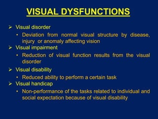
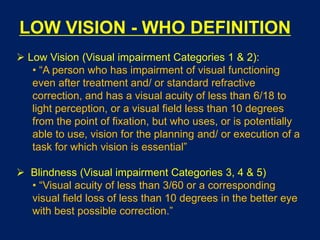
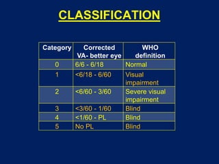
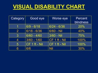
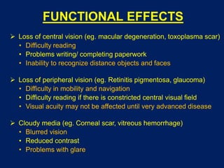
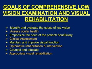
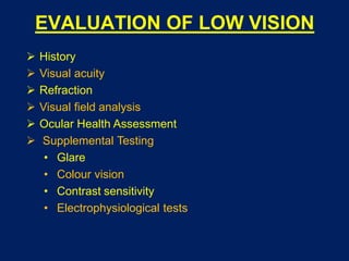
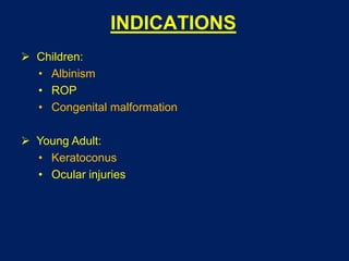
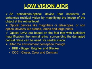
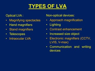
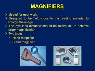
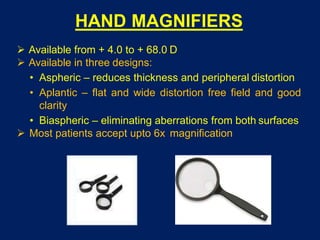
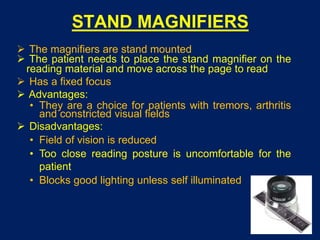
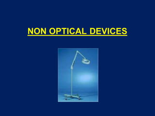
LOW VISION AIDS
Dr Saurabh Kushwaha
Resident (Ophthalmology)
SCOPE
 Visual Dysfunctions
 WHO Definition - Low Vision
 Classification
 Goals of management
 Indications
 Low vision aids
 Types of LVA
VISUAL DYSFUNCTIONS
 Visual disorder
• Deviation from normal visual structure by disease,
injury or anomaly affecting vision
 Visual impairment
• Reduction of visual function results from the visual
disorder
 Visual disability
• Reduced ability to perform a certain task
 Visual handicap
• Non-performance of the tasks related to individual and
social expectation because of visual disability
LOW VISION - WHO DEFINITION
 Low Vision (Visual impairment Categories 1 & 2):
• “A person who has impairment of visual functioning
even after treatment and/ or standard refractive
correction, and has a visual acuity of less than 6/18 to
light perception, or a visual field less than 10 degrees
from the point of fixation, but who uses, or is potentially
able to use, vision for the planning and/ or execution of a
task for which vision is essential”
 Blindness (Visual impairment Categories 3, 4 & 5)
• “Visual acuity of less than 3/60 or a corresponding
visual field loss of less than 10 degrees in the better eye
with best possible correction.”
CLASSIFICATION
Category Corrected
VA- better eye
WHO
definition
0 6/6 - 6/18 Normal
1 <6/18 - 6/60 Visual
impairment
2 <6/60 - 3/60 Severe visual
impairment
3 <3/60 - 1/60 Blind
4 <1/60 - PL Blind
5 No PL Blind
VISUAL DISABILITY CHART
Category Good eye Worse eye Percent
blindness
1 6/9 - 6/18 6/24 - 6/36 20%
2 6/18 - 6/36 6/60 - Nil 40%
3 6/60 - 4/60 3/60 - Nil 75%
4 3/60 - 1/60 CF 1 ft - Nil 100%
5 CF 1 ft - Nil CF 1 ft - Nil 100%
6 6/6 Nil 30%
FUNCTIONAL EFFECTS
 Loss of central vision (eg. macular degeneration, toxoplasma scar)
• Difficulty reading
• Problems writing/ completing paperwork
• Inability to recognize distance objects and faces
 Loss of peripheral vision (eg. Retinitis pigmentosa, glaucoma)
• Difficulty in mobility and navigation
• Difficulty reading if there is constricted central visual field
• Visual acuity may not be affected until very advanced disease
 Cloudy media (eg. Corneal scar, vitreous hemorrhage)
• Blurred vision
• Reduced contrast
• Problems with glare
GOALS OF COMPREHENSIVE LOW
VISION EXAMINATION AND VISUAL
REHABILITATION
 Identify and evaluate the cause of low vision
 Assess ocular health
 Emphasize the need of the patient/ beneficiary
 Clinical Assessment
 Maintain and improve visual function
 Optometric rehabilitation & intervention
 Counsel and educate
 Appropriate visual rehabilitation
EVALUATION OF LOW VISION
 History
 Visual acuity
 Refraction
 Visual field analysis
 Ocular Health Assessment
 Supplemental Testing
• Glare
• Colour vision
• Contrast sensitivity
• Electrophysiological tests
INDICATIONS
 Children:
• Albinism
• ROP
• Congenital malformation
 Young Adult:
• Keratoconus
• Ocular injuries
 Old age:
• Glaucoma
• ARMD
• Diabetic maculopathy
• Macular degeneration
• Retinal degeneration
• Chorioretinitis
• Optic atrophy
• Myopic degeneration
LOW VISION AIDS
 An optical/non-optical device that improves or
enhances residual vision by magnifying the image of the
object at the retinal level.
 Optical devices like magnifiers or telescopes, or non
optical devices like stands, lamps and large prints.
 Optical LVAs are based on the fact that with sufficient
magnification, the normal retina surrounding the damaged
central retina can be used for central vision.
 Alter the environment perception through
• BBB - Bigger, Brighter and Blacker
• CCC - Closer, Color and Contrast
TYPES OF LVA
Optical LVA :
• Magnifying spectacles
• Hand magnifiers
• Stand magnifiers
• Telescopes
• Intraocular LVA
Non-optical devices:
• Approach magnification
• Lighting
• Contrast enhancement
• Increased size object
• Electronic magnifiers (CCTV,
LVIS, V-max)
• Communication and writing
devices
TYPES OF MAGNIFICATION
 Low vision aids make use of angular magnifications by
increasing :
• Relative size
• Relative distance
 Angular : it is the apparent size of the object compared
with true size of the object seen without the device. eg.
Telescopic system
Angular magnification M = ω’/ ω
 Relative size: by making the object appear bigger (no
accommodation required) eg. CCTV
 Relative distance: by bringing the object closer (requires
good accommodation) eg. magnifiers
LOW VISION OPTICAL DEVICES
FOR NEAR
MAGNIFYING SPECTACLES
 High plus reading glasses to magnify the images
 Given as an add to the best distance refraction
 Reading distance is calculated by 100 divided by add
 Magnification is 1/4th the power of the lens.
 Used for near work
 Amount of add needed depends on the accommodation
and the reading distance
 Reading add can be predicted using the Kestenbaum
rule i.e the amount of add needed to read 1M print is the
inverse of the visual acuity fraction
 However usually greater add is required than predicted
as the patient also has reduced contrast sensitivity
 If the patient is monocular, the poorer eye may be
occluded if it improves the functioning
 When binocular corrections are needed :
• Base in prisms are added to compensate for
convergence angle.
• Optical center may be decentred
 Aspheric lenses may be used to reduce lenticular
distortion
 Advantages:
• Hands are free
• Field of view larger when compared to telescope
• Greater reading speed
• Can be given in both monocular and binocular forms
• More portable
• Cosmetically acceptable
 Disadvantages:
• Higher the power, closer the reading distance
• Close reading distance causes fatigue and
unacceptable posture
• Patients with eccentric fixation are unable to fix
through these glasses
MAGNIFIERS
 Useful for near work
 Designed to be held close to the reading material to
enlarge the image
 The eye lens distance should be minimum to achieve
larger magnification
 Two types:
• Hand magnifier
• Stand magnifier
HAND MAGNIFIERS
 Available from + 4.0 to + 68.0 D
 Available in three designs:
• Aspheric – reduces thickness and peripheral distortion
• Aplantic – flat and wide distortion free field and good
clarity
• Biaspheric – eliminating aberrations from both surfaces
 Most patients accept upto 6x magnification
 Advantages:
• The eye to lens distance can be varied
• Patient can maintain normal reading distance
• Work well with patients with eccentric viewing
• Some have light source which further enhances vision
• Easily available, over the counter
 Disadvantages:
• It occupies both hands
• Patients with tremors, arthritis etc have difficulty
holding the magnifier
• Maintaining focus is a problem especially for elderly
• Field of vision is limited
STAND MAGNIFIERS
 The magnifiers are stand mounted
 The patient needs to place the stand magnifier on the
reading material and move across the page to read
 Has a fixed focus
 Advantages:
• They are a choice for patients with tremors, arthritis
and constricted visual fields
 Disadvantages:
• Field of vision is reduced
• Too close reading posture is uncomfortable for the
patient
• Blocks good lighting unless self illuminated
CLOSED CIRCUIT TELEVISION
SYSTEM
 Closed circuit television system (CCTV) consists of a
monitor, a camera and a platform to place the reading text
 It has control for brightness, contrast and change of
polarity
 Magnification varies from 3X to 60X
LOW VISION OPTICAL DEVICES
FOR DISTANCE
TELESCOPES
 Work on the principle of angular magnification
 Telescopes with magnification power from 2x to 10x are
prescribed
 They can be prescribed for near, intermediate and
distant tasks
 Field of view decreases with magnification
 Types:
• Hand held monocular
• Clip on design
• Bioptic design: mounted on a pair of eyeglasses
 Principal
• Telescopes consist of two lenses (in practice two
optical systems) mounted such that the focal point of
the objective coincides with the focal point of the ocular.
• Objective lens is a converging lens
Galilean telescope Keplerian telescope
The eye piece is a negative
lens and the objective is a
positive lens
Both eye piece and objective
are positive lens
Resultant image is virtual and
erect
Resultant image is real and
inverted. Prisms are
incorporated to erect the image
Loss of light reduces
brightness of the image
Loss of light is more in this
system
Field quality is poor Field quality is relatively good
 Magnification of a telescope is given by the formula
M = fo/fe
 Telescopes can be used to focus near objects by
• Changing the distance between objective and
ocular lens
• Increasing the power of the objective lens
 Advantages:
• Only possible device to enhance distant vision
 Disadvantage:
• Restriction of the field of view
• Appearance and apprehension
• Expensive and costly
• Depth perception is distorted
NON OPTICAL DEVICES
ILLUMINATION
 Positioning
• Light source should be to the side of better eye
• Moving light closer will yield higher illumination
 Higher levels of illumination is needed in patients with
• Lost cone functions (macular degeneration)
• Glaucoma
• Diabetic retinopathy
• Retinitis pigmentosa, Chorioretinitis
 Reduced illumination
• Albinism
• Aniridia
READING STAND
 Easy comfortable posture to the patient
WRITING GUIDE
 Black cards with rectangular cut outs horizontally along the
card
 The patient can feel the empty cut out spaces and write
SIGNATURE GUIDE
TYPOSCOPE / READING GUIDE
 Masking device with a line cut out from an opaque, non
reflecting black plastic or thick paper.
 Reduces glare and controls contrast.
NOTEX
 It is a rectangular piece of cardboard with steps on top
right corner which helps in identifying the currency of
the note
 1st cut indicates Rs. 500, 2nd cut indicates Rs.100, 3rd
cut indicates Rs 50 and so on.
RELATIVE SIZE DEVICES
 Larger object subtends a larger visual angle at the eye
and is thus easier to resolve
• Large print material
• Large type playing cards, computer keyboards
• Enlarged clocks, telephones, calendars
COMPUTER SOFTWARE
 Jaws screen reading software
 Connect out loud internet and email software
 Magic 8.0 screen magnification software and speech
GLARE REDUCING DEVICES
 Glare is described as unwanted light
 It is disabling in patients with cataracts, corneal opacities,
albinism, retinitis pigmentosa
 Devices to prevent glare:
• Sunglasses
• Caps
• Umbrella
• Polaroid glasses
• NoIR filters
• Corning photochromic filters (CPF glasses)
CPF GLASEES
 Attenuate 100% of UVB wavelengths.
 Block 99% of UVA wavelengths.
 The blue light portion of the visible spectrum is most
likely to scatter in the eye, causing discomfort and hazy
illusion.
 Attenuate 98% of high-energy blue light, with exception
of CPF 450, which attenuates 96% of high-energy blue
light.
 The number of the CPF glasses correspond to
wavelength in nanometers above which light is
transmitted
CPF® 550
(red)
Lens colour varies from
orange-red when lightened
to brown when darkened.
Retinitis pigmentosa
Albinism
CPF® 527
(orange)
Orange-amber lens darkens
to brown in sunlight, giving
individuals better visual
function and reduced glare
Retinitis pigmentosa
Diabetic retinopathy
CPF® 450
(yellow)
Enhances contrast and
helps control glare indoors
Optic atrophy
Albinism
Pseudophakia
CPF® 511
(yellow
orange)
Provides moderate blue
light filtering medium-range
Filter
Glaucoma
Aphakia
Pseudophakia
Optic atrophy
Developing cataracts
NoIR FILTERS
 Absorbs the short wavelengths of the visible spectrum
that can scatter within the ocular media,
 Also absorbs ultraviolet light (upto 4000 nm) and
infrared light
 Manages overall visible light transmission (VLT) to allow
the proper amount of light energy to reach the eyes.
Includes a full range of lenses (spanning 90% to 1%
VLT)
• 2% dark amber: 100% UV, infrared and blue light
protection, helpful on very bright days
• 13% standard grey: good for postoperative cataract,
glaucoma, diabetics and those who had corneal
transplants
• 20% medium plum: good in low light situations and
can be worn indoors
• 58% light grey: reduce indoor glare especially under
fluorescent light
• 65% yellow: retinitis pigmentosa and macular
dgeneration
COLOR AND CONTRAST
ENHANCEMENT
 Maximize contrast by using a light color against black
or dark color
 Choose colors in the room or working area which have
high contrast
PINHOLE GLASSES
 Multiple holes of approximately 1mm size are made in
the glasses
 The distance between the holes should be atleast 3-3.5
mm or approximately the size of the pupil
 Used in patients with corneal opacities or conditions
with irregular reflexes
 Not used in patients with central field defects as it
reduces illumination and visual acuity
MOBILITY ASSISTING DEVICES
 Patients with low vision suffer a major problem of mobility
• Long canes
• Strong portable lights
FIELD EXPANDING DEVICES
 As the magnification increases, the field of view
decreases
 Three methods of increasing the field:
• Compress the existing image to include more of
available area
• Provide prisms that relocates the image from a non
seeing to a seeing area
• Use a mirror to reflect an image from a non seeing
area
 Reverse telescopes: they are usually not accepted due
to minification
 Fresnel lenses with power of 10-15D with base in the
direction of field loss
FUTURE
BIONIC EYE
Designed for patients who are blind due to diseases like
retinitis pigmentosa or AMD
Can also be tried for those with severe vision loss
Relies on patient having a healthy optic nerve and a
developed visual cortex
Cannot be used for people who were born blind
The prosthesis consists of :
• A digital camera built into a pair of glasses
• A video processing microchip built into a hand held unit
• A radio transmitter on the glasses
• A receiver implanted above the ear
• A retinal implant with electrodes on a chip behind the
retina
Camera captures an image
Send image to microchip
Convert image to electrical impulse of light and dark pixels
Send image to radiotansmitter
Transmits pulses wirelessly to the receiver
Sends impulses to the retinal implant by a hair thin implanted wire
The stimulated electrodes generate electrical signals that travel to
the visual cortex
THANK YOU

Low vision aids

  • 1.
    LOW VISION AIDS DrSaurabh Kushwaha Resident (Ophthalmology)
  • 2.
    SCOPE  Visual Dysfunctions WHO Definition - Low Vision  Classification  Goals of management  Indications  Low vision aids  Types of LVA
  • 3.
    VISUAL DYSFUNCTIONS  Visualdisorder • Deviation from normal visual structure by disease, injury or anomaly affecting vision  Visual impairment • Reduction of visual function results from the visual disorder  Visual disability • Reduced ability to perform a certain task  Visual handicap • Non-performance of the tasks related to individual and social expectation because of visual disability
  • 4.
    LOW VISION -WHO DEFINITION  Low Vision (Visual impairment Categories 1 & 2): • “A person who has impairment of visual functioning even after treatment and/ or standard refractive correction, and has a visual acuity of less than 6/18 to light perception, or a visual field less than 10 degrees from the point of fixation, but who uses, or is potentially able to use, vision for the planning and/ or execution of a task for which vision is essential”  Blindness (Visual impairment Categories 3, 4 & 5) • “Visual acuity of less than 3/60 or a corresponding visual field loss of less than 10 degrees in the better eye with best possible correction.”
  • 5.
    CLASSIFICATION Category Corrected VA- bettereye WHO definition 0 6/6 - 6/18 Normal 1 <6/18 - 6/60 Visual impairment 2 <6/60 - 3/60 Severe visual impairment 3 <3/60 - 1/60 Blind 4 <1/60 - PL Blind 5 No PL Blind
  • 6.
    VISUAL DISABILITY CHART CategoryGood eye Worse eye Percent blindness 1 6/9 - 6/18 6/24 - 6/36 20% 2 6/18 - 6/36 6/60 - Nil 40% 3 6/60 - 4/60 3/60 - Nil 75% 4 3/60 - 1/60 CF 1 ft - Nil 100% 5 CF 1 ft - Nil CF 1 ft - Nil 100% 6 6/6 Nil 30%
  • 7.
    FUNCTIONAL EFFECTS  Lossof central vision (eg. macular degeneration, toxoplasma scar) • Difficulty reading • Problems writing/ completing paperwork • Inability to recognize distance objects and faces  Loss of peripheral vision (eg. Retinitis pigmentosa, glaucoma) • Difficulty in mobility and navigation • Difficulty reading if there is constricted central visual field • Visual acuity may not be affected until very advanced disease  Cloudy media (eg. Corneal scar, vitreous hemorrhage) • Blurred vision • Reduced contrast • Problems with glare
  • 8.
    GOALS OF COMPREHENSIVELOW VISION EXAMINATION AND VISUAL REHABILITATION  Identify and evaluate the cause of low vision  Assess ocular health  Emphasize the need of the patient/ beneficiary  Clinical Assessment  Maintain and improve visual function  Optometric rehabilitation & intervention  Counsel and educate  Appropriate visual rehabilitation
  • 9.
    EVALUATION OF LOWVISION  History  Visual acuity  Refraction  Visual field analysis  Ocular Health Assessment  Supplemental Testing • Glare • Colour vision • Contrast sensitivity • Electrophysiological tests
  • 10.
    INDICATIONS  Children: • Albinism •ROP • Congenital malformation  Young Adult: • Keratoconus • Ocular injuries
  • 11.
     Old age: •Glaucoma • ARMD • Diabetic maculopathy • Macular degeneration • Retinal degeneration • Chorioretinitis • Optic atrophy • Myopic degeneration
  • 12.
    LOW VISION AIDS An optical/non-optical device that improves or enhances residual vision by magnifying the image of the object at the retinal level.  Optical devices like magnifiers or telescopes, or non optical devices like stands, lamps and large prints.  Optical LVAs are based on the fact that with sufficient magnification, the normal retina surrounding the damaged central retina can be used for central vision.  Alter the environment perception through • BBB - Bigger, Brighter and Blacker • CCC - Closer, Color and Contrast
  • 13.
    TYPES OF LVA OpticalLVA : • Magnifying spectacles • Hand magnifiers • Stand magnifiers • Telescopes • Intraocular LVA Non-optical devices: • Approach magnification • Lighting • Contrast enhancement • Increased size object • Electronic magnifiers (CCTV, LVIS, V-max) • Communication and writing devices
  • 14.
    TYPES OF MAGNIFICATION Low vision aids make use of angular magnifications by increasing : • Relative size • Relative distance  Angular : it is the apparent size of the object compared with true size of the object seen without the device. eg. Telescopic system Angular magnification M = ω’/ ω
  • 15.
     Relative size:by making the object appear bigger (no accommodation required) eg. CCTV
  • 16.
     Relative distance:by bringing the object closer (requires good accommodation) eg. magnifiers
  • 17.
    LOW VISION OPTICALDEVICES FOR NEAR
  • 18.
    MAGNIFYING SPECTACLES  Highplus reading glasses to magnify the images  Given as an add to the best distance refraction  Reading distance is calculated by 100 divided by add  Magnification is 1/4th the power of the lens.  Used for near work  Amount of add needed depends on the accommodation and the reading distance
  • 19.
     Reading addcan be predicted using the Kestenbaum rule i.e the amount of add needed to read 1M print is the inverse of the visual acuity fraction  However usually greater add is required than predicted as the patient also has reduced contrast sensitivity  If the patient is monocular, the poorer eye may be occluded if it improves the functioning  When binocular corrections are needed : • Base in prisms are added to compensate for convergence angle. • Optical center may be decentred  Aspheric lenses may be used to reduce lenticular distortion
  • 20.
     Advantages: • Handsare free • Field of view larger when compared to telescope • Greater reading speed • Can be given in both monocular and binocular forms • More portable • Cosmetically acceptable  Disadvantages: • Higher the power, closer the reading distance • Close reading distance causes fatigue and unacceptable posture • Patients with eccentric fixation are unable to fix through these glasses
  • 21.
    MAGNIFIERS  Useful fornear work  Designed to be held close to the reading material to enlarge the image  The eye lens distance should be minimum to achieve larger magnification  Two types: • Hand magnifier • Stand magnifier
  • 22.
    HAND MAGNIFIERS  Availablefrom + 4.0 to + 68.0 D  Available in three designs: • Aspheric – reduces thickness and peripheral distortion • Aplantic – flat and wide distortion free field and good clarity • Biaspheric – eliminating aberrations from both surfaces  Most patients accept upto 6x magnification
  • 23.
     Advantages: • Theeye to lens distance can be varied • Patient can maintain normal reading distance • Work well with patients with eccentric viewing • Some have light source which further enhances vision • Easily available, over the counter  Disadvantages: • It occupies both hands • Patients with tremors, arthritis etc have difficulty holding the magnifier • Maintaining focus is a problem especially for elderly • Field of vision is limited
  • 24.
    STAND MAGNIFIERS  Themagnifiers are stand mounted  The patient needs to place the stand magnifier on the reading material and move across the page to read  Has a fixed focus  Advantages: • They are a choice for patients with tremors, arthritis and constricted visual fields  Disadvantages: • Field of vision is reduced • Too close reading posture is uncomfortable for the patient • Blocks good lighting unless self illuminated
  • 25.
    CLOSED CIRCUIT TELEVISION SYSTEM Closed circuit television system (CCTV) consists of a monitor, a camera and a platform to place the reading text  It has control for brightness, contrast and change of polarity  Magnification varies from 3X to 60X
  • 26.
    LOW VISION OPTICALDEVICES FOR DISTANCE
  • 27.
    TELESCOPES  Work onthe principle of angular magnification  Telescopes with magnification power from 2x to 10x are prescribed  They can be prescribed for near, intermediate and distant tasks  Field of view decreases with magnification  Types: • Hand held monocular • Clip on design • Bioptic design: mounted on a pair of eyeglasses
  • 28.
     Principal • Telescopesconsist of two lenses (in practice two optical systems) mounted such that the focal point of the objective coincides with the focal point of the ocular. • Objective lens is a converging lens Galilean telescope Keplerian telescope The eye piece is a negative lens and the objective is a positive lens Both eye piece and objective are positive lens Resultant image is virtual and erect Resultant image is real and inverted. Prisms are incorporated to erect the image Loss of light reduces brightness of the image Loss of light is more in this system Field quality is poor Field quality is relatively good
  • 29.
     Magnification ofa telescope is given by the formula M = fo/fe  Telescopes can be used to focus near objects by • Changing the distance between objective and ocular lens • Increasing the power of the objective lens
  • 30.
     Advantages: • Onlypossible device to enhance distant vision  Disadvantage: • Restriction of the field of view • Appearance and apprehension • Expensive and costly • Depth perception is distorted
  • 31.
  • 32.
    ILLUMINATION  Positioning • Lightsource should be to the side of better eye • Moving light closer will yield higher illumination  Higher levels of illumination is needed in patients with • Lost cone functions (macular degeneration) • Glaucoma • Diabetic retinopathy • Retinitis pigmentosa, Chorioretinitis  Reduced illumination • Albinism • Aniridia
  • 33.
    READING STAND  Easycomfortable posture to the patient
  • 34.
    WRITING GUIDE  Blackcards with rectangular cut outs horizontally along the card  The patient can feel the empty cut out spaces and write
  • 35.
  • 36.
    TYPOSCOPE / READINGGUIDE  Masking device with a line cut out from an opaque, non reflecting black plastic or thick paper.  Reduces glare and controls contrast.
  • 37.
    NOTEX  It isa rectangular piece of cardboard with steps on top right corner which helps in identifying the currency of the note  1st cut indicates Rs. 500, 2nd cut indicates Rs.100, 3rd cut indicates Rs 50 and so on.
  • 38.
    RELATIVE SIZE DEVICES Larger object subtends a larger visual angle at the eye and is thus easier to resolve • Large print material • Large type playing cards, computer keyboards • Enlarged clocks, telephones, calendars
  • 39.
    COMPUTER SOFTWARE  Jawsscreen reading software  Connect out loud internet and email software  Magic 8.0 screen magnification software and speech
  • 40.
    GLARE REDUCING DEVICES Glare is described as unwanted light  It is disabling in patients with cataracts, corneal opacities, albinism, retinitis pigmentosa  Devices to prevent glare: • Sunglasses • Caps • Umbrella • Polaroid glasses • NoIR filters • Corning photochromic filters (CPF glasses)
  • 41.
    CPF GLASEES  Attenuate100% of UVB wavelengths.  Block 99% of UVA wavelengths.  The blue light portion of the visible spectrum is most likely to scatter in the eye, causing discomfort and hazy illusion.  Attenuate 98% of high-energy blue light, with exception of CPF 450, which attenuates 96% of high-energy blue light.  The number of the CPF glasses correspond to wavelength in nanometers above which light is transmitted
  • 42.
    CPF® 550 (red) Lens colourvaries from orange-red when lightened to brown when darkened. Retinitis pigmentosa Albinism CPF® 527 (orange) Orange-amber lens darkens to brown in sunlight, giving individuals better visual function and reduced glare Retinitis pigmentosa Diabetic retinopathy CPF® 450 (yellow) Enhances contrast and helps control glare indoors Optic atrophy Albinism Pseudophakia CPF® 511 (yellow orange) Provides moderate blue light filtering medium-range Filter Glaucoma Aphakia Pseudophakia Optic atrophy Developing cataracts
  • 43.
    NoIR FILTERS  Absorbsthe short wavelengths of the visible spectrum that can scatter within the ocular media,  Also absorbs ultraviolet light (upto 4000 nm) and infrared light  Manages overall visible light transmission (VLT) to allow the proper amount of light energy to reach the eyes.
  • 44.
    Includes a fullrange of lenses (spanning 90% to 1% VLT) • 2% dark amber: 100% UV, infrared and blue light protection, helpful on very bright days • 13% standard grey: good for postoperative cataract, glaucoma, diabetics and those who had corneal transplants • 20% medium plum: good in low light situations and can be worn indoors • 58% light grey: reduce indoor glare especially under fluorescent light • 65% yellow: retinitis pigmentosa and macular dgeneration
  • 45.
    COLOR AND CONTRAST ENHANCEMENT Maximize contrast by using a light color against black or dark color  Choose colors in the room or working area which have high contrast
  • 46.
    PINHOLE GLASSES  Multipleholes of approximately 1mm size are made in the glasses  The distance between the holes should be atleast 3-3.5 mm or approximately the size of the pupil  Used in patients with corneal opacities or conditions with irregular reflexes  Not used in patients with central field defects as it reduces illumination and visual acuity
  • 47.
    MOBILITY ASSISTING DEVICES Patients with low vision suffer a major problem of mobility • Long canes • Strong portable lights
  • 48.
    FIELD EXPANDING DEVICES As the magnification increases, the field of view decreases  Three methods of increasing the field: • Compress the existing image to include more of available area • Provide prisms that relocates the image from a non seeing to a seeing area • Use a mirror to reflect an image from a non seeing area  Reverse telescopes: they are usually not accepted due to minification  Fresnel lenses with power of 10-15D with base in the direction of field loss
  • 49.
  • 50.
    BIONIC EYE Designed forpatients who are blind due to diseases like retinitis pigmentosa or AMD Can also be tried for those with severe vision loss Relies on patient having a healthy optic nerve and a developed visual cortex Cannot be used for people who were born blind The prosthesis consists of : • A digital camera built into a pair of glasses • A video processing microchip built into a hand held unit • A radio transmitter on the glasses • A receiver implanted above the ear • A retinal implant with electrodes on a chip behind the retina
  • 51.
    Camera captures animage Send image to microchip Convert image to electrical impulse of light and dark pixels Send image to radiotansmitter Transmits pulses wirelessly to the receiver Sends impulses to the retinal implant by a hair thin implanted wire The stimulated electrodes generate electrical signals that travel to the visual cortex
  • 52.