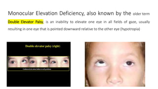
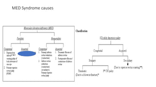
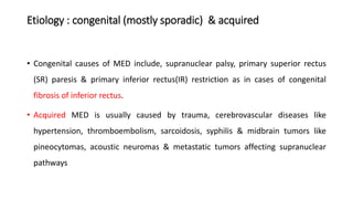
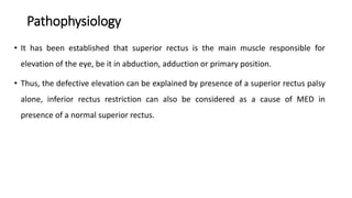
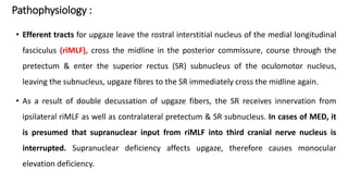
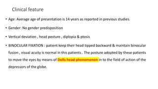
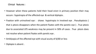
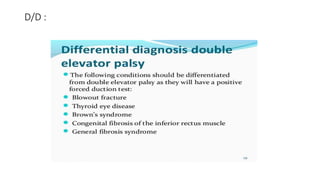
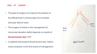
SlideShare a Scribd company logo
DOUBLE ELEVATOR PALSY
(MED: MONOCULAR ELEVATION DEFICIENCY SYNDROME)
Dr. VINIT KUMAR
SCEH, LAHAN, NEPAL
PEDIATRIC OPHTHALMOLOGY
Monocular Elevation Deficiency, also known by the older term
Double Elevator Palsy, is an inability to elevate one eye in all fields of gaze, usually
resulting in one eye that is pointed downward relative to the other eye (hypotropia)
Disease
• Monocular Elevation Deficiency (MED) is the limitation of elevation of the affected
eye from any position of gaze with normal ductions in all other gazes.
• MED is a frequent cause of hypotropia & can be associated with
ptosis/pseudoptosis.
• Superior rectus palsy, inferior rectus restriction & supranuclear lesions have been
found to be contributory to MED.
MED Syndrome causes
Etiology : congenital (mostly sporadic) & acquired
• Congenital causes of MED include, supranuclear palsy, primary superior rectus
(SR) paresis & primary inferior rectus(IR) restriction as in cases of congenital
fibrosis of inferior rectus.
• Acquired MED is usually caused by trauma, cerebrovascular diseases like
hypertension, thromboembolism, sarcoidosis, syphilis & midbrain tumors like
pineocytomas, acoustic neuromas & metastatic tumors affecting supranuclear
pathways
Pathology : Ziffer classification
• Type 1 includes patients with primary IR restriction or fibrosis
• Forced duction test (FDT) demonstrates a tight IR. The upward saccades are usually
normal until stopped by tight IR , preventing further upgaze. Bells phenomenon is
usually poor.
• In Type 2 MED, cases of primary SR palsy are included. FDT is free & upward saccades
are slow both below & above midline.
• Bell’s phenomenon is usually absent.
• Type 3 MED is supranuclear type & is usually congenital.
• It is characterized by intact or mildly reduced vertical saccadic velocity below
midline but abnormal or absent velocity above midline & FDT is free.
Pathophysiology
• It has been established that superior rectus is the main muscle responsible for
elevation of the eye, be it in abduction, adduction or primary position.
• Thus, the defective elevation can be explained by presence of a superior rectus palsy
alone, inferior rectus restriction can also be considered as a cause of MED in
presence of a normal superior rectus.
Pathophysiology :
• Efferent tracts for upgaze leave the rostral interstitial nucleus of the medial longitudinal
fasciculus (riMLF), cross the midline in the posterior commissure, course through the
pretectum & enter the superior rectus (SR) subnucleus of the oculomotor nucleus,
leaving the subnucleus, upgaze fibres to the SR immediately cross the midline again.
• As a result of double decussation of upgaze fibers, the SR receives innervation from
ipsilateral riMLF as well as contralateral pretectum & SR subnucleus. In cases of MED, it
is presumed that supranuclear input from riMLF into third cranial nerve nucleus is
interrupted. Supranuclear deficiency affects upgaze, therefore causes monocular
elevation deficiency.
Clinical feature
• Age: Average age of presentation is 14 years as reported in previous studies.
• Gender: No gender predisposition
• Vertical deviation , head posture , diplopia & ptosis
• BINOCULAR FIXATION : patient keep their head tipped backward & maintain binocular
fusion , visual acuity is normal in this patients . The posture adopted by these patients
to move the eyes by means of Dolls head phenomenon in to the field of action of the
depressors of the globe.
Clinical features :
• However when these patients hold their head erect in primary position their may
occure : hypotropia of the affected eye & vertical diplopia .
• Fixation with uninvolved eye : shows hypotropia in involved eye . Pseudoptosis (
that is ptosis disappears when the patient fixates with the paretic eye ) . True ptosis
due to associated LPS weakness may be present in 50% of cases . True ptosis does
not resolve when patient fixates with paretic eye .
• Amblyopia of the affected eye with visual acuity of 6/60 or less
• Diplopia is absent .
Chief Complaints
• Vertical misalignment of the eyes
• Diplopia
• Abnormal head posture
• Inability to elevate the affected eye
• Drooping of eyelid
• Decrease vision
• Fixation with involved eye : shows following features : hypertropia of normal eye
which is greater due to secondary deviation , visual acuity in the normal eye may be
reduced , & true ptosis due to associated weakness of LPS may be present .
CLINICAL FEATURES :
• Limitation of elevation ( in adduction as well as abduction, both on version & ductions )
beyond midline is main characteristic of this condition .
• Other clinical characteristics :
• An extra or deep lower lid fold on the affected side may be seen in patients with IR
muscle restriction.
• Bells phenomenon : is poor / absent with IR muscle restriction .
• Positional tonometry : with applanation tonometer shows a rise in IOP of more than 3mm Hg in 15
degree upward gaze as compared to that in primary gaze in patient with restrictive limitations . But no
such significant increase in IOP occurs in paretic weakness
Signs
• Ptosis/Pseudoptosis: Ptosis can be associated with hypotropia because of fascial
attachments between levator palpebrae superioris & superior rectus muscle..
Pseudoptosis & true ptosis both the components can be present in the patient.
• In these cases when the patient is made to fix with the affected eye, ptosis partly
recovers. True ptosis when present is usually complex type & is associated with
Marcus Gunn Jaw Winking phenomenon (MGJWP).
• Amblyopia may be present if there is a constant deviation of affected eye or
associated ptosis, or anisometropia .
• Abnormal Head Posture : Most commonly patient has a chin elevation if
binocular fusion is present. Abnormal head posture may not be present if the
affected eye is amblyopic.
• Upward Saccades: Upward saccades are slow/floating in cases of SR palsy, absent
above the midline in supranuclear palsy & intact (coming to an abrupt halt) in
cases associated with IR restriction .
• Bell’s phenomenon is usually absent in cases of IR restriction & SR palsy as they
limit the upward rotation of globe, but Bell's is usually present in cases of MED
resulting from supranuclear defects.
• Deviation: There is hypotropia of the affected eye when the normal eye is fixing , but as the
affected eye takes up fixation there is hypertropia of the normal eye. MED cases may also have
co-existent horizontal deviations.
Diagnostic procedures
• Forced duction test
• Forced duction test is a clinical test which should be done pre-operatively to reveal
any restriction.
• The test is typically performed under topical anaesthesia, conjunctiva is held at
limbus with Pierce Hoskin forceps or globe holding forceps, & patient is asked to look
in opposite direction of testing muscle to relax the concerned muscle. A positive
Forced Duction Test (FDT) is seen in patients of IR restriction.
• Active Force Generation Test
• Active force generation test is done under topical anaesthesia by asking the
patient to look in the direction of muscle being tested, presence of a tug is
suggestive of residual SR action.
• Diplopia Charting
• Red & green glasses over right & left eyes respectively, dissociate the two images
& would be seen double.
• Test can be done both for distance & near, with the help of illuminated slit target.
The slit is used vertically for charting horizontal deviations & horizontally for
charting vertical deviations. The seperation between two images is recorded & is
useful in diagnosis & follow-up.
Hess Screen & Lees Screen
• Hess Screen & Lees Screen is used to document relative incomitance & underactions
& is based on haploscopic principle. Two test objects are shown to the patient which
is seen as one localisation.
• Hess Screen consist of a three and a half feet black cloth marked with a series of red
lines forming a square of 5 degree each. From the central zero point three squares
15 degree marks the inner square and outer square has excursion of 30 degree.
• The outer square is used for mild incomitances not detected on inner square
charting. Patient wears a red green glass, with red glass in front of right eye and
green glass in front of left eye, patient is given a green pointer which he aligns on
the red dots and the observer records the points.
• Lees Screen is also similar but does not require red green glass instead uses a
mirror septa to cause dissociation of two eye.
Differential diagnosis
• Brown’s Syndrome is characterised by limitation of elevation in adduction, with
divergence in upgaze caused by slippage of globe resulting in characterstic Y pattern .
• Vertical Duane Syndrome can be diagnosed by the presence of globe retraction in
downgaze along with limitation of elevation in upgaze.
• Congenital fibrosis of extra ocular muscles initially affecting inferior recti can be
misdiagnosed as MED.
• …….Bilateral presentation involvement of other extraocular muscles are features that
help distinguish it.
• Partial third nerve palsy involving the superior division of oculomotor nerve may
simulate the findings of MED.
• Progressive external ophthalmoplegia involves multiple extra ocular muscles.
• Orbital inflammatory disease and orbital cellulitis present with a tender inflamed
globe.
D/D :
Management
• Management of MED includes both non-surgical & surgical components.
• Non-surgical management
• The correction of underlying refractive error is to be done & amblyopia therapy is
given if needed, followed by surgery whenever indicated.
Indications of surgery ?? ??
• Which include …….
• Significant vertical deviation in primary gaze
• Significant abnormal head posture/ AHP
• Deviation-induced amblyopia
• Diplopia in primary gaze, & restricted binocular fields.
GOAL OF SURGERY …
• The goal of surgery is to improve the position of
the affected eye in primary gaze & to increase
binocular field of vision.
• The surgery of choice in the management of
monocular elevation deficit depends on results of
forced duction test. FDT
• In patients with positive forced duction test, inferior
rectus recession is the first choice of management.
• In patients with negative forced duction test
but a positive active force generation test,
superior rectus resection can be done.
• In patients with negative FDT /forced duction
test & a negative active force generation test
/ATGT Knapp's procedure is preferred.
• Knapp's procedure
• Knapp’s procedure is commonly practiced & is now a well-established treatment
for MED syndrome . In the Knapp’s procedure, the tendons of the medial & lateral
rectus muscles are transposed to the insertion of the superior rectus muscle.
• Knapp’s in his work studied 15 patients over a period of eight years. The
procedure showed marked variability correcting 21-55 PD (average 38 PD)
hypotropia .
• Modifications of Knapp's procedure
• For management of MED cases with associated horizontal deviation partial
tendon Knapp’s (Modifed Knapp’s) procedure has been described, in which the
superior part of equally divided horizontal muscles is placed near the superior
rectus muscle insertion, allowing the correction of horizontal deviation with the
untouched inferior half.
• Augmented Knapp's procedure, Knapp's procedure can be combined with
posterior fixation sutures on the horizontal recti, known as Augmented Knapp's.
• Following the surgical correction of hypotropia & improvement in Bell’s
phenomenon, associated ptosis and/or Marcus Gunn phenomenon be addressed.
• Contralateral superior rectus recession
• Contralateral superior rectus recession can be an option of management in the
patients with residual hypotropia after inferior rectus recession.
• Modified Nishida's for monocular elevation deficit / MED syndrome
• No split no tenotomy technique by Nishida et al has originally been described for
abducen nerve palsy, the procedure has been found to be useful in the
management of monocular elevation deficit by Murthy et al and can help in
correction of upto 30 pd of vertical deviation with low risk of anterior segment
ischemia
Management of Acquired MED
• As there are wide variety of causes of acquired MED, thorough investigations
(general, ophthalmologic, neurologic, neuro-ophthalmologic & orthoptic (ocular
motility work up) should be undertaken, & management varies according to the
underlying cause.
Complications
• Ptosis can lead to refractive error & amblyopia, also if ptosis surgery is done before the strabismus
surgery it can lead to exposure keratopathy as most of the patients have poor Bell's
phenomenon.
• Surgical complications :
• Anterior segment ischemia : presenting with corneal edema, thinning, non
pigmented keratin precipitates, iris atrophy & anterior chamber flare.
• Anterior segment ischemia can result if three or more adjacent recti surgery has
been done in one eye, adults with atherosclerosis, previous radiation therapy &
retinal detachment surgery are more vulnerable.
• The complication can be avoided if two vertical recti along with one horizontal
recti are not operated in the same sitting.
• Management includes, systemic & local corticosteroid .
• Overcorrections & undercorrections.
• Tenon's prolapse & conjunctival cyst can result due to improper apposition of the
conjunctiva which allows Tenon's to prolapse & interferes with healing process.
• Corneal dellen formation can occur due to disruption of tear film & local
dehydration.
• Other intraoperative complications like surgery on wrong eye, wrong muscle,
hemorrhages, scleral perforation,slipped muscle, bradycardia, infections may also
occur in patients undergoing surgery.
IN Summary ………
Points to remember
• Recession of IR muscle : useful for presence of inferior restriction for a vertical deviation up to
18-20 D
• Recession of the IR muscle plus resection of SR is indicated when in the presence of inferior
restriction & the deviation is more than 18 to 20 D
• KNAPPS procedure usually corrects 20 to 35 PD of hypotropia in primary position. Knapp after
IR recession usually has greater effect
• Augmented transposition using posterior fixation suture should be done for large hypotropia >
35 PD with no IR restriction
• Modified Knapp’s : if horizontal deviation is present then recess- resect & also transpose the
MR & LR muscles .
Double  elevator  palsy

More Related Content

What's hot

Intermitent exotropia
Intermitent exotropiaIntermitent exotropia
Intermitent exotropia
SSSIHMS-PG
 
Evaluation of squint - The Basics
Evaluation of squint - The BasicsEvaluation of squint - The Basics
Evaluation of squint - The Basics
drindeevarmishra
 
Hess chart
Hess chartHess chart
Optical coherence tomography in glaucoma - Dr Shylesh Dabke
Optical coherence tomography in glaucoma - Dr Shylesh DabkeOptical coherence tomography in glaucoma - Dr Shylesh Dabke
Optical coherence tomography in glaucoma - Dr Shylesh Dabke
Shylesh Dabke
 
Amblyopia Management
Amblyopia ManagementAmblyopia Management
Amblyopia Managementsiraj safi
 
Accommodation/ Accommodation of Eye, Measurement of Accommodation of Eye (hea...
Accommodation/ Accommodation of Eye, Measurement of Accommodation of Eye (hea...Accommodation/ Accommodation of Eye, Measurement of Accommodation of Eye (hea...
Accommodation/ Accommodation of Eye, Measurement of Accommodation of Eye (hea...
Bikash Sapkota
 
Ptosis workup
Ptosis workupPtosis workup
Ptosis workup
Azizul Islam
 
Specular microscopy
Specular microscopySpecular microscopy
Specular microscopy
Ruchi sood
 
Microtropia
MicrotropiaMicrotropia
Microtropia
DrAzmat Ali
 
Subjective refraction by optom praveen
Subjective refraction by optom praveenSubjective refraction by optom praveen
Subjective refraction by optom praveen
optom.praveen p
 
Hess chart
Hess chartHess chart
Hess chart
Jagdish Dukre
 
Presbyopia/ Methods of Presbyopic Addition Determination (healthkura.com)
Presbyopia/ Methods of Presbyopic Addition Determination (healthkura.com)Presbyopia/ Methods of Presbyopic Addition Determination (healthkura.com)
Presbyopia/ Methods of Presbyopic Addition Determination (healthkura.com)
Bikash Sapkota
 
Prescription of glasses in children
Prescription of glasses in childrenPrescription of glasses in children
Prescription of glasses in children
Eranda Wannigama
 
Brown's syndrome
Brown's syndromeBrown's syndrome
Brown's syndrome
DrAzmat Ali
 
Principles of strabismus surgery
Principles of strabismus surgeryPrinciples of strabismus surgery
Principles of strabismus surgery
punnukmc
 
AC/A
AC/AAC/A
AC/A
zarin45
 
Peripheral fundus & its disorders
Peripheral fundus & its disordersPeripheral fundus & its disorders
Peripheral fundus & its disorders
Rohit Rao
 
Sensory evaluation of squint
Sensory  evaluation of squintSensory  evaluation of squint
Sensory evaluation of squint
Dr Laltanpuia Chhangte
 
Exodeviations , Exotropia
Exodeviations , ExotropiaExodeviations , Exotropia
Exodeviations , Exotropia
Vivek Chaudhary
 

What's hot (20)

Intermitent exotropia
Intermitent exotropiaIntermitent exotropia
Intermitent exotropia
 
Paralytic strabismus
Paralytic strabismusParalytic strabismus
Paralytic strabismus
 
Evaluation of squint - The Basics
Evaluation of squint - The BasicsEvaluation of squint - The Basics
Evaluation of squint - The Basics
 
Hess chart
Hess chartHess chart
Hess chart
 
Optical coherence tomography in glaucoma - Dr Shylesh Dabke
Optical coherence tomography in glaucoma - Dr Shylesh DabkeOptical coherence tomography in glaucoma - Dr Shylesh Dabke
Optical coherence tomography in glaucoma - Dr Shylesh Dabke
 
Amblyopia Management
Amblyopia ManagementAmblyopia Management
Amblyopia Management
 
Accommodation/ Accommodation of Eye, Measurement of Accommodation of Eye (hea...
Accommodation/ Accommodation of Eye, Measurement of Accommodation of Eye (hea...Accommodation/ Accommodation of Eye, Measurement of Accommodation of Eye (hea...
Accommodation/ Accommodation of Eye, Measurement of Accommodation of Eye (hea...
 
Ptosis workup
Ptosis workupPtosis workup
Ptosis workup
 
Specular microscopy
Specular microscopySpecular microscopy
Specular microscopy
 
Microtropia
MicrotropiaMicrotropia
Microtropia
 
Subjective refraction by optom praveen
Subjective refraction by optom praveenSubjective refraction by optom praveen
Subjective refraction by optom praveen
 
Hess chart
Hess chartHess chart
Hess chart
 
Presbyopia/ Methods of Presbyopic Addition Determination (healthkura.com)
Presbyopia/ Methods of Presbyopic Addition Determination (healthkura.com)Presbyopia/ Methods of Presbyopic Addition Determination (healthkura.com)
Presbyopia/ Methods of Presbyopic Addition Determination (healthkura.com)
 
Prescription of glasses in children
Prescription of glasses in childrenPrescription of glasses in children
Prescription of glasses in children
 
Brown's syndrome
Brown's syndromeBrown's syndrome
Brown's syndrome
 
Principles of strabismus surgery
Principles of strabismus surgeryPrinciples of strabismus surgery
Principles of strabismus surgery
 
AC/A
AC/AAC/A
AC/A
 
Peripheral fundus & its disorders
Peripheral fundus & its disordersPeripheral fundus & its disorders
Peripheral fundus & its disorders
 
Sensory evaluation of squint
Sensory  evaluation of squintSensory  evaluation of squint
Sensory evaluation of squint
 
Exodeviations , Exotropia
Exodeviations , ExotropiaExodeviations , Exotropia
Exodeviations , Exotropia
 

Similar to Double elevator palsy

Diplopia
Diplopia Diplopia
Diplopia
Anisha Rathod
 
Duane Retraction syndrome.pptx
Duane Retraction syndrome.pptxDuane Retraction syndrome.pptx
Duane Retraction syndrome.pptx
Nasrinsultanarmc
 
Duane syndrome 2
Duane syndrome 2Duane syndrome 2
Duane syndrome 2
huda alhashimy
 
Hypermetropia/Hyperopia
Hypermetropia/HyperopiaHypermetropia/Hyperopia
Hypermetropia/Hyperopia
Suraj Shil
 
The patient with diplopia
The patient with diplopia  The patient with diplopia
The patient with diplopia
siraj safi
 
Second lecture neuro ophthalmology
Second lecture neuro ophthalmologySecond lecture neuro ophthalmology
Second lecture neuro ophthalmology
Anisur Rahman
 
Examination of a case of strabismus
Examination of a case of strabismusExamination of a case of strabismus
Examination of a case of strabismus
Anisha Rathod
 
Anomalies Of Convergence
Anomalies Of ConvergenceAnomalies Of Convergence
Anomalies Of Convergence
mahendra singh
 
Duane Retraction Syndrome- Congenital Cranial Dysinnervation Disorder (Muscul...
Duane Retraction Syndrome- Congenital Cranial Dysinnervation Disorder (Muscul...Duane Retraction Syndrome- Congenital Cranial Dysinnervation Disorder (Muscul...
Duane Retraction Syndrome- Congenital Cranial Dysinnervation Disorder (Muscul...
Tanvi Gupta
 
Strabismus stdents 2
Strabismus stdents 2Strabismus stdents 2
Strabismus stdents 2
د.الأحمدي السمان
 
Hypermetropia and Aphakia
Hypermetropia and AphakiaHypermetropia and Aphakia
Hypermetropia and Aphakia
KumarSingh44
 
Myopia
Myopia Myopia
Hypermetropia
HypermetropiaHypermetropia
Hypermetropia
Dr.Siddharth Gautam
 
Strabismus american acadamy of ophthalmology.pptx
Strabismus american acadamy of ophthalmology.pptxStrabismus american acadamy of ophthalmology.pptx
Strabismus american acadamy of ophthalmology.pptx
fajrimohammed
 
Suppression third yr arya
Suppression third yr aryaSuppression third yr arya
Suppression third yr arya
arya das
 
Lecture on Pupillary Reflexes; Common Abnormalities For 4th Year MBBS Undergr...
Lecture on Pupillary Reflexes; Common Abnormalities For 4th Year MBBS Undergr...Lecture on Pupillary Reflexes; Common Abnormalities For 4th Year MBBS Undergr...
Lecture on Pupillary Reflexes; Common Abnormalities For 4th Year MBBS Undergr...
DrHussainAhmadKhaqan
 
Orbital complications of zygomaticomaxillary complex fracture
Orbital complications of zygomaticomaxillary complex fracture Orbital complications of zygomaticomaxillary complex fracture
Orbital complications of zygomaticomaxillary complex fracture
mrinalini123456789
 
Refractive errors (eye condions)
Refractive errors (eye condions)Refractive errors (eye condions)
Refractive errors (eye condions)
NehaNupur8
 
Strabismus-Clinical Examinations
Strabismus-Clinical ExaminationsStrabismus-Clinical Examinations
Strabismus-Clinical Examinations
Burdwan Medical College and Hospital
 
Congenital cranial dysinnervation disorder
Congenital cranial dysinnervation disorderCongenital cranial dysinnervation disorder
Congenital cranial dysinnervation disorder
Anju Nagar
 

Similar to Double elevator palsy (20)

Diplopia
Diplopia Diplopia
Diplopia
 
Duane Retraction syndrome.pptx
Duane Retraction syndrome.pptxDuane Retraction syndrome.pptx
Duane Retraction syndrome.pptx
 
Duane syndrome 2
Duane syndrome 2Duane syndrome 2
Duane syndrome 2
 
Hypermetropia/Hyperopia
Hypermetropia/HyperopiaHypermetropia/Hyperopia
Hypermetropia/Hyperopia
 
The patient with diplopia
The patient with diplopia  The patient with diplopia
The patient with diplopia
 
Second lecture neuro ophthalmology
Second lecture neuro ophthalmologySecond lecture neuro ophthalmology
Second lecture neuro ophthalmology
 
Examination of a case of strabismus
Examination of a case of strabismusExamination of a case of strabismus
Examination of a case of strabismus
 
Anomalies Of Convergence
Anomalies Of ConvergenceAnomalies Of Convergence
Anomalies Of Convergence
 
Duane Retraction Syndrome- Congenital Cranial Dysinnervation Disorder (Muscul...
Duane Retraction Syndrome- Congenital Cranial Dysinnervation Disorder (Muscul...Duane Retraction Syndrome- Congenital Cranial Dysinnervation Disorder (Muscul...
Duane Retraction Syndrome- Congenital Cranial Dysinnervation Disorder (Muscul...
 
Strabismus stdents 2
Strabismus stdents 2Strabismus stdents 2
Strabismus stdents 2
 
Hypermetropia and Aphakia
Hypermetropia and AphakiaHypermetropia and Aphakia
Hypermetropia and Aphakia
 
Myopia
Myopia Myopia
Myopia
 
Hypermetropia
HypermetropiaHypermetropia
Hypermetropia
 
Strabismus american acadamy of ophthalmology.pptx
Strabismus american acadamy of ophthalmology.pptxStrabismus american acadamy of ophthalmology.pptx
Strabismus american acadamy of ophthalmology.pptx
 
Suppression third yr arya
Suppression third yr aryaSuppression third yr arya
Suppression third yr arya
 
Lecture on Pupillary Reflexes; Common Abnormalities For 4th Year MBBS Undergr...
Lecture on Pupillary Reflexes; Common Abnormalities For 4th Year MBBS Undergr...Lecture on Pupillary Reflexes; Common Abnormalities For 4th Year MBBS Undergr...
Lecture on Pupillary Reflexes; Common Abnormalities For 4th Year MBBS Undergr...
 
Orbital complications of zygomaticomaxillary complex fracture
Orbital complications of zygomaticomaxillary complex fracture Orbital complications of zygomaticomaxillary complex fracture
Orbital complications of zygomaticomaxillary complex fracture
 
Refractive errors (eye condions)
Refractive errors (eye condions)Refractive errors (eye condions)
Refractive errors (eye condions)
 
Strabismus-Clinical Examinations
Strabismus-Clinical ExaminationsStrabismus-Clinical Examinations
Strabismus-Clinical Examinations
 
Congenital cranial dysinnervation disorder
Congenital cranial dysinnervation disorderCongenital cranial dysinnervation disorder
Congenital cranial dysinnervation disorder
 

More from Vinitkumar MJ

Geometric optics
Geometric opticsGeometric optics
Geometric optics
Vinitkumar MJ
 
Reduced eye
Reduced eyeReduced eye
Reduced eye
Vinitkumar MJ
 
Assessment of ocular alignment
Assessment of ocular alignmentAssessment of ocular alignment
Assessment of ocular alignment
Vinitkumar MJ
 
Techniques of fundus
Techniques of fundusTechniques of fundus
Techniques of fundus
Vinitkumar MJ
 
@Blood sugar level venipuncture
@Blood sugar level venipuncture@Blood sugar level venipuncture
@Blood sugar level venipuncture
Vinitkumar MJ
 
Dry eye ( investiigations & basic )
Dry eye ( investiigations & basic )Dry eye ( investiigations & basic )
Dry eye ( investiigations & basic )
Vinitkumar MJ
 
Visual acuity in child converted
Visual acuity in child convertedVisual acuity in child converted
Visual acuity in child converted
Vinitkumar MJ
 
Entropion o.a claa 2nd year
Entropion o.a claa 2nd yearEntropion o.a claa 2nd year
Entropion o.a claa 2nd year
Vinitkumar MJ
 
Enucleation, evisceration, exenteration
Enucleation, evisceration, exenterationEnucleation, evisceration, exenteration
Enucleation, evisceration, exenteration
Vinitkumar MJ
 
Eyelid surgery
Eyelid surgeryEyelid surgery
Eyelid surgery
Vinitkumar MJ
 
Cataract surgery
Cataract surgeryCataract surgery
Cataract surgery
Vinitkumar MJ
 
Strabismus o.a class
Strabismus o.a classStrabismus o.a class
Strabismus o.a class
Vinitkumar MJ
 
Gonorrhea converted
Gonorrhea convertedGonorrhea converted
Gonorrhea converted
Vinitkumar MJ
 
Std syphilis- (1)
Std syphilis- (1)Std syphilis- (1)
Std syphilis- (1)
Vinitkumar MJ
 
Rubella
RubellaRubella
Rubella
Vinitkumar MJ
 
Toxoplasmosis
ToxoplasmosisToxoplasmosis
Toxoplasmosis
Vinitkumar MJ
 
Hypertensive retinopathy converted
Hypertensive retinopathy  convertedHypertensive retinopathy  converted
Hypertensive retinopathy converted
Vinitkumar MJ
 
Scientific session
Scientific sessionScientific session
Scientific session
Vinitkumar MJ
 
Keratoconus, lenticonus, lentiglobus , microspherophakia &
Keratoconus, lenticonus, lentiglobus , microspherophakia &Keratoconus, lenticonus, lentiglobus , microspherophakia &
Keratoconus, lenticonus, lentiglobus , microspherophakia &
Vinitkumar MJ
 
Cscr ( central serous chorioretinopathy )
Cscr ( central serous chorioretinopathy )Cscr ( central serous chorioretinopathy )
Cscr ( central serous chorioretinopathy )
Vinitkumar MJ
 

More from Vinitkumar MJ (20)

Geometric optics
Geometric opticsGeometric optics
Geometric optics
 
Reduced eye
Reduced eyeReduced eye
Reduced eye
 
Assessment of ocular alignment
Assessment of ocular alignmentAssessment of ocular alignment
Assessment of ocular alignment
 
Techniques of fundus
Techniques of fundusTechniques of fundus
Techniques of fundus
 
@Blood sugar level venipuncture
@Blood sugar level venipuncture@Blood sugar level venipuncture
@Blood sugar level venipuncture
 
Dry eye ( investiigations & basic )
Dry eye ( investiigations & basic )Dry eye ( investiigations & basic )
Dry eye ( investiigations & basic )
 
Visual acuity in child converted
Visual acuity in child convertedVisual acuity in child converted
Visual acuity in child converted
 
Entropion o.a claa 2nd year
Entropion o.a claa 2nd yearEntropion o.a claa 2nd year
Entropion o.a claa 2nd year
 
Enucleation, evisceration, exenteration
Enucleation, evisceration, exenterationEnucleation, evisceration, exenteration
Enucleation, evisceration, exenteration
 
Eyelid surgery
Eyelid surgeryEyelid surgery
Eyelid surgery
 
Cataract surgery
Cataract surgeryCataract surgery
Cataract surgery
 
Strabismus o.a class
Strabismus o.a classStrabismus o.a class
Strabismus o.a class
 
Gonorrhea converted
Gonorrhea convertedGonorrhea converted
Gonorrhea converted
 
Std syphilis- (1)
Std syphilis- (1)Std syphilis- (1)
Std syphilis- (1)
 
Rubella
RubellaRubella
Rubella
 
Toxoplasmosis
ToxoplasmosisToxoplasmosis
Toxoplasmosis
 
Hypertensive retinopathy converted
Hypertensive retinopathy  convertedHypertensive retinopathy  converted
Hypertensive retinopathy converted
 
Scientific session
Scientific sessionScientific session
Scientific session
 
Keratoconus, lenticonus, lentiglobus , microspherophakia &
Keratoconus, lenticonus, lentiglobus , microspherophakia &Keratoconus, lenticonus, lentiglobus , microspherophakia &
Keratoconus, lenticonus, lentiglobus , microspherophakia &
 
Cscr ( central serous chorioretinopathy )
Cscr ( central serous chorioretinopathy )Cscr ( central serous chorioretinopathy )
Cscr ( central serous chorioretinopathy )
 

Recently uploaded

Charaka Samhita Sutra sthana Chapter 15 Upakalpaniyaadhyaya
Charaka Samhita Sutra sthana Chapter 15 UpakalpaniyaadhyayaCharaka Samhita Sutra sthana Chapter 15 Upakalpaniyaadhyaya
Charaka Samhita Sutra sthana Chapter 15 Upakalpaniyaadhyaya
Dr KHALID B.M
 
Ocular injury ppt Upendra pal optometrist upums saifai etawah
Ocular injury  ppt  Upendra pal  optometrist upums saifai etawahOcular injury  ppt  Upendra pal  optometrist upums saifai etawah
Ocular injury ppt Upendra pal optometrist upums saifai etawah
pal078100
 
POST OPERATIVE OLIGURIA and its management
POST OPERATIVE OLIGURIA and its managementPOST OPERATIVE OLIGURIA and its management
POST OPERATIVE OLIGURIA and its management
touseefaziz1
 
Cervical & Brachial Plexus By Dr. RIG.pptx
Cervical & Brachial Plexus By Dr. RIG.pptxCervical & Brachial Plexus By Dr. RIG.pptx
Cervical & Brachial Plexus By Dr. RIG.pptx
Dr. Rabia Inam Gandapore
 
HOT NEW PRODUCT! BIG SALES FAST SHIPPING NOW FROM CHINA!! EU KU DB BK substit...
HOT NEW PRODUCT! BIG SALES FAST SHIPPING NOW FROM CHINA!! EU KU DB BK substit...HOT NEW PRODUCT! BIG SALES FAST SHIPPING NOW FROM CHINA!! EU KU DB BK substit...
HOT NEW PRODUCT! BIG SALES FAST SHIPPING NOW FROM CHINA!! EU KU DB BK substit...
GL Anaacs
 
ARTIFICIAL INTELLIGENCE IN HEALTHCARE.pdf
ARTIFICIAL INTELLIGENCE IN  HEALTHCARE.pdfARTIFICIAL INTELLIGENCE IN  HEALTHCARE.pdf
ARTIFICIAL INTELLIGENCE IN HEALTHCARE.pdf
Anujkumaranit
 
Pulmonary Thromboembolism - etilogy, types, medical- Surgical and nursing man...
Pulmonary Thromboembolism - etilogy, types, medical- Surgical and nursing man...Pulmonary Thromboembolism - etilogy, types, medical- Surgical and nursing man...
Pulmonary Thromboembolism - etilogy, types, medical- Surgical and nursing man...
VarunMahajani
 
micro teaching on communication m.sc nursing.pdf
micro teaching on communication m.sc nursing.pdfmicro teaching on communication m.sc nursing.pdf
micro teaching on communication m.sc nursing.pdf
Anurag Sharma
 
Alcohol_Dr. Jeenal Mistry MD Pharmacology.pdf
Alcohol_Dr. Jeenal Mistry MD Pharmacology.pdfAlcohol_Dr. Jeenal Mistry MD Pharmacology.pdf
Alcohol_Dr. Jeenal Mistry MD Pharmacology.pdf
Dr Jeenal Mistry
 
Superficial & Deep Fascia of the NECK.pptx
Superficial & Deep Fascia of the NECK.pptxSuperficial & Deep Fascia of the NECK.pptx
Superficial & Deep Fascia of the NECK.pptx
Dr. Rabia Inam Gandapore
 
Surat @ℂall @Girls ꧁❤8527049040❤꧂@ℂall @Girls Service Vip Top Model Safe
Surat @ℂall @Girls ꧁❤8527049040❤꧂@ℂall @Girls Service Vip Top Model SafeSurat @ℂall @Girls ꧁❤8527049040❤꧂@ℂall @Girls Service Vip Top Model Safe
Surat @ℂall @Girls ꧁❤8527049040❤꧂@ℂall @Girls Service Vip Top Model Safe
Savita Shen $i11
 
heat stroke and heat exhaustion in children
heat stroke and heat exhaustion in childrenheat stroke and heat exhaustion in children
heat stroke and heat exhaustion in children
SumeraAhmad5
 
BENIGN PROSTATIC HYPERPLASIA.BPH. BPHpdf
BENIGN PROSTATIC HYPERPLASIA.BPH. BPHpdfBENIGN PROSTATIC HYPERPLASIA.BPH. BPHpdf
BENIGN PROSTATIC HYPERPLASIA.BPH. BPHpdf
DR SETH JOTHAM
 
Prix Galien International 2024 Forum Program
Prix Galien International 2024 Forum ProgramPrix Galien International 2024 Forum Program
Prix Galien International 2024 Forum Program
Levi Shapiro
 
MANAGEMENT OF ATRIOVENTRICULAR CONDUCTION BLOCK.pdf
MANAGEMENT OF ATRIOVENTRICULAR CONDUCTION BLOCK.pdfMANAGEMENT OF ATRIOVENTRICULAR CONDUCTION BLOCK.pdf
MANAGEMENT OF ATRIOVENTRICULAR CONDUCTION BLOCK.pdf
Jim Jacob Roy
 
Physiology of Chemical Sensation of smell.pdf
Physiology of Chemical Sensation of smell.pdfPhysiology of Chemical Sensation of smell.pdf
Physiology of Chemical Sensation of smell.pdf
MedicoseAcademics
 
Couples presenting to the infertility clinic- Do they really have infertility...
Couples presenting to the infertility clinic- Do they really have infertility...Couples presenting to the infertility clinic- Do they really have infertility...
Couples presenting to the infertility clinic- Do they really have infertility...
Sujoy Dasgupta
 
Ozempic: Preoperative Management of Patients on GLP-1 Receptor Agonists
Ozempic: Preoperative Management of Patients on GLP-1 Receptor Agonists  Ozempic: Preoperative Management of Patients on GLP-1 Receptor Agonists
Ozempic: Preoperative Management of Patients on GLP-1 Receptor Agonists
Saeid Safari
 
Maxilla, Mandible & Hyoid Bone & Clinical Correlations by Dr. RIG.pptx
Maxilla, Mandible & Hyoid Bone & Clinical Correlations by Dr. RIG.pptxMaxilla, Mandible & Hyoid Bone & Clinical Correlations by Dr. RIG.pptx
Maxilla, Mandible & Hyoid Bone & Clinical Correlations by Dr. RIG.pptx
Dr. Rabia Inam Gandapore
 
Ophthalmology Clinical Tests for OSCE exam
Ophthalmology Clinical Tests for OSCE examOphthalmology Clinical Tests for OSCE exam
Ophthalmology Clinical Tests for OSCE exam
KafrELShiekh University
 

Recently uploaded (20)

Charaka Samhita Sutra sthana Chapter 15 Upakalpaniyaadhyaya
Charaka Samhita Sutra sthana Chapter 15 UpakalpaniyaadhyayaCharaka Samhita Sutra sthana Chapter 15 Upakalpaniyaadhyaya
Charaka Samhita Sutra sthana Chapter 15 Upakalpaniyaadhyaya
 
Ocular injury ppt Upendra pal optometrist upums saifai etawah
Ocular injury  ppt  Upendra pal  optometrist upums saifai etawahOcular injury  ppt  Upendra pal  optometrist upums saifai etawah
Ocular injury ppt Upendra pal optometrist upums saifai etawah
 
POST OPERATIVE OLIGURIA and its management
POST OPERATIVE OLIGURIA and its managementPOST OPERATIVE OLIGURIA and its management
POST OPERATIVE OLIGURIA and its management
 
Cervical & Brachial Plexus By Dr. RIG.pptx
Cervical & Brachial Plexus By Dr. RIG.pptxCervical & Brachial Plexus By Dr. RIG.pptx
Cervical & Brachial Plexus By Dr. RIG.pptx
 
HOT NEW PRODUCT! BIG SALES FAST SHIPPING NOW FROM CHINA!! EU KU DB BK substit...
HOT NEW PRODUCT! BIG SALES FAST SHIPPING NOW FROM CHINA!! EU KU DB BK substit...HOT NEW PRODUCT! BIG SALES FAST SHIPPING NOW FROM CHINA!! EU KU DB BK substit...
HOT NEW PRODUCT! BIG SALES FAST SHIPPING NOW FROM CHINA!! EU KU DB BK substit...
 
ARTIFICIAL INTELLIGENCE IN HEALTHCARE.pdf
ARTIFICIAL INTELLIGENCE IN  HEALTHCARE.pdfARTIFICIAL INTELLIGENCE IN  HEALTHCARE.pdf
ARTIFICIAL INTELLIGENCE IN HEALTHCARE.pdf
 
Pulmonary Thromboembolism - etilogy, types, medical- Surgical and nursing man...
Pulmonary Thromboembolism - etilogy, types, medical- Surgical and nursing man...Pulmonary Thromboembolism - etilogy, types, medical- Surgical and nursing man...
Pulmonary Thromboembolism - etilogy, types, medical- Surgical and nursing man...
 
micro teaching on communication m.sc nursing.pdf
micro teaching on communication m.sc nursing.pdfmicro teaching on communication m.sc nursing.pdf
micro teaching on communication m.sc nursing.pdf
 
Alcohol_Dr. Jeenal Mistry MD Pharmacology.pdf
Alcohol_Dr. Jeenal Mistry MD Pharmacology.pdfAlcohol_Dr. Jeenal Mistry MD Pharmacology.pdf
Alcohol_Dr. Jeenal Mistry MD Pharmacology.pdf
 
Superficial & Deep Fascia of the NECK.pptx
Superficial & Deep Fascia of the NECK.pptxSuperficial & Deep Fascia of the NECK.pptx
Superficial & Deep Fascia of the NECK.pptx
 
Surat @ℂall @Girls ꧁❤8527049040❤꧂@ℂall @Girls Service Vip Top Model Safe
Surat @ℂall @Girls ꧁❤8527049040❤꧂@ℂall @Girls Service Vip Top Model SafeSurat @ℂall @Girls ꧁❤8527049040❤꧂@ℂall @Girls Service Vip Top Model Safe
Surat @ℂall @Girls ꧁❤8527049040❤꧂@ℂall @Girls Service Vip Top Model Safe
 
heat stroke and heat exhaustion in children
heat stroke and heat exhaustion in childrenheat stroke and heat exhaustion in children
heat stroke and heat exhaustion in children
 
BENIGN PROSTATIC HYPERPLASIA.BPH. BPHpdf
BENIGN PROSTATIC HYPERPLASIA.BPH. BPHpdfBENIGN PROSTATIC HYPERPLASIA.BPH. BPHpdf
BENIGN PROSTATIC HYPERPLASIA.BPH. BPHpdf
 
Prix Galien International 2024 Forum Program
Prix Galien International 2024 Forum ProgramPrix Galien International 2024 Forum Program
Prix Galien International 2024 Forum Program
 
MANAGEMENT OF ATRIOVENTRICULAR CONDUCTION BLOCK.pdf
MANAGEMENT OF ATRIOVENTRICULAR CONDUCTION BLOCK.pdfMANAGEMENT OF ATRIOVENTRICULAR CONDUCTION BLOCK.pdf
MANAGEMENT OF ATRIOVENTRICULAR CONDUCTION BLOCK.pdf
 
Physiology of Chemical Sensation of smell.pdf
Physiology of Chemical Sensation of smell.pdfPhysiology of Chemical Sensation of smell.pdf
Physiology of Chemical Sensation of smell.pdf
 
Couples presenting to the infertility clinic- Do they really have infertility...
Couples presenting to the infertility clinic- Do they really have infertility...Couples presenting to the infertility clinic- Do they really have infertility...
Couples presenting to the infertility clinic- Do they really have infertility...
 
Ozempic: Preoperative Management of Patients on GLP-1 Receptor Agonists
Ozempic: Preoperative Management of Patients on GLP-1 Receptor Agonists  Ozempic: Preoperative Management of Patients on GLP-1 Receptor Agonists
Ozempic: Preoperative Management of Patients on GLP-1 Receptor Agonists
 
Maxilla, Mandible & Hyoid Bone & Clinical Correlations by Dr. RIG.pptx
Maxilla, Mandible & Hyoid Bone & Clinical Correlations by Dr. RIG.pptxMaxilla, Mandible & Hyoid Bone & Clinical Correlations by Dr. RIG.pptx
Maxilla, Mandible & Hyoid Bone & Clinical Correlations by Dr. RIG.pptx
 
Ophthalmology Clinical Tests for OSCE exam
Ophthalmology Clinical Tests for OSCE examOphthalmology Clinical Tests for OSCE exam
Ophthalmology Clinical Tests for OSCE exam
 

Double elevator palsy

  • 1. DOUBLE ELEVATOR PALSY (MED: MONOCULAR ELEVATION DEFICIENCY SYNDROME) Dr. VINIT KUMAR SCEH, LAHAN, NEPAL PEDIATRIC OPHTHALMOLOGY
  • 2. Monocular Elevation Deficiency, also known by the older term Double Elevator Palsy, is an inability to elevate one eye in all fields of gaze, usually resulting in one eye that is pointed downward relative to the other eye (hypotropia)
  • 3.
  • 4. Disease • Monocular Elevation Deficiency (MED) is the limitation of elevation of the affected eye from any position of gaze with normal ductions in all other gazes. • MED is a frequent cause of hypotropia & can be associated with ptosis/pseudoptosis. • Superior rectus palsy, inferior rectus restriction & supranuclear lesions have been found to be contributory to MED.
  • 6. Etiology : congenital (mostly sporadic) & acquired • Congenital causes of MED include, supranuclear palsy, primary superior rectus (SR) paresis & primary inferior rectus(IR) restriction as in cases of congenital fibrosis of inferior rectus. • Acquired MED is usually caused by trauma, cerebrovascular diseases like hypertension, thromboembolism, sarcoidosis, syphilis & midbrain tumors like pineocytomas, acoustic neuromas & metastatic tumors affecting supranuclear pathways
  • 7. Pathology : Ziffer classification • Type 1 includes patients with primary IR restriction or fibrosis • Forced duction test (FDT) demonstrates a tight IR. The upward saccades are usually normal until stopped by tight IR , preventing further upgaze. Bells phenomenon is usually poor. • In Type 2 MED, cases of primary SR palsy are included. FDT is free & upward saccades are slow both below & above midline. • Bell’s phenomenon is usually absent.
  • 8. • Type 3 MED is supranuclear type & is usually congenital. • It is characterized by intact or mildly reduced vertical saccadic velocity below midline but abnormal or absent velocity above midline & FDT is free.
  • 9. Pathophysiology • It has been established that superior rectus is the main muscle responsible for elevation of the eye, be it in abduction, adduction or primary position. • Thus, the defective elevation can be explained by presence of a superior rectus palsy alone, inferior rectus restriction can also be considered as a cause of MED in presence of a normal superior rectus.
  • 10. Pathophysiology : • Efferent tracts for upgaze leave the rostral interstitial nucleus of the medial longitudinal fasciculus (riMLF), cross the midline in the posterior commissure, course through the pretectum & enter the superior rectus (SR) subnucleus of the oculomotor nucleus, leaving the subnucleus, upgaze fibres to the SR immediately cross the midline again. • As a result of double decussation of upgaze fibers, the SR receives innervation from ipsilateral riMLF as well as contralateral pretectum & SR subnucleus. In cases of MED, it is presumed that supranuclear input from riMLF into third cranial nerve nucleus is interrupted. Supranuclear deficiency affects upgaze, therefore causes monocular elevation deficiency.
  • 11. Clinical feature • Age: Average age of presentation is 14 years as reported in previous studies. • Gender: No gender predisposition • Vertical deviation , head posture , diplopia & ptosis • BINOCULAR FIXATION : patient keep their head tipped backward & maintain binocular fusion , visual acuity is normal in this patients . The posture adopted by these patients to move the eyes by means of Dolls head phenomenon in to the field of action of the depressors of the globe.
  • 12. Clinical features : • However when these patients hold their head erect in primary position their may occure : hypotropia of the affected eye & vertical diplopia . • Fixation with uninvolved eye : shows hypotropia in involved eye . Pseudoptosis ( that is ptosis disappears when the patient fixates with the paretic eye ) . True ptosis due to associated LPS weakness may be present in 50% of cases . True ptosis does not resolve when patient fixates with paretic eye . • Amblyopia of the affected eye with visual acuity of 6/60 or less • Diplopia is absent .
  • 13. Chief Complaints • Vertical misalignment of the eyes • Diplopia • Abnormal head posture • Inability to elevate the affected eye • Drooping of eyelid • Decrease vision • Fixation with involved eye : shows following features : hypertropia of normal eye which is greater due to secondary deviation , visual acuity in the normal eye may be reduced , & true ptosis due to associated weakness of LPS may be present .
  • 14. CLINICAL FEATURES : • Limitation of elevation ( in adduction as well as abduction, both on version & ductions ) beyond midline is main characteristic of this condition . • Other clinical characteristics : • An extra or deep lower lid fold on the affected side may be seen in patients with IR muscle restriction. • Bells phenomenon : is poor / absent with IR muscle restriction . • Positional tonometry : with applanation tonometer shows a rise in IOP of more than 3mm Hg in 15 degree upward gaze as compared to that in primary gaze in patient with restrictive limitations . But no such significant increase in IOP occurs in paretic weakness
  • 15. Signs • Ptosis/Pseudoptosis: Ptosis can be associated with hypotropia because of fascial attachments between levator palpebrae superioris & superior rectus muscle.. Pseudoptosis & true ptosis both the components can be present in the patient. • In these cases when the patient is made to fix with the affected eye, ptosis partly recovers. True ptosis when present is usually complex type & is associated with Marcus Gunn Jaw Winking phenomenon (MGJWP). • Amblyopia may be present if there is a constant deviation of affected eye or associated ptosis, or anisometropia .
  • 16. • Abnormal Head Posture : Most commonly patient has a chin elevation if binocular fusion is present. Abnormal head posture may not be present if the affected eye is amblyopic. • Upward Saccades: Upward saccades are slow/floating in cases of SR palsy, absent above the midline in supranuclear palsy & intact (coming to an abrupt halt) in cases associated with IR restriction .
  • 17. • Bell’s phenomenon is usually absent in cases of IR restriction & SR palsy as they limit the upward rotation of globe, but Bell's is usually present in cases of MED resulting from supranuclear defects. • Deviation: There is hypotropia of the affected eye when the normal eye is fixing , but as the affected eye takes up fixation there is hypertropia of the normal eye. MED cases may also have co-existent horizontal deviations.
  • 18. Diagnostic procedures • Forced duction test • Forced duction test is a clinical test which should be done pre-operatively to reveal any restriction. • The test is typically performed under topical anaesthesia, conjunctiva is held at limbus with Pierce Hoskin forceps or globe holding forceps, & patient is asked to look in opposite direction of testing muscle to relax the concerned muscle. A positive Forced Duction Test (FDT) is seen in patients of IR restriction.
  • 19. • Active Force Generation Test • Active force generation test is done under topical anaesthesia by asking the patient to look in the direction of muscle being tested, presence of a tug is suggestive of residual SR action.
  • 20. • Diplopia Charting • Red & green glasses over right & left eyes respectively, dissociate the two images & would be seen double. • Test can be done both for distance & near, with the help of illuminated slit target. The slit is used vertically for charting horizontal deviations & horizontally for charting vertical deviations. The seperation between two images is recorded & is useful in diagnosis & follow-up.
  • 21. Hess Screen & Lees Screen • Hess Screen & Lees Screen is used to document relative incomitance & underactions & is based on haploscopic principle. Two test objects are shown to the patient which is seen as one localisation. • Hess Screen consist of a three and a half feet black cloth marked with a series of red lines forming a square of 5 degree each. From the central zero point three squares 15 degree marks the inner square and outer square has excursion of 30 degree.
  • 22. • The outer square is used for mild incomitances not detected on inner square charting. Patient wears a red green glass, with red glass in front of right eye and green glass in front of left eye, patient is given a green pointer which he aligns on the red dots and the observer records the points. • Lees Screen is also similar but does not require red green glass instead uses a mirror septa to cause dissociation of two eye.
  • 23. Differential diagnosis • Brown’s Syndrome is characterised by limitation of elevation in adduction, with divergence in upgaze caused by slippage of globe resulting in characterstic Y pattern . • Vertical Duane Syndrome can be diagnosed by the presence of globe retraction in downgaze along with limitation of elevation in upgaze. • Congenital fibrosis of extra ocular muscles initially affecting inferior recti can be misdiagnosed as MED. • …….Bilateral presentation involvement of other extraocular muscles are features that help distinguish it.
  • 24. • Partial third nerve palsy involving the superior division of oculomotor nerve may simulate the findings of MED. • Progressive external ophthalmoplegia involves multiple extra ocular muscles. • Orbital inflammatory disease and orbital cellulitis present with a tender inflamed globe.
  • 25. D/D :
  • 26. Management • Management of MED includes both non-surgical & surgical components. • Non-surgical management • The correction of underlying refractive error is to be done & amblyopia therapy is given if needed, followed by surgery whenever indicated.
  • 27. Indications of surgery ?? ?? • Which include ……. • Significant vertical deviation in primary gaze • Significant abnormal head posture/ AHP • Deviation-induced amblyopia • Diplopia in primary gaze, & restricted binocular fields.
  • 28. GOAL OF SURGERY … • The goal of surgery is to improve the position of the affected eye in primary gaze & to increase binocular field of vision. • The surgery of choice in the management of monocular elevation deficit depends on results of forced duction test. FDT • In patients with positive forced duction test, inferior rectus recession is the first choice of management.
  • 29. • In patients with negative forced duction test but a positive active force generation test, superior rectus resection can be done. • In patients with negative FDT /forced duction test & a negative active force generation test /ATGT Knapp's procedure is preferred.
  • 30.
  • 31. • Knapp's procedure • Knapp’s procedure is commonly practiced & is now a well-established treatment for MED syndrome . In the Knapp’s procedure, the tendons of the medial & lateral rectus muscles are transposed to the insertion of the superior rectus muscle. • Knapp’s in his work studied 15 patients over a period of eight years. The procedure showed marked variability correcting 21-55 PD (average 38 PD) hypotropia .
  • 32. • Modifications of Knapp's procedure • For management of MED cases with associated horizontal deviation partial tendon Knapp’s (Modifed Knapp’s) procedure has been described, in which the superior part of equally divided horizontal muscles is placed near the superior rectus muscle insertion, allowing the correction of horizontal deviation with the untouched inferior half.
  • 33. • Augmented Knapp's procedure, Knapp's procedure can be combined with posterior fixation sutures on the horizontal recti, known as Augmented Knapp's. • Following the surgical correction of hypotropia & improvement in Bell’s phenomenon, associated ptosis and/or Marcus Gunn phenomenon be addressed.
  • 34. • Contralateral superior rectus recession • Contralateral superior rectus recession can be an option of management in the patients with residual hypotropia after inferior rectus recession.
  • 35. • Modified Nishida's for monocular elevation deficit / MED syndrome • No split no tenotomy technique by Nishida et al has originally been described for abducen nerve palsy, the procedure has been found to be useful in the management of monocular elevation deficit by Murthy et al and can help in correction of upto 30 pd of vertical deviation with low risk of anterior segment ischemia
  • 36. Management of Acquired MED • As there are wide variety of causes of acquired MED, thorough investigations (general, ophthalmologic, neurologic, neuro-ophthalmologic & orthoptic (ocular motility work up) should be undertaken, & management varies according to the underlying cause.
  • 37. Complications • Ptosis can lead to refractive error & amblyopia, also if ptosis surgery is done before the strabismus surgery it can lead to exposure keratopathy as most of the patients have poor Bell's phenomenon. • Surgical complications : • Anterior segment ischemia : presenting with corneal edema, thinning, non pigmented keratin precipitates, iris atrophy & anterior chamber flare. • Anterior segment ischemia can result if three or more adjacent recti surgery has been done in one eye, adults with atherosclerosis, previous radiation therapy & retinal detachment surgery are more vulnerable.
  • 38. • The complication can be avoided if two vertical recti along with one horizontal recti are not operated in the same sitting. • Management includes, systemic & local corticosteroid . • Overcorrections & undercorrections. • Tenon's prolapse & conjunctival cyst can result due to improper apposition of the conjunctiva which allows Tenon's to prolapse & interferes with healing process.
  • 39. • Corneal dellen formation can occur due to disruption of tear film & local dehydration. • Other intraoperative complications like surgery on wrong eye, wrong muscle, hemorrhages, scleral perforation,slipped muscle, bradycardia, infections may also occur in patients undergoing surgery.
  • 41. Points to remember • Recession of IR muscle : useful for presence of inferior restriction for a vertical deviation up to 18-20 D • Recession of the IR muscle plus resection of SR is indicated when in the presence of inferior restriction & the deviation is more than 18 to 20 D • KNAPPS procedure usually corrects 20 to 35 PD of hypotropia in primary position. Knapp after IR recession usually has greater effect • Augmented transposition using posterior fixation suture should be done for large hypotropia > 35 PD with no IR restriction • Modified Knapp’s : if horizontal deviation is present then recess- resect & also transpose the MR & LR muscles .