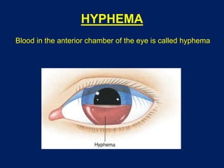
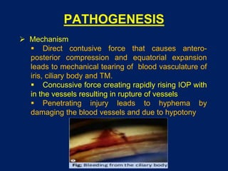
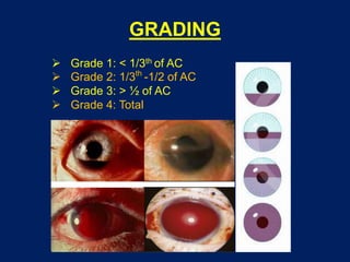
HYPHEMA
Dr Saurabh Kushwaha
Resident Ophthalmology
SCOPE
 Introduction
 Etiology
 Pathogenesis
 Grading
 Clinical features
 Work-up
 Complications
 Management
 Follow-up
 Prognosis
HYPHEMA
Blood in the anterior chamber of the eye is called hyphema
ETIOLOGY
 Traumatic
 Blunt trauma, penetrating injury
 Strenuous conditions
 Whooping cough, Asthma
 Blood dyscraias
 Aplastic anaemia, Leukaemia, haemophilia
 Neovascularisation (Rubeosis iridis)
 DM, BRVO, CRVO
 Miscellaneous
 Herpetic keratouvetitis
 Intraocular tumours (Retinoblastoma, iris melanoma)
 Vascular anomaly (Juvenile xanthogranuloma)
 Secondary to ocular surgery or laser
 Medications with anticoagulant properties (NSAIDs,
Aspirin, Warfarin, Clopidogrel)
PATHOGENESIS
 Mechanism
 Direct contusive force that causes antero-
posterior compression and equatorial expansion
leads to mechanical tearing of blood vasculature of
iris, ciliary body and TM.
 Concussive force creating rapidly rising IOP with
in the vessels resulting in rupture of vessels
 Penetrating injury leads to hyphema by
damaging the blood vessels and due to hypotony
GRADING
 Grade 1: < 1/3th of AC
 Grade 2: 1/3th
-1/2 of AC
 Grade 3: > ½ of AC
 Grade 4: Total
CLINICAL FEATURES
 Blurring of vision
 Photophobia
 Pain
 Headache
 Blood or clot or both in the AC
 Mean duration of elevated IOP is 6 days
 Duration in uncomplicated hyphema is 5-6 days
WORK-UP
 History
 Mechanism of injury
 Time of injury
 H/o medication (Aspirin, Warfarin)
 H/o Sickle cell disease
 H/o Coagulopathy – bleeding gums, epistaxis
 Examination
 Rule out any rupture globe or penetrating injuries
 Visual acuity
 IOP
 Slit lamp
 B scan (gently)
 CT scan - suspected orbital floor # or IOFB
COMPLICATIONS
 Increased IOP:
 Occlusion by clot, inflammatory cells, erythrocytic debris
 Pupillary block due collar button shaped clot
 Peripharal anterior synechiae: if hyphema > 01 week
 Optic atrophy:
 IOP > 50 mmHg for 5 days or >35 mmHg for 7 days.
 Contusion to the optic nerve
 Secondary to damage to short posterior ciliary arteries.
 Sickle cell disease even at normal IOP
 Secondary haemorrhage (rebleed): usually occurs after
day 4
 Increase in size of hyphema
 Layer of fresh RBCs over previous clot
 Change of colour from dark red to bright red.
COMPLICATIONS
 Corneal blood staining:
Factors which influence corneal blood staining:
 Rebleeding
 Prolonged clot duration
 Sustained increase in IOP
 Corneal endothelial dysfunction
Two main risk factors of corneal blood staining:
 IOP >25 mm of Hg
 > 6 days duration
Earliest signs:
 straw coloured discoloration of deep stroma
 presence of tiny yellow granules in posterior
1/3rd
of the cornea
MANAGEMENT
 Quick absorption of blood
 Prevention of complications
 Avoidance of recurrence
 Discontinuation of anti coagulation therapy
 Limiting activities
 Rest in semi upright position and head end elevation
 Patching
OUTPATIENT MANAGEMENT
 Walton et al:
 no associated injury
 hyphema < ½ of AC volume
 satisfactory IOP
 no blood dyscrasia
 safe home environment
 good patient compliance
 good follow up
 no time delay at presentation
MEDICAL MANAGEMENT
 Anti inflammatory drugs - NSAIDs
 Cycloplegics
 Topical, oral and systemic anti glaucoma drugs
 Steroids
 Anti fibrinolytic drugs - tranexamic acid, TPA,
aminocaproic acid
 Laser photocoagulation of bleeding points by
goino prism
 Anti fibrinolytic agents: epsilon amino caproic acid
(EACA) and Tranexamic acid are used to prevent
rebleed
 EACA (amicar) binds to lysine molecules in the
clot via lysine binding site and inhibits fibrin clot
digestion. Its dose is 100mg/kg every 4 hours to a
maximum dose of 30g/day by mouth for 5 days.
 Tranexamic acid also has similar mechanism.
 The results of various studies indicate that both
amicar and tranexamic acid have beneficial effect
on rate of secondary haemorrhage but none of
them had improved the final visual outcome.
 Corticosteroids:
 Stabilizes the blood-ocular barrier
 Directly inhibits fibrinolysis
 Decrease incidence of secondary haemorrhage.
 Prednisolone (0.6 mg/kg) is effective in reducing
the incidence of rebleed
 Statistical analysis indicates that coticosteroids
decreases the incidence of rebleeding without
having any effect on the long term visual outcome.
 Cycloplegics:
 Studies advocate use of Cycloplegics as they
relieve ciliary spasm and prevents formation of
PAS
SURGICAL MANAGEMENT
 Indications (Walton et al):
 Microscopic corneal blood staining
 IOP > 60 mm of Hg for 2 days
 IOP > 50 mm of Hg for >4 days
 IOP > 35 mm of Hg for 7 days
 Pre-existing glaucomatous optic atrophy
 Risk of corneal blood staining (e.g 4 days after the
onset of glaucoma, > ½ - total hyphema with IOP >
25mmHg > 6 days
 Risk of synechiae formation (e.g > 50% hyphema for
> 8 days
 Visual obstruction in children at risk of amblyopia
 Sickle cell disease if IOP >25 mm of Hg for >24 hours
or spikes repeatedly >30 mm of Hg
SURGICAL MANAGEMENT
 Simple paracentesis - allow the clot to retract
 Irrigation and aspiration
 Through a large limbal incision: manual expression,
cryoextraction, ultrasonic emulsification
 Clot expression and limbal delivery is usually
advocated 4 to 7 days after the initial injury, when clot
consolidation and retraction is at its peak.
 Clot irrigation with trabeculectomy is reserved for
total hyphema
FOLLOW-UP
 Hospitalised patient - Daily V/A, IOP, Slit lamp exam
 After discharge - next follow up after 2-3 days
 1 - 2 weeks as per severity
 After 4 weeks - gonioscopy and fundus exam
PROGNOSIS
 Determinants:
 Amount of associated damage to other ocular
structures like choroidal rupture, macular scarring
 Secondary hemorrahge (rebleed)
 Complications of Glaucoma, corneal blood
staining or optic atrophy
 Recovery of V/A is good in approx 75% of patients:
 Hyphema <1/3 of AC -- V/A 6/12 or better in
80% cases
 Hyphema >1/2 - 2/3 of AC -- V/A 6/12 or better
in 60% cases
 Hyphema Grade IV -- V/A 6/12 or better in 35%
cases
THANK YOU

Hyphema

  • 1.
  • 2.
    SCOPE  Introduction  Etiology Pathogenesis  Grading  Clinical features  Work-up  Complications  Management  Follow-up  Prognosis
  • 3.
    HYPHEMA Blood in theanterior chamber of the eye is called hyphema
  • 4.
    ETIOLOGY  Traumatic  Blunttrauma, penetrating injury  Strenuous conditions  Whooping cough, Asthma  Blood dyscraias  Aplastic anaemia, Leukaemia, haemophilia  Neovascularisation (Rubeosis iridis)  DM, BRVO, CRVO  Miscellaneous  Herpetic keratouvetitis  Intraocular tumours (Retinoblastoma, iris melanoma)  Vascular anomaly (Juvenile xanthogranuloma)  Secondary to ocular surgery or laser  Medications with anticoagulant properties (NSAIDs, Aspirin, Warfarin, Clopidogrel)
  • 5.
    PATHOGENESIS  Mechanism  Directcontusive force that causes antero- posterior compression and equatorial expansion leads to mechanical tearing of blood vasculature of iris, ciliary body and TM.  Concussive force creating rapidly rising IOP with in the vessels resulting in rupture of vessels  Penetrating injury leads to hyphema by damaging the blood vessels and due to hypotony
  • 6.
    GRADING  Grade 1:< 1/3th of AC  Grade 2: 1/3th -1/2 of AC  Grade 3: > ½ of AC  Grade 4: Total
  • 7.
    CLINICAL FEATURES  Blurringof vision  Photophobia  Pain  Headache  Blood or clot or both in the AC  Mean duration of elevated IOP is 6 days  Duration in uncomplicated hyphema is 5-6 days
  • 8.
    WORK-UP  History  Mechanismof injury  Time of injury  H/o medication (Aspirin, Warfarin)  H/o Sickle cell disease  H/o Coagulopathy – bleeding gums, epistaxis  Examination  Rule out any rupture globe or penetrating injuries  Visual acuity  IOP  Slit lamp  B scan (gently)  CT scan - suspected orbital floor # or IOFB
  • 9.
    COMPLICATIONS  Increased IOP: Occlusion by clot, inflammatory cells, erythrocytic debris  Pupillary block due collar button shaped clot  Peripharal anterior synechiae: if hyphema > 01 week  Optic atrophy:  IOP > 50 mmHg for 5 days or >35 mmHg for 7 days.  Contusion to the optic nerve  Secondary to damage to short posterior ciliary arteries.  Sickle cell disease even at normal IOP  Secondary haemorrhage (rebleed): usually occurs after day 4  Increase in size of hyphema  Layer of fresh RBCs over previous clot  Change of colour from dark red to bright red.
  • 10.
    COMPLICATIONS  Corneal bloodstaining: Factors which influence corneal blood staining:  Rebleeding  Prolonged clot duration  Sustained increase in IOP  Corneal endothelial dysfunction Two main risk factors of corneal blood staining:  IOP >25 mm of Hg  > 6 days duration Earliest signs:  straw coloured discoloration of deep stroma  presence of tiny yellow granules in posterior 1/3rd of the cornea
  • 11.
    MANAGEMENT  Quick absorptionof blood  Prevention of complications  Avoidance of recurrence  Discontinuation of anti coagulation therapy  Limiting activities  Rest in semi upright position and head end elevation  Patching
  • 12.
    OUTPATIENT MANAGEMENT  Waltonet al:  no associated injury  hyphema < ½ of AC volume  satisfactory IOP  no blood dyscrasia  safe home environment  good patient compliance  good follow up  no time delay at presentation
  • 13.
    MEDICAL MANAGEMENT  Antiinflammatory drugs - NSAIDs  Cycloplegics  Topical, oral and systemic anti glaucoma drugs  Steroids  Anti fibrinolytic drugs - tranexamic acid, TPA, aminocaproic acid  Laser photocoagulation of bleeding points by goino prism
  • 14.
     Anti fibrinolyticagents: epsilon amino caproic acid (EACA) and Tranexamic acid are used to prevent rebleed  EACA (amicar) binds to lysine molecules in the clot via lysine binding site and inhibits fibrin clot digestion. Its dose is 100mg/kg every 4 hours to a maximum dose of 30g/day by mouth for 5 days.  Tranexamic acid also has similar mechanism.  The results of various studies indicate that both amicar and tranexamic acid have beneficial effect on rate of secondary haemorrhage but none of them had improved the final visual outcome.
  • 15.
     Corticosteroids:  Stabilizesthe blood-ocular barrier  Directly inhibits fibrinolysis  Decrease incidence of secondary haemorrhage.  Prednisolone (0.6 mg/kg) is effective in reducing the incidence of rebleed  Statistical analysis indicates that coticosteroids decreases the incidence of rebleeding without having any effect on the long term visual outcome.  Cycloplegics:  Studies advocate use of Cycloplegics as they relieve ciliary spasm and prevents formation of PAS
  • 16.
    SURGICAL MANAGEMENT  Indications(Walton et al):  Microscopic corneal blood staining  IOP > 60 mm of Hg for 2 days  IOP > 50 mm of Hg for >4 days  IOP > 35 mm of Hg for 7 days  Pre-existing glaucomatous optic atrophy  Risk of corneal blood staining (e.g 4 days after the onset of glaucoma, > ½ - total hyphema with IOP > 25mmHg > 6 days  Risk of synechiae formation (e.g > 50% hyphema for > 8 days  Visual obstruction in children at risk of amblyopia  Sickle cell disease if IOP >25 mm of Hg for >24 hours or spikes repeatedly >30 mm of Hg
  • 17.
    SURGICAL MANAGEMENT  Simpleparacentesis - allow the clot to retract  Irrigation and aspiration  Through a large limbal incision: manual expression, cryoextraction, ultrasonic emulsification  Clot expression and limbal delivery is usually advocated 4 to 7 days after the initial injury, when clot consolidation and retraction is at its peak.  Clot irrigation with trabeculectomy is reserved for total hyphema
  • 18.
    FOLLOW-UP  Hospitalised patient- Daily V/A, IOP, Slit lamp exam  After discharge - next follow up after 2-3 days  1 - 2 weeks as per severity  After 4 weeks - gonioscopy and fundus exam
  • 19.
    PROGNOSIS  Determinants:  Amountof associated damage to other ocular structures like choroidal rupture, macular scarring  Secondary hemorrahge (rebleed)  Complications of Glaucoma, corneal blood staining or optic atrophy  Recovery of V/A is good in approx 75% of patients:  Hyphema <1/3 of AC -- V/A 6/12 or better in 80% cases  Hyphema >1/2 - 2/3 of AC -- V/A 6/12 or better in 60% cases  Hyphema Grade IV -- V/A 6/12 or better in 35% cases
  • 20.