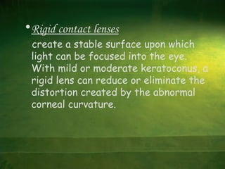
Keratoconus
SABARISABARI
Nomenclature
From Greek
• kerato = cornea
• conus = cone-shape
Definition
• Keratoconus (KC)
Is a non-inflammatory ectatic
condition of the cornea in which
there is progressive central thinning
of the cornea changing it from dome-
shaped to cone-shaped.
Normal Vision
KeratoconusVisionKeratoconusVision
Pathophysiology
• All layers of the cornea are believed to
be affected by KC, although the most
notable features are the thinning of the
corneal stroma, the ruptures in the
Bowman layer, and the deposition of iron
in the basal epithelial cells, forming the
Fleischer ring.
• Breaks in and folds close to the
Descemet membrane result in acute
hydrops and striae, respectively.
Changes occuring in the cornea
1. Thinning in corneal stroma
2. Breaks in bowmans membrane
3. Iron deposit in the basal layer of
the epithelium
4. Variable amounts of diffuse scarring
Breaks in Bowman’s layer
Onset of keratoconus
• It can be anywhere between the ages
of 8 and 45.
• In the majority of cases, it becomes
apparent between the ages of 16 and
30 years.
• It affects males and females equally
• Keratoconus is present in both eyes in
over 90% of cases.
• The onset is not always at the same
time or to the same magnitude.
Keratoconus has been associated with
conditions such as hay fever, asthma,
eczema, Down's syndrome, Marfan's
syndrome and mitral valve prolapse.
Classification
• Based on severity of curvature
• Based on the shape
Based on severity of curvature
Mild : less than 45.00D
Moderate : 45.00 to 52.00D
Advanced : 52.00 to 62.00D
Severe : more than 62.00D
Based on the shape
• Nipple cones (Small size 5mm )
• Oval cones (larger (5-6mm) ellipsoid)
• Globus cones (Largest >6mm ,may involve
over 75% of cornea. )
Etiology
1. Heredity
2. Eye rubbing
3. Contact lenses wear
4. Hormonal change
Heredity
• There is less than one in ten
chances that a blood relative of
keratoconic patient will have KC.
Eye rubbing
• especially children due to some disease as
vernal keratoconjunctivitis
• So corneal curvature will be changed
• Eye rubbing among keratoconic patient
has been reported 66% to 73%.
Hormonal change
• that KC is often first develops
around puberty.
• or advances during pregnancy .
Symptoms and Signs
• The symptoms of keratoconus usually
start in puberty (in the teens) and
may progress for the next 10 to 20
years.
• Nearsightedness
• Astigmatism
• Blurred vision - even when wearing
glasses and contact lenses
• Glare at night
• Light sensitivity
• Frequent prescription changes in
glasses and contact lenses
• Eye rubbing
• Diplopia or polyopia.
• Sometimes keratoconus is diagnosed
when sudden swelling (hydrops)
develops.
This occurs most often in patients
with Down’s syndrome.
The classic signs of keratoconus
Physical examination with
a slit lamp is done and
showed the following:
• Fleischer's ring (an iron
colored ring surrounding the
cone)
• Vogt's striae (stress
lines caused by
corneal thinning)
• Apical scarring
(scarring at the apex
of the cone).
• Corneal thinning :
In advanced cases,
the thinning of the
central cornea can
be seen on
examination.
Diagnosis
Diagnosing keratoconus in its early
stages is more difficult. It requires
a thorough history and examination
Visual Acuity :
 Reduced visual acuity due to oblique
astigmatism
Retinoscopy :
 scissor reflex
Ophthalmoscopy :
 Oil droplet
Munson's sign:
 It’s an angulation of the lower lid
during inferior gaze due to corneal
protrusion
Reduced intraocular pressure:
Due to corneal thinning or reduced
scleral rigidity.
The topography of keratoconus:
The photokeratoscope or placido disc
can provide an overview of the
cornea and can show the relative
steepness of any corneal area.
• Photokeratoscope with normal round
curvature
Distorted pattern of the rings
An Orbscan is the most advanced
topography unit ,
This unit can simultaneously measure
the curvature and thickness of the
cornea over the entire surface.
Treatments
•Glasses:
In mild or early keratoconus, glasses
can be used for vision correction.
Unfortunately, as keratoconus
progresses, the irregular shape of
the cornea cannot be corrected with
glasses.
•Contact Lenses:
 Soft contact lenses
Rigid contact lenses
(hard and gas permeable)
•Contact Lenses:
 Soft contact lenses
may be worn with early or mild keratoconus.
Since soft contact lenses conform to the
shape of the cornea.
They are not able to eliminate visual
distortion created by the irregular shape.
For this reason, patients usually require the
use of a rigid contact lens.
•Rigid contact lenses
create a stable surface upon which
light can be focused into the eye.
With mild or moderate keratoconus, a
rigid lens can reduce or eliminate the
distortion created by the abnormal
corneal curvature.
Surgical Intervention
• A variety of surgical procedures are
available for patients with keratoconus.
Penetrating keratoplastyPenetrating keratoplasty is the most
common.
• Recently, less invasive procedures, such as
Intacs, have been developed
• Penetrating keratoplasty:
In this procedure, the central area of the
cornea is excised and a full-thickness
corneal button is sutured into the recipient.
• Lamellar keratoplasty:
A partial thickness portion of the cornea
is excised and a partial thickness donor
corneal button is sutured into the recipient
transplant.
• Intacs: Intrastromal Corneal Ring
Two arc shaped segments of inert plastic
are inserted into the peripheral (outer)
cornea. These segments add volume to the
thinned cornea. This flattens the steep
curvature and reduces the irregular shape.
Intrastromal Corneal Ring
Thank You

Keratoconus

  • 1.
  • 2.
    Nomenclature From Greek • kerato= cornea • conus = cone-shape
  • 3.
    Definition • Keratoconus (KC) Is anon-inflammatory ectatic condition of the cornea in which there is progressive central thinning of the cornea changing it from dome- shaped to cone-shaped.
  • 4.
  • 5.
    Pathophysiology • All layersof the cornea are believed to be affected by KC, although the most notable features are the thinning of the corneal stroma, the ruptures in the Bowman layer, and the deposition of iron in the basal epithelial cells, forming the Fleischer ring.
  • 6.
    • Breaks inand folds close to the Descemet membrane result in acute hydrops and striae, respectively.
  • 7.
    Changes occuring inthe cornea 1. Thinning in corneal stroma 2. Breaks in bowmans membrane 3. Iron deposit in the basal layer of the epithelium 4. Variable amounts of diffuse scarring
  • 8.
  • 9.
    Onset of keratoconus •It can be anywhere between the ages of 8 and 45. • In the majority of cases, it becomes apparent between the ages of 16 and 30 years.
  • 10.
    • It affectsmales and females equally • Keratoconus is present in both eyes in over 90% of cases. • The onset is not always at the same time or to the same magnitude.
  • 11.
    Keratoconus has beenassociated with conditions such as hay fever, asthma, eczema, Down's syndrome, Marfan's syndrome and mitral valve prolapse.
  • 12.
    Classification • Based onseverity of curvature • Based on the shape
  • 13.
    Based on severityof curvature Mild : less than 45.00D Moderate : 45.00 to 52.00D Advanced : 52.00 to 62.00D Severe : more than 62.00D
  • 14.
    Based on theshape • Nipple cones (Small size 5mm ) • Oval cones (larger (5-6mm) ellipsoid) • Globus cones (Largest >6mm ,may involve over 75% of cornea. )
  • 15.
    Etiology 1. Heredity 2. Eyerubbing 3. Contact lenses wear 4. Hormonal change
  • 16.
    Heredity • There isless than one in ten chances that a blood relative of keratoconic patient will have KC.
  • 17.
    Eye rubbing • especiallychildren due to some disease as vernal keratoconjunctivitis • So corneal curvature will be changed • Eye rubbing among keratoconic patient has been reported 66% to 73%.
  • 18.
    Hormonal change • thatKC is often first develops around puberty. • or advances during pregnancy .
  • 19.
    Symptoms and Signs •The symptoms of keratoconus usually start in puberty (in the teens) and may progress for the next 10 to 20 years.
  • 20.
    • Nearsightedness • Astigmatism •Blurred vision - even when wearing glasses and contact lenses • Glare at night
  • 21.
    • Light sensitivity •Frequent prescription changes in glasses and contact lenses • Eye rubbing • Diplopia or polyopia.
  • 22.
    • Sometimes keratoconusis diagnosed when sudden swelling (hydrops) develops. This occurs most often in patients with Down’s syndrome.
  • 23.
    The classic signsof keratoconus Physical examination with a slit lamp is done and showed the following: • Fleischer's ring (an iron colored ring surrounding the cone)
  • 24.
    • Vogt's striae(stress lines caused by corneal thinning) • Apical scarring (scarring at the apex of the cone).
  • 25.
    • Corneal thinning: In advanced cases, the thinning of the central cornea can be seen on examination.
  • 26.
    Diagnosis Diagnosing keratoconus inits early stages is more difficult. It requires a thorough history and examination
  • 27.
    Visual Acuity : Reduced visual acuity due to oblique astigmatism Retinoscopy :  scissor reflex Ophthalmoscopy :  Oil droplet
  • 28.
    Munson's sign:  It’san angulation of the lower lid during inferior gaze due to corneal protrusion
  • 29.
    Reduced intraocular pressure: Dueto corneal thinning or reduced scleral rigidity. The topography of keratoconus: The photokeratoscope or placido disc can provide an overview of the cornea and can show the relative steepness of any corneal area.
  • 30.
    • Photokeratoscope withnormal round curvature Distorted pattern of the rings
  • 31.
    An Orbscan isthe most advanced topography unit , This unit can simultaneously measure the curvature and thickness of the cornea over the entire surface.
  • 32.
    Treatments •Glasses: In mild orearly keratoconus, glasses can be used for vision correction. Unfortunately, as keratoconus progresses, the irregular shape of the cornea cannot be corrected with glasses.
  • 33.
    •Contact Lenses:  Softcontact lenses Rigid contact lenses (hard and gas permeable)
  • 34.
    •Contact Lenses:  Softcontact lenses may be worn with early or mild keratoconus. Since soft contact lenses conform to the shape of the cornea. They are not able to eliminate visual distortion created by the irregular shape. For this reason, patients usually require the use of a rigid contact lens.
  • 35.
    •Rigid contact lenses createa stable surface upon which light can be focused into the eye. With mild or moderate keratoconus, a rigid lens can reduce or eliminate the distortion created by the abnormal corneal curvature.
  • 36.
    Surgical Intervention • Avariety of surgical procedures are available for patients with keratoconus. Penetrating keratoplastyPenetrating keratoplasty is the most common. • Recently, less invasive procedures, such as Intacs, have been developed
  • 37.
    • Penetrating keratoplasty: Inthis procedure, the central area of the cornea is excised and a full-thickness corneal button is sutured into the recipient.
  • 38.
    • Lamellar keratoplasty: Apartial thickness portion of the cornea is excised and a partial thickness donor corneal button is sutured into the recipient transplant. • Intacs: Intrastromal Corneal Ring Two arc shaped segments of inert plastic are inserted into the peripheral (outer) cornea. These segments add volume to the thinned cornea. This flattens the steep curvature and reduces the irregular shape.
  • 39.
  • 40.