
More Related Content
What's hot
What's hot (20)
Similar to Lesions of retina
Similar to Lesions of retina (20)
More from abhishek ghelani
More from abhishek ghelani (20)
Recently uploaded
Recently uploaded (20)
Lesions of retina
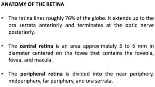
- 1. ANATOMY OF THE RETINA • The retina lines roughly 76% of the globe. It extends up to the ora serrata anteriorly and terminates at the optic nerve posteriorly. • The central retina is an area approximately 5 to 6 mm in diameter centered on the fovea that contains the foveola, fovea, and macula. • The peripheral retina is divided into the near periphery, midperiphery, far periphery, and ora serrata.
- 2. umbo Parafovea (0.5 mm) Perifovea(1.5 mm) Macula (5.5 mm) Fovea (1.5 mm) Foveola(0.35 mm)
- 4. • Clinically the peripheral retina is defined as the zone from the equator to the ora serrata and is approximately three to four disk diameters in width. • The vortex veins represent the equator. • a 1.5-mm ring peripheral to the temporal major vascular arcades called the near periphery. • The retina around the equator is called the equatorial retina, and the region anterior to it is called the peripheral retina
- 5. • The midperiphery consists of an annular area 3 mm wide surrounding the near peripheral retina. • The far peripheral retina extends in width 9 to 10 mm beyond the midperipheral retina temporally and 16 mm nasally.
- 6. Examination • Photograph of a normal disk. • Points to note while examining the disk are its • color, • margins, • the cup, • neuroretinal rim, • the vessels and • the peripapillary area
- 7. • Normal arterioles and venules. • The arterioles are thinner, red in color and show a prominent golden reflex. • The venules are broader, flatter, and darker red in color. • The arterioles and venules run alongside each other for some distance and their branches often cross each other
- 8. • At arteriovenous crossings the arterioles may cross over the venules • or the venules may cross over the arterioles • Though the former is more common
- 9. • The retina has an orangish color which varies with the degree of pigmentation in the pigment epithelium. • Some normal variations of the pigment in the RPE can allow the orange stripes of choroidal vessels to be seen giving it a tigroid appearance. • These vessels are better seen in the mid-periphery and beyond. • Choroidal vessels seen as orange tape like criss-crossing bands
- 10. The ampulla of a vortex vein is seen as an orange bulb with choroidal veins draining into it
- 11. • Junction between the temporal ora serrata and the pars plana • the nasal retina has more prominant dentate processes than the temporal retina
- 12. Variations of normal optic nerve head A normal disk in which the cup cannot be appreciated A normal disk with a small cup
- 13. Physiologically enlarged cup showing a uniform thinning of the entire neuroretinal rim A normal disk with blurred margins and absent cup giving a false impression of disk edema
- 14. A hyperemic disk with a small cup and blurred margins in a hypermetropic eye Blurring of the disk margins (initially the superior and inferior) with absent cup raises suspicions of early papilledema. Presence of venous pulsations or the ability to induce them, suggests absence of it.
- 15. • papilledema characterized by a hyperemic protuberant disk with blurred margins and obscured cup. • The blood vessels on the disk appear to climb down from the elevated margins towards the surrounding retina and at places appear to be hidden by the swollen fibers. • The veins of the retina are engorged. • There is surrounding peripapillary edema
- 16. Established papilledema with blurring of the entire margin of the optic disk with some elevation, dilated turgid and tortuous veins, soft exudates and few retinal hemorrhages Chronic papilledema with blurring of the entire margin of an elevated optic disk with deposition of hard exudates
- 17. Arteriosclerotic changes seen as increased reflex from the wall of the arterioles making them appear as silver wires In hypertensives the vessels may become tortuous
- 18. In Hypertensive optic neuropathy, optic nerve head edema is present along with other signs of hypertensive retinopathy—retinal hemorrhages, cotton-wools spots, macular edema, macular star, arteriovenous crossing changes. These changes are bilateral
- 19. In diabetic papillopathy, unilateral or more commonly bilateral hyperemic swollen disks maybe present in association with mild to moderate visual loss. Other changes of diabetic retinopathy may be absent.
- 20. • Hyperemic disks with blurred margins in patients, who have been addicted to alcohol and tobacco over a prolonged period of time, must be suspected to have (tobacco-alcohol) toxic optic neuropathy. • The disk swelling is less and the vascular changes of papilledema are not seen. • Later in the course of the disease the disks become pale and excavated. • A dramatic and bilateral loss of vision with bilateral swollen hyperemic disks along with marked attenuation of the blood vessels maybe seen in methyl alcohol poisoning. • These disks rapidly turn pale. In the majority, by the time the patient presents the disks are invariably pale.
- 21. • Bilateral hyperemic disks with blurred margins may be seen in young patients, usually male, in Leber’s hereditary optic neuropathy. • The optic disks look hyperemic with tortuous and telangiectatic vessels and blurred margins. However, the disk prominence is mild. • A peculiar swelling of the fibers around the disk has also been described (pseudoedema). The two eyes are involved in rapid succession which then proceeds to optic atrophy • Patients on chronic therapy with the antiarrhythmic drug Amiodarone may develop an optic neuropathy showing hyperemic disks with blurred margins. • The cornea shows superficial brown deposits of the drug in characteristic whorls.
- 22. • Hyperemic swollen disks are the hallmark of optic neuritis which is usually unilateral in adults but can be bilateral in children. • If it is associated with paraplegia or other neurological defects that may occur before or after the optic neuritis it is likely to be neuromyelitis optica. • Bilateral optic neuritis in adults is rare and usually associated with extensive sinusitis and syphilitic meningitis.
- 23. Glittering yellow deposits of synchiasis scintillans which fly around within the liquefied vitreous. At rest, they settle down only to be scattered like a ‘golden shower ‘with the next eye movement Multiple small yellowish- white deposits of Asteroid hyalosis suspended in a formed vitreous gel and hence move very little with movements of the eye
- 24. • Mild optic disk edema associated with retinal edema and hard exudates deposited as and incomplete macular star in Neuroretinitis. • The disk edema is relatively milder than in optic neuritis and the macular star appears early • Long standing neuroretinitis showing features of chronic disk and macular edema, resolving macular star, dilated epipapillary vessels and retinal striae (Paton’s lines)
- 25. Optic nerve meningiomas distorting the globe and causing inferior disk edema Hyperemic disk edema associated with posterior uveitis (Vogt Koyanagi Harada syndrome). Multiple patches of choroiditis are seen at the posterior pole
- 26. Patches of active choroiditis seen as moderate sized yellow lesions with fuzzy margins. Several pigmented healed scars are also present
- 27. Progressive cytomegalovirus retinitis with some retinal hemorrhages and vasculitis CMV retinitis in the peripapillary region with involvement of the optic nerve head
- 28. • Healed CMV retinitis leaving an area of atrophic retina. The vessels are sclerosed • The atrophic retina easily develops atrophic holes frequently resulting in rhegmatogenous retinal detachment
- 29. • An extensive lesion comprising of fluffy dense whitening of the retina with scattered retinal hemorrhages CMV retinitis (fulminant form). This appearance is called a ‘pizza pie’ or ‘cottage cheese with ketchup’ appearance. The lesions are sharply demarcated with small isolated satellite lesions at the advancing edge. The retinitis often occurs first in relation to the vessels of the posterior pole as one or more foci which then coalesce and progress. • The unaffected retina looks normal
- 30. • Large confluent, yellow white, granular looking areas in acute retinal necrosis (ARN). The patches appear granular and are more deeply placed. Initially the disease process starts as multiple small lesions in the mid-peripheral retina that coalesce and spread towards the periphery as well as posteriorly. • The macula remains spared till late
- 31. Deep retinal opacification present all around in the mid-peripheral retina with characteristic clearing of the retinal opacification around the blood vessels (perivenular) (black arrows) giving a ‘cracked mud’ appearance in progressive outer retinal necrosis (PORN)
- 32. White, cobweb like subretinal fibrosis secondary to long standing choroiditis
- 33. Bilateral, subretinal gliotic sheets that have rapidly encompassed the entire posterior pole, suspicious of subretinal fibrosis and uveitis
- 34. Multiple scars of variable size scattered randomly anywhere in the retina following multifocal choroiditis Patches of healed choroiditis can vary in their appearance (here there is no pigmentation and the lesions are confluent)
- 35. Hyperemic disk edema associated with central retinal vein occlusion Papillophlebitis. There are multiple soft exudates, some hemorrhages and patches of retinal edema especially in the inferior retina
- 36. • Optic nerve head studded with refractile globular deposits of varying size that reflect light brightly, in a patient with optic nerve head drusen • The drusen exhibit autofluorescence. • This feature clinches the diagnosis
- 37. • A pale swollen disk in anterior ischemic optic neuropathy (the fuzzy looking swollen tissue along with pallor gives the disk a ‘milky appearance’). On FFA the disk shows patches of hypoflourescence due to nonperfusion (arrows)
- 38. • Atrophy of the papillomacular bundle results in temporal pallor and commonly occurs following retrobulbar neuritis and bilaterally in pituitary tumors. • A band or bowtie pallor with sparing of the superior and inferior parts of the disk implies optic tract involvement.
- 39. • ‘Disk at risk’ is a disk that is crowded with nerve fibers that decrease the size of the cup. • Such disks are predisposed to anterior ischemic optic neuropathy. The other eye of this patient has earlier suffered an ischemic optic neuropathy.
- 40. • In patients with acute lymphocytic leukemia, lymphocytic infiltration of the retrolaminar portion of the optic nerve results in a pale disk edema. Presence of whitish fluffy infiltrates on the surface of the disk, obscuring its details is suggestive of prelaminar infiltration. • Similar infiltration may occur in lymphoma, metastatic carcinoma, and nasopharyngeal carcinoma
- 41. • Waxy yellow pallor of the disk with attenuated arterioles and bone corpuscle shaped pigment deposits (consecutive Optic atrophy secondary to Retinitis Pigmentosa) • Pigment clumping seen in the posterior pole, bilaterally, with minimal pigment disturbance in the peripheral retina in inverse RP or pericentral retinitis pigmentosa. • The arterioles are attenuated and the disk shows a waxy pallor. The features are present bilaterally
- 42. • classic ‘bone corpuscle’ shaped pigmentation in retinitis pigmentosa • Attenuation of the retinal vessels along with a fine mottling of the RPE in the mid-peripheral retina but no discernible pigment clumping referred to as retinitis pigmentosa sine pigmento
- 43. Sparing of the macula inspite of extensive involvement of the mid- peripheral retina in retinitis pigmentosa classic ‘bone corpuscle’ shaped pigmentation in retinitis pigmentosa Geographic atrophy of the macula in retinitis pigmentosa
- 44. Multiple small white spots in the mid peripheral retina with an atrophic patch at the macula and attenuated arterioles in retinitis punctata albescens Densely packed multiple yellow white spots of fundus albipunctatus in a patient complaining of night blindness.
- 45. • A total lack of chorioretinal pigment in albinism resulting in a completely hypopigmented fundus. All levels of the choroidal vessels are visible. The fovea is indistinct • There is complete lack of pigment till the retinal periphery
- 46. Almost complete atrophy of the choroid with only small patches of intact choroid, seen in an elderly male is likely to be the end stages of choroideremia FFA highlights the small island of normal choroidal tissue remaining at the macula
- 47. • Highly enlarged disk bearing a coloboma • Morning Glory anomaly wherein the optic disk appears to be embedded in the center of a funnel shaped excavation of the entire peripapillary region. The disk is covered with varying amounts of grayish white glial tissue which envelops the retinal vessels. • The vessels are greater in number and arise more peripherally. They are abnormally narrow and straight, spreading radially giving the appearance of the ‘morning glory flower’. The abnormal disk is surrounded by a halo of chorioretinal atrophy.
- 48. Size of the coloboma can vary (a small choroidal coloboma) A large coloboma involving the disk and macula
- 49. • Melanocytoma of the optic nerve head appearing as a black mass obscuring the disk • Linear hemorrhages in the shape of splinters of wood on the disk surface are called Splinter hemorrhages and are characteristically found in > uncontrolled glaucoma, > anterior ischemic optic neuropathy, > central retinal vein occlusion
- 50. • A choroidal nevus present inferotemporal to the disk, seen as an ill-defined, deeply located, pigmented lesion. • The margins of the nevus are feathery • Dark pigmented lesion on the optic nerve head (Melanocytoma)
- 51. • New vessels on the disk (NVD) appear as a network of fine vessels arborising on the surface of the disk- the rest of the retina shows features of the underlying disease such as retinal hemorrhages, exudates • Collaterals on the optic disk appear as small clumps of vessels that are tortuous and curly
- 52. • A choroidal melanoma seen as a brown, elevated, globular mass below the retina
- 53. • Feathery white patches fanning out from the optic disk on to the retinal surface following the pattern of the nerve fibers is likely to be a patch of myelinated nerve fibers—they can vary greatly in their extent • Small grayish wisp of tissue attached to the disk surface is likely to be an embryonic remnant of the hyaloid system referred to as a Bergmeister’s papilla
- 54. • Persistent hyperplastic primary vitreous is seen as a membrane that fans out from the optic nerve head extending anteriorly and is often associated with a fold of the retina
- 55. • Fibrovascular proliferation on the optic disk look like grayish white membranes situated on the disk surface and growing into the vitreous. Proliferating vessels are seen within the membrane. These vessels can disappear as the proliferation regresses leaving a sheet of dense fibrous tissue. • The rest of the fundus shows features of the causative retinal pathology
- 56. • A small, oval disk that is abnormally tilted such that the long axis of the disk lies obliquely is called a tilted disk. • There is thinning and hypopigmentation of the inferonasal retinal pigment epithelium and choroid • A severely tilted disk in which the disk appears to be almost horizontally placed. • Obliquity in the anteroposterior axis causes the disk to look small as seen ‘end on’
- 57. • Grayish looking thickened macula with cherry red spot due to ischemic macular edema in a patient with central retinal artery occlusion • The patient also had advanced glaucomatous cupping of the disc • Glassy appearance of an edematous macula in a patient with small branch vein occlusion. • There are several retinal hemorrhages and the edematous area is bordered by hard exudates
- 58. • Multiple, small, ‘red colored’ cysts arranged in a rosette around the fovea is the characteristic appearance of cystoid macular edema • It is commonly seen postoperatively after cataract surgery or in association with uveitis, central retinal vein occlusion, diabetic retinopathy, retinitis pigmentosa or with a choroidal neovascular membrane • Bilateral CME may occur in association with pars planitis and the use of certain drugs such as latanoprost, epinephrine, nicotinic acid, tamoxifen.
- 59. • A star shaped radial arrangement of linear yellow hard exudates centered on the fovea called a ‘macular star’ seen in a patient with neuroretinitis • A complete macular star along with retinal hemorrhages, soft exudates and disk edema in Grade 4 hypertensive retinopathy
- 60. • A well circumscribed round to oval area of neurosensory elevation centered at the fovea in central serous chorioretinopathy (CSCR) • A small CSCR can be easily overlooked
- 61. • Deposition of subretinal fibrin within the cavity of CSCR
- 62. • Retinal pigment epithelial alterations at the macula and as a linear track (arrow) below the macula called ‘CSCR tracks’. • These lesions are better seen by FFA as transmission defects (arrow)
- 63. • A small, well defined area of retinal elevation looking like an ‘orange blister’ is the characteristic appearance of a pigment epithelial detachment • A moderate sized pigment epithelium detachment
- 64. • Thickened macula along with subretinal hemorrhage due to an underlying choroidal neovascular membrane • Subretinal blood that is darker in color with well defined margins suggests that the blood may be below the pigment epithelium
- 65. • Presence of a subretinal grayish green membranous lesion that resembles a wet tissue and thickens the macula unevenly is likely to be a choroidal neovascular membrane (CNVM). • The thickening is also due to the presence of the membrane, serous elevation of the retina and intraretinal edema.
- 66. • Scar consisting of subretinal gliosis, pigmentation and chorioretinal anastamoses secondary to a choroidal neovascular membrane • Scar consisting mainly of white retinal gliosis with no pigmentation
- 67. • Some scars have characteristic features which help to identify the cause. • Scars following congenital toxoplasmosis have a predilection for the macula causing a focal necrotizing retinitis. • But most of these lesions are detected at a later stage when the macula shows a deep punched out pigmented scar. • Active toxoplasmosis with significant vitreous inflammation ‘headlight in fog’ appearance
- 68. • Scars following choroidal tear appear as multiple curved linear scars concentric to the disk margin.
- 69. Patch of retinitis adjacent to an old scar suggests the possibility of toxoplasmosis. The vitreous may show cells specially overlying the active lesions A focus of active retinitis looks like a fluffy white lesion as the involved retina becomes thickened, elevated and edematous
- 70. Scars secondary to a posterior pole toxocara granuloma also have a characteristic appearance
- 71. • Wrinkles (fine retinal folds) across the macula in a patient with a shallow retinal detachment • A better defined ERM causing tortuosity of the blood vessels and ILM folds
- 72. • A contracting epiretinal membrane can cause the clivus to be drawn towards the center deepening the foveal depression. This causes the fovea to look darker red and when seen in contrast to the whitish membrane it can falsely look like a macular hole, actually a pseudohole • A macular hole is seen as a circular or oval almost punched out red colored defect in the macular tissue with a gray well defined edge
- 73. • Fluid that seeps in through a full thickness macular hole appears as a cuff of subretinal fluid around the hole • A large macular hole with urrounding cuff of fluid
- 74. • A macular cyst can be mistaken for a macular hole • A pseudohole due to an epiretinal membrane can be mistaken for a macular hole
- 75. Horseshoe retinal tear appears as a red colored ‘V’ shaped break in the retina, looking like a piece of cloth that is torn when caught in barbed wire Retinal hole with surrounding subretinal fluid
- 76. Difficult to see large horseshoe tear that can easily be missed on cursory examination. Such tears are better visualized by indentation
- 77. A giant retinal tear with retinal detachment and a ‘rolled out’ edge
- 78. Gray dome like elevation of the retina in the peripheral retina commonly in the inferotemporal quadrant and bilateral is likely to be peripheral retinoschisis Fibrovascular fronds causing tractional retinal detachment
- 79. The peripheral retina looks grayish and elevated with a well defined linear posterior edge due to indentation by a scleral buckle—‘the buckle effect’
- 80. In a retinal dialysis the retina appears to be disinserted from the ora and there is no retinal tissue between the break and the ora
- 81. Snowflake degeneration appears as tightly packed small white specks looking like snowflakes scattered in the peripheral retina
- 82. • A well defined red lesion at the macula due to the presence of a post traumatic macular hemorrhage (subretinal). There is another patch of subretinal hemorrhage nasally • Massive ‘subretinal’ hemorrhage, fresh (red) and altered (decolored)
- 83. • An area of subretinal hemorrhage can appear as a dark blackish- maroon lesion of the fundus mimicking pigmented lesions. • Dark subretinal blood with areas of white altered blood at the macula
- 84. • Normally at an AV crossing the venule crosses the arteriole obliquely but with arteriolar sclerosis the venule is seen to be deflected in such a manner that it crosses the arteriole at right angles (Salus sign) • All caliber changes occurring at arteriovenous crossings are collectively referred to as ‘AV nicking’ seen here at multiple sites (arrows)
- 85. Compression of the vein (arrow) results in an apparent impediment to the flow of blood in the vein across the crossing resulting in ‘banking’ and swelling of the peripheral part of the vein (Gunn’s sign)
- 86. • Multiple ‘intraretinal’ macular hemorrhages following a branch vein occlusion • Preretinal macular hemorrhage obscures the underlying retina
- 87. Sheathing of a vessel with an adjacent patch of retinal hemorrhage due to active vasculitis A sheathed occluded vessel as a result of old vasculitis
- 88. Active vasculitis with perivascular infiltration and adjacent retinal hemorrhages
- 89. Resolving vasculitis with decreasing perivascular infiltration, retinal hemorrhages and retinal edema
- 90. • Frosted branch angiitis is a severe form of vasculitis that affects almost the entire vasculature. • The profuse perivascular infiltration causes the blood vessels to look like frosted branches
- 92. • Cherry red spot secondary to traumatic macular edema (Berlin’s edema) • Post-traumatic edema (Berlin’s edema) involving large areas of the retina along with retinal and subretinal hemorrhage. • This type of opacification is usually at a deeper level of the retina and is well defined and more glistening. There is associated macular edema, choroidal tears and subretinal hemorrhage
- 93. • Prominent choroidal folds in a patient with choroidal effusion syndrome • Sharply demarcated areas of atrophic pigment epithelium and choriocapillaris uncovering the larger choroidal vessels in central areolar choroidal dystrophy
- 94. • Bulls eye maculopathy secondary to chloroquine toxicity—early changes include stippling or mottling and blunting of the foveal reflex. • Later there is central irregular pigmentation surrounded by a concentric zone of hypopigmentation • Bulls eye maculopathy seen in a patient with advanced cone dystrophy. • A pigmented area in the center is surrounded by a complete or incomplete parafoveal ring of depigmentation
- 95. • Lattice (reticular/net) like pattern at the macula in the rare Sjogren’s reticular dystrophy
- 96. • Bilateral pigment mottling in a glistening, orizontally ovoid area giving a metallic beaten bronze appearance is highly suggestive of Stargardt’s disease. • The mottling can occupy an area of 2 disk diameters. • A ring of yellow flecksoften surrounds this area. The flecks are peculiarly shaped and are curved esembling fish or comma or crescent like shapes. • They can extend up to the mid peripheral retina but never beyond. These changes are then referred to as fundus flavimaculatus.
- 97. In fundus flavimaculatus the yellowish flecks are in small curvilinear shapes resembling commas or small fish occurring bilaterally and symmetrically in the posterior pole up to the midperiphery. Patches of RPE atrophy at the macula (Stargardt’s dystrophy) maybe present
- 98. • In the initial stages of the disease the fovea shows a granular appearance and gives the impression of being covered by a varnish. • These changes are picked up earlier by FFA which shows a mottled hypo and hyperfluorescence. • Fundus flavimaculatus, if present shows characteristic ‘choroidal silence’ on FFA in which the choroidal circulation is masked and the retinal capillaries stand out in contrast
- 99. • Patch of pigmentation just temporal to the fovea bilaterally, seen in late stages of Idiopathic macular telangiectasias (also called parafoveal or juxtafoveal telangiectasias)
- 100. • Bilaterally symmetrical idiopathic macular telengiectasia (IMT) also called juxtafoveal or parafoveal telengiectasia. • The capillaries in this area exhibit mild irregular dilatation with adjacent graying due to edema of the surrounding retina. • On FFA the parafoveal vessels are dilated and telangiectatic and leak gently in the late phases especially in the temporal network
- 101. Retinal edema, crystalline deposits and dilated ectatic vessels seen in the temporal parafoveal region suggestive of idiopathic macular telangiectasias (formally referred to as idiopathic juxtafoveal telangiectasias/parafoveal telangiectasias)
- 102. • small red dots scattered across the posterior pole are likely to be microaneurysms (black arrows) whereas retinal hemorrhages are usually larger (white arrow) • Often a bunch of microaneurysms are seen at the center of a ring of retinal thickening and hard exudates (circinate girdle)
- 103. • Punctate or rounded hemorrhages, looking like ‘dots and blots’ are hemorrhages in deeper layers of the retina • Preretinal hemorrhages (arrow head) • multiple ‘Roth’s spots (arrows) in anemic retinopathy
- 104. Venous reduplication (black arrow) is a sign of increasing retinal ischemia as seen in severe NPDR along with other signs such as soft exudates and retinal hemorrhages in multiple quadrants A network of arborising or broom like vessels lying superficially on the disk or on the retinal surface is suggestive of neovascularisation
- 105. NVE form at the junction of ischemic and non-ischemic retina New vessels growing in a radial pattern also referred to as ‘sea-fan’ neovascularization.
- 106. Over time, delicate fibrous tissue becomes evident along the new vessels. Initially this tissue is translucent but later it becomes opaque and white and the complex is now referred to as a fibrovascular proliferation (FVP) Traction from a detaching posterior vitreous can cause a fibrovascular frond to bleed resulting in vitreous hemorrhage
- 107. A contracting fibrovascular frond is seen to exert traction at its points of attachment to the retina causing a drag on the retinal vessels NVD fanning out into the vitreous body
- 108. • Multiple, round white spots, that are regularly spaced, in a specific pattern due to recent laser application • Photocoagulation scars appear as multiple, regularly arranged, circular scars scattered in a pattern
- 109. Multiple cotton-wool spots, retinal hemorrhages, disk edema in the posterior pole seen in hypertensive retinopathy Boat-shaped sub-hyaloid hemorrhage
- 110. • Angioid streaks (breaks in the Bruch’s membrane) seen as dark red lines radiating from the optic disk in all directions that lie deep in the retina and resemble cracks
- 111. • The breaks allow the choriocapillaris to show through. They are associated with mottling of the RPE at the macula
- 112. • Rounded, sharply demarcated, yellowish white areas with varying amounts of pigmentation seen in a myopic eye represent focal areas of chorioretinal atrophy in myopia • As the choriocapillaris atrophies the larger choroidal vessels are seen to cross these areas • Criss-crossing yellow colored choroidal vessels at the macula due to choroidal sclerosis. A ‘laquer crack’ seen as a fine linear reddish line is also present
- 113. Geographical patches of chorioretinal atrophy exposing the underlying sclera in severe degenerative myopia
- 114. • The cracks in the Bruchs membrane allow the growth of fine choroidal neovascular membranes which become more obvious when they bleed. • These blotches of hemorrhages are often referred to as Fuchs’ spot and rapidly disappear leaving an area of pigmentation and scarring.
- 115. • Multiple yellow, pale or white punctate round deposits of varying size at the posterior pole (drusen)
- 116. • Small (less than 63 microns) flat drusen with well defined margins are called hard drusen • Large drusen (greater than 125 microns) with less distinct, fuzzy margins that are elevated or dome shaped are called soft drusen. • They tend to become confluent in various shapes as they coalesce
- 117. Still larger confluent, soft drusen (500 microns) may have a pool of serous fluid around them and are referred to as drusenoid PEDs (Drusenoid pigment epithelial defects) Regressing drusen are whiter and refractile in appearance. Their margins become irregular and areas of calcification start to appear. Associated RPE atrophy becomes prominent
- 118. • Sharply demarcated, round to oval area of depigmentation, showing the underlying choroidal vessels is an area of geographic atrophy in Dry AMD
- 119. • A sharply demarcated hypopigmented spot due to loss of pigment from the retinal pigment epithelium may be a result of exposure to ultraviolet rays (photic retinopathy). • At times there maybe clumps of pigment adjacent to the hypopigmented lesion. • If examined soon after exposure the fovea shows a focal whitish gray lesion resembling a photocoagulation spot of varying intensity.
- 120. • Bilateral yellow colored cyst like lesion under the macula is the unmistakable appearance of vitelliform macular dystrophy. • A variant called adult vitelliform dystrophy occurs in middle-aged individuals and is often unilateral. The lesions are smaller and the EOG is normal. • On FFA these lesions are characteristically ‘autofluorescent’. The cyst does not fill and in fact masks the underlying choroidal fluorescence.
- 121. • The vitelliform contents disintegrate and liquefy showing a fluid level giving the appearance of a hypopyon • A small collection of altered subretinal blood under the fovea.
- 122. A subretinal cystic lesion that is translucent white in color along with a characteristic white dot within the lesion (scolex) is the characteristic appearance of a subretinal Cysticercus
- 123. White bands of subretinal fibrosis in a self settled retinal detachment
- 124. • A well defined area of blotchy pigmentation involving a sector or a large segment of the retina with well defined convex margins due to spontaneous resolution of retinal detachment • A case of spontaneous resolution of a superior retinal detachment with blotches of coarse pigmentation and a subretinal bandr running near the edge
- 125. A pair of dilated and tortuous vessels emerging from a tumor is likely to be the efferent and afferent vessels of a peripheral capillary hemangioma Highly dilated and tortuous vessels, secondary to peripheral arteriovenous communications in Wyburn-Mason syndrome
- 126. A lattice palisade degeneration or dystrophy appears as a linear, spindle/cigar shaped lesion parallel to the ora made up of yellow spots with white lines of sclerotic vessels criss-crossing across
- 127. A lattice harboring a retinal holeA large tear present at the edge of a pigmented lattice. Such tears can sometimes unzip the entire lattice resulting in a giant tear