Acquired macular diseases: AMD, RAP, PED, ERM, MH, CSR, CME, IMT
•Download as PPTX, PDF•
6 likes•1,169 views

Early macular signs are usually subtle, be armed with other clinical tools.

Recommended
More Related Content
What's hot
What's hot (20)
Similar to Acquired macular diseases: AMD, RAP, PED, ERM, MH, CSR, CME, IMT
Similar to Acquired macular diseases: AMD, RAP, PED, ERM, MH, CSR, CME, IMT (20)
More from Samhaa Mohammed
More from Samhaa Mohammed (20)
Recently uploaded
Recently uploaded (20)
Acquired macular diseases: AMD, RAP, PED, ERM, MH, CSR, CME, IMT
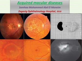
- 1. Acquired macular diseases Samhaa Mohammed Abd El Moneim Zagazig Ophthalmology Hospital, 2018 Samhaa Mohammed
- 2. Acquired macular diseases AMD(dry&wetCNV)√ RAP,PCV,PED √ Interfacedisorders(ERM,MH,VMT) CSR CME Idiopathic maculartelangectasia Degenerativemyopia Angioidstreaks Choroidalfolds,hypotony maculopathy,solarretinopathy,focalchoroidal excavation Samhaa Mohammed
- 3. Macular assessment tests VA (distant & near) Amsler grid Contrast sensitivity (↓ in ON) Pupil & color (↓ in ON) Photostress test Samhaa Mohammed
- 4. AMD ↓ VA + AMD - Drusen - RPE changes - GA Dry - CNV - Soft drusen Wet Samhaa Mohammed
- 5. AMD Risk factors Age Race (white, caucasian) Hereditary (CFH gene↓) - Smoking - Dietary Fat - HTN, CVD - F > M - Hypermet ropia - Cat. Surgery AMD Samhaa Mohammed
- 6. Drusen • Alone Not equal AMD • Extracellular deposits (lipid, lipofuscin like!) between PRE & Bruch̛s RF (drusen convert into AMD): • Size of drusen (small, intermediate, large) • Type (hard or soft) • GA, RPE changes • Age, FH, other eye Samhaa Mohammed
- 7. Drusen - < 63 mic (<1/2 v width) - Well defined - Dry AMD Small (hard) - 63-125 mic Intermediate - > 125 mic - One large drusen ↑risk of Wet AMD Large (soft) Hard Soft Confluent Calcific DD Doyne honey comb retinal dystrophy (AD drusen, Malattia leventine): 40 y Cuticular drusen (stars in sky): 20y, small, PED Type 2 membrano-proliferate GN: older children Samhaa Mohammed
- 9. AMD ↓ VA + AMD - Drusen - RPE changes - GA Dry - CNV Wet Samhaa Mohammed
- 10. Dry AMD • Hyperfl: RPE atrophy (window) or late staining • Hypofl: high lipid content (masking)FFA • Hyperreflective between RPE & Bruchs membrane. • Outer retinal layers corrugation: basal laminar (between RPE & RPE BM), basal linear (between RPE & bruch inner collagenous layer) OCT Prevention (RF, Antioxidant?) Amsler grid Investigation LVA AMD work up Samhaa Mohammed
- 11. Dry AMD Small, intermediate drusen Small, intermediate drusen, pigmentary changes Small, intermediate drusen, GA Pigmentary changes, GA Samhaa Mohammed
- 13. Dry AMD TTT Indication Extensive intermediate confluent drusen One large drusen GA in one/ both eyes Late AMD in one eye Daily TTT regimen/d Vit E 400 IU Vit c 500mg Lutein 10mg Zeaxanthin 2mg (instead of B carotene, vit A) → lung cancer in smokers. Zinc (25- 80mg) ↓ dose to ↓ SE (genitourinary problems) Copper (2mg) With high zinc dose Treat RF Samhaa Mohammed
- 14. AMD ↓ VA + AMD - Drusen - RPE changes - GA Dry - CNV Wet Samhaa Mohammed
- 15. CNV • NVs from choroid → penetrate bruch membrane to retina. • C/P: • Acute, subacute or chronic drop of vision. • Metamorphopsia (amsler chart). • Associated drusen, cause, haemorhage, scar, subretinal fluid. • Greyish green, pinkish yellow lesion. • Poor prognosis. Type 1 • Sub RPE Type 2 • Sub retinal Samhaa Mohammed
- 16. CNV causes Degenerative (AMD, myopia, RAP, PCV, angioid streaks) Inflamatory (post uveitis, POHS, toxoplasmosis) Trauma (choroidal rpture, extensive laser) Tumours (choroidal naevus, haemangioma) Samhaa Mohammed
- 17. Wet AMD Grey to green subfoveal CNV Intra, sub retinal hge Extensive lipid deposition Disciforrm scar Samhaa Mohammed
- 18. CNV • Classic: early hyper f ↑ size & intensity, starts well, ends ill defined. • Occult: late leakage with undetermined cause.FFA • Dx: layers disruption, SRF, scarring, RPE & Bruchs membrane. • Mointor TTT response.OCT Samhaa Mohammed
- 20. CNV TTT regimen Anti VEGF (main role) (treat & extend/ m for 3m then ↑ interval duration) - Avastin, Bevacuzimab (/m) - Lucentis, Ranbizumab (/m) - Elyea, Aflipercept (/2m) Laser PDT SE (genitourinary problems, CU ↓) Not available in Egypt • Treatment is effective in active CNV rather than disciform fibrotic CNV. • Activity signs (SRF, haemorhage, enlarging membrane, progressive decreasing vision). • Follow up with VA, OCT macular thickness.Samhaa Mohammed
- 21. PED 1. Serous PED: RPE problem Immune related! Pooling Samhaa Mohammed
- 22. PED 2. Fibrovascular PED 3. Drusenoid PED 4. Haemorhagic PED Worst prognosis (fibrovascular, hgic) 2 4Samhaa Mohammed
- 23. RPE Tear IVI, Laser Loss of dome shape of PED Crescent shape hypo + hyper fl. Rolled up RPE Dedunded RPE Dedunded RPE Rolled up RPE Corrugated elevated RPE Samhaa Mohammed
- 24. PCV, RAP PCV Late middle age, unilateral sudden visual drop Multiple serous PED, reddish nodules 50% have spontaneous resolution Anti VEGF has lower effect than CNV RAP Stage 1 IRN Stage 2 SRN → PED Stage 3 CNV with RCA FA: similar to occult or minimally classic CNV OCT hyper reflective NV Samhaa Mohammed
- 25. Acquired macular diseases AMD(dry&wetCNV)√ RAP,PCV,PED √ Interfacedisorders(ERM,MH,VMT) √ CSR CME Idiopathic maculartelangectasia Degenerativemyopia Angioidstreaks Choroidalfolds, hypotony maculopathy,solarretinopathy, focalchoroidal excavation Samhaa Mohammed
- 26. ERM • PVD → glial fibrocellular tissue proliferation → ERM→ macular pucker. Causes: • Idiopathic • 2ry to Vascular (DR, CRVO) Inflamation (IU, PU) Trauma Retinal surgery, laser, cryo TTT indication: • High visual requirements (occupation, young age) • Duration of visual loss < 6 months • VA < 6/12 • Well-defined ERM edge • No associated CME PPV + peeling Samhaa Mohammed
- 27. ERM Advanced membrane ERM ERM in OCT pseudoholeSamhaa Mohammed
- 29. MH • ERM, VMT …. MH • Unilateral, female, old age • FTMH • Lamellar MH • Pseudo hole • Investigation: • Amsler grid: scotoma. • Watze Allen sign: broken slit beam • OCT: definite dx • FFA: window defect in late stage (RPE atrophy) • FAF: hyper autoflurescenceSamhaa Mohammed
- 30. MH Stage 1a Impending MH Photoreceptors detach from inner retina, schesis Stage 1b Occult MH Photoreceptors with centrifugal displacement Stage 2 Small FTMH FTMH <400 mic Stage 3 Large FTMH FTMH > 400mic without Complete PVD Stage 4 Large FTMH FTMH > 400 MIC Complete PVD Samhaa Mohammed
- 32. MH TTT options • Observation (50% of stage 1 spontaneously closed). • PPV with ILM peeling + gas tamponade (face down). • Vitreolysis with ocriplasmin (in AP or equatorial traction). Indication • FTMH (success 90% in stage 2). • High visual needs. • Young age. • < 6m. • Decrease vision of the other eye. Examine the other eye: MH of ther eye will be 1% if PVD, 10% if no PVD.Samhaa Mohammed
- 33. Acquired macular diseases AMD(dry&wetCNV)√ RAP,PCV,PED √ Interfacedisorders(ERM,MH,VMT) √ CSR √ CME Idiopathic maculartelangectasia Degenerativemyopia Angioidstreaks Choroidalfolds,hypotonymaculopathy,solarretinopathy,focalchoroidal excavation Samhaa Mohammed
- 34. CSR • RPE pump dysfunction → loss of outer RBB. • Unilateral VA 6/12 (impove with weak plus lens)/ or biateral. • Micropsia. • Patchy RPE atrophy/ hyperplasia, NSD (in chronic CSR, after resolution). Course : • Spontneous resolution: in 80% within 6 m • Recurrence : in 50% • Chronic or multiple recurrence: RPE, photoreceptors degeneration investigation: • OCT: NSR elevation, RPE changes, associated RPE detachment. • FFA: ink plot, smoke stack hyperfuorescence. • Amsler grid Samhaa Mohammed
- 36. CSR Smoke stack Ink blot CSR, PED Samhaa Mohammed
- 37. CSR TTT : • Observation. • Stop steroid, modify patient life style. • Micropulse laser: to RPE at site of leakage to speed fluid absorption only. • Argon laser: < 10 burns, 0.1 sec., 50-200mic, mild burn at leakage site • Recently, anti-aldesterone; Emiplerone (tensoplerone): 25mg tab (twice/d) has a promising effect over > 6m. • Intervention TTT indication: • High visual requirement • Persistent leakage > 6m • Recurrence with decrease in vision • Fellow eye with CSR and decrease vision Samhaa Mohammed
- 38. Acquired macular diseases AMD(dry&wetCNV)√ RAP,PCV,PED √ Interfacedisorders(ERM,MH,VMT),MH√ CSR √ CME√ Idiopathic maculartelangectasia Degenerativemyopia Angioidstreaks Choroidalfolds,hypotony maculopathy, solarretinopathy,focalchoroidal excavation Samhaa Mohammed
- 39. CME • Lost inner BRB, inflamatory or mechanical factors. • Fluid accumuation in OPL, INL → cyst-like spaces → loss of mullers cells and cyst coalescence → MH. • Investigation: • OCT: intraretinal cysts, retinal thickening. • FFA: flower petal appearance hyperfuorescence. • TTT: • According to the cause. Samhaa Mohammed
- 41. CME • Laser • Anti VEGF, steroid IVI Vascular(PDR, RVO) • Systemic steroid, immunesuppressiveInflammatory (PU, IU) • CAI (systemic), NSAIDS (oral, systemic)Surgical (Irvine-Gass), laser • PPV+ relief of tractionVMT, ERM • Drug cessationDrug induced (PG) • CAIRetinal dystrophies (RPE) Samhaa Mohammed
- 42. Acquired macular diseases AMD(dry&wetCNV)√ RAP,PCV,PED √ Interfacedisorders(ERM,MH,VMT),MH√ CSR √ CME√ Idiopathic maculartelangectasia√ Degenerativemyopia Angioidstreaks Choroidalfolds,hypotonymaculopathy,solarretinopathy,focalchoroidal excavation Samhaa Mohammed
- 43. Idiopathic macular telangectasia IMT, MacTel 1. Aneurysmal telangectasia (Leber miliary aneurysm): • Like coats disease, middle age male. • Unilateral cysts, early temporal central then peripheral. Mild ↓ VA • FFA: leakage (CME, Tel, microaneurysm) • OCT: CME, ERD, thickening, photoreceptors disruption, hyperreflective Tel, pigment clumbs • FAF: loss of foveal hypoflurescence then hyperreflectivity 2. Perifoveal telangectasia: • M=F, bilatral, like RAP, more common, worse than type 1 & better than CNV 3. Occlusve telangectasia: • According to the cause, poor prognosis, occlusion of parafoveal capillaries with aneurysmal dilatation of terminal ones. The worst Early detection (red free light, FAF) Aneurysmal Tel Perifoveal Tel Occlusive Tel 2ry CNV Samhaa Mohammed
- 44. Idiopathic macular telangectasia IMT, MacTel Early type 1 with red free FA microaneurysm FA Early phase FA late leakage Samhaa Mohammed
- 45. Idiopathic macular telangectasia IMT, MacTel Type 2 greyish loss of parafoveal temporal transparency Crystals in early type 2 TEL Pigment plaques OCT subfoveal cyst Samhaa Mohammed
- 46. Acquired macular diseases AMD(dry&wetCNV)√ RAP,PCV,PED√ Interfacedisorders(ERM,MH,VMT)√ CSR√ CME√ Idiopathic maculartelangectasia √ Degenerativemyopia Angioidstreaks Choroidal folds, hypotony maculopathy,solarretinopathy, focalchoroidal excavation Samhaa Mohammed
- 47. Degenerative myopia Systemic association Down ROP Stickler Ehler Danlos Marfan Pierre Robin High myopia: > -6pd, AL > 26mm. Pathological myopia: degenerative progressive ++ of AP diameter with 2ry ocular changes 2ry to mechanical stretch. Samhaa Mohammed
- 48. Degenerative myopia Dx Tasellated (tigroid) fundus Coin shaped hge Chorioretinal atrophy CNV, Fuchs spot ON head anomaly (tilted, PPA) Post staphyloma Lattice degeneration Intrachoroidal excavation Lacquer cracks RRD, retinoschesis, MH Samhaa Mohammed
- 49. Degenerative myopia Tessellated Chorioretinal atrophy Tilted disc, focal atrophy Lacquer cracks Samhaa Mohammed
- 50. Degenerative myopia Coin shaped hge Fuchs spot Post staphyloma in axial CT Post staphyloma in OCT Samhaa Mohammed
- 51. Degenerative myopia Dome shaped macula with foveoschesis Intrachoroidal excavation Intrachoroidal excavation Macular hole Samhaa Mohammed
- 52. Acquired macular diseases AMD(dry&wetCNV)√ RAP,PCV,PED √ Interfacedisorders(ERM,MH,VMT),MH√ CSR √ CME√ Idiopathic maculartelangectasia √ Degenerativemyopia√ Angioidstreaks√ Choroidalfolds,hypotony maculopathy, solarretinopathy, focalchoroidal excavation Samhaa Mohammed
- 53. Angioid streaks • Thickenned brittle brucks membrane leads to cracks in it. • Grey linear, irregular serrated edges from OD, may intercommunicate. • Peau d̛orange fundus. • OD drusen. • Complication: CNV, choroidal rupture(trivial trauma). • Investigation: • Red free photo, FAF: demonstrate streaks than clinical ex. • FFA: window defect, or hypo fl (RPE hyperplasia). • TTT: • Prevention • CNV Samhaa Mohammed
- 54. Angioid streaks OD drusen Peau d̛orangeSamhaa Mohammed
- 55. Anigiod streaks • Thickenned brittle brucks membrane leads to cracks in it. Systemic association (PEPSI) Pseudoxanthoma elasticum (elastic fibers, skin, eye, CVS) Ehler Danlos (Collagen, AD) Paget disease (Ca deposition, bone fracture, eye) Sickle cell disease Idiopathic Samhaa Mohammed
- 56. Acquired macular diseases AMD(dry&wetCNV)√ RAP,PCV,PED√ Interfacedisorders(ERM,MH,VMT)√ CSR√ CME√ Idiopathic maculartelangectasia √ Degenerativemyopia√ Angioidstreaks√ Choroidalfolds,hypotony maculopathy, solarretinopathy,focalchoroidal excavation√ Samhaa Mohammed
- 57. Choroidal folds • Scleral & choroidal compression. • Causes: • Idiopathic: high hyperopia (exclude ICT) • Papillodema • Orbital diseases: TED, IOID • Ocular causes: post scleritis, choroidal tumours, hypotony, posterior uveitis, buckle • Investigation: • OCT: differentiate retinal, chorioretinal, choroidal folds • FAF: demonstrate folds • FFA: hyperfl CRESTS through stretched RPE, hypofl TROUGH through compressed corrugated RPE • CT, MRI: cause Samhaa Mohammed
- 59. Solar maculopathy • Photochemical retinal injury due to prolonged unprotected gaze at the sun. • ↓ VA, returns over the course of months. • Small yellow to white foveal spot. • OCT: well-defined defect at IS/OS photoreceptor layer (outer retinal microhole!). • Good prognosis. Samhaa Mohammed
- 60. Focal choroidal excavation • Bilateral, middle age in eastern asian, no Hx of ocular disease. • Variable vision, ↓ with CNV, PCV, CSR formation. • OCT: IS/OS follow outward indentation of excavation (Conforming FCE), but seperation of it from RPE (Non-Conforming of FCE). Conforming FCE Non- Conforming Samhaa Mohammed
Editor's Notes
- Obstruction ,enothelial wall يتخن , يتقفل ,,, or pericytes in capi تضعف وتعمل microaneurysm. Artery wall < vein Leakage
- Obstruction ,enothelial wall يتخن , يتقفل ,,, or pericytes in capi تضعف وتعمل microaneurysm. Artery wall < vein Leakage
- Obstruction ,enothelial wall يتخن , يتقفل ,,, or pericytes in capi تضعف وتعمل microaneurysm. Artery wall < vein Leakage
- Obstruction ,enothelial wall يتخن , يتقفل ,,, or pericytes in capi تضعف وتعمل microaneurysm. Artery wall < vein Leakage
- Obstruction ,enothelial wall يتخن , يتقفل ,,, or pericytes in capi تضعف وتعمل microaneurysm. Artery wall < vein Leakage
- Obstruction ,enothelial wall يتخن , يتقفل ,,, or pericytes in capi تضعف وتعمل microaneurysm. Artery wall < vein Leakage
- Obstruction ,enothelial wall يتخن , يتقفل ,,, or pericytes in capi تضعف وتعمل microaneurysm. Artery wall < vein Leakage
- Obstruction ,enothelial wall يتخن , يتقفل ,,, or pericytes in capi تضعف وتعمل microaneurysm. Artery wall < vein Leakage