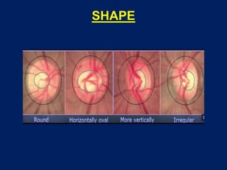
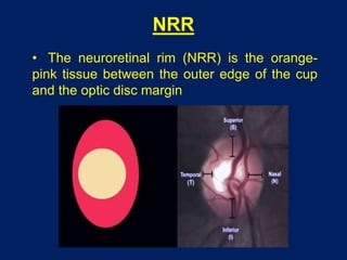
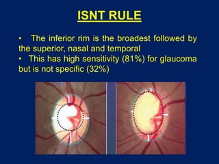
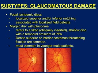
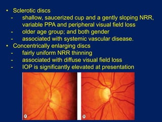
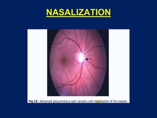
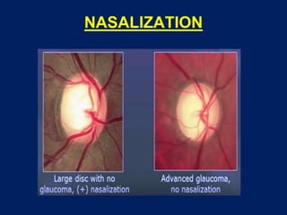
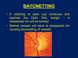
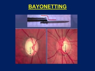
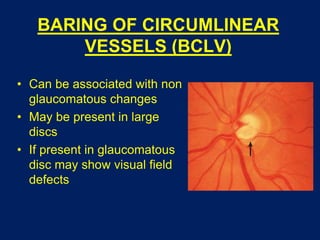
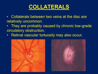
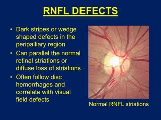
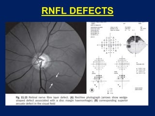
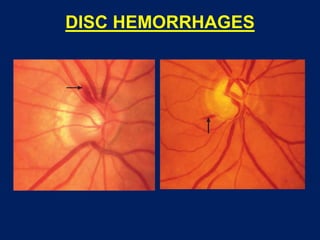
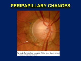
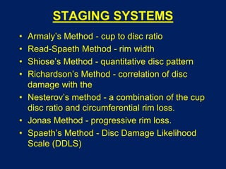
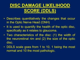
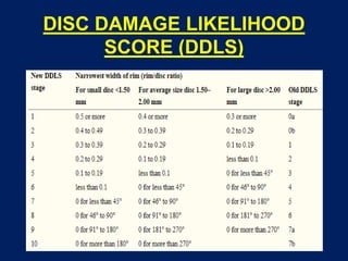
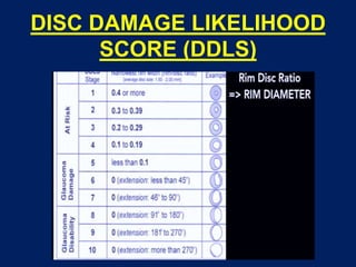
CLINICAL EVALUATION OF
OPTIC DISC CHANGES
Dr Saurabh Kushwaha
Resident (Ophthalmology)
SCOPE
• Papillary changes
• Vascular changes
• Peripapillary changes
CLINICAL EVALUATION
• Slit-lamp funduscopy
• Monocular direct ophthalmoscope examination
• Photographs of the ONH
• Clinician’s disc drawings
PAPILLARY CHANGES
• Size
• Shape
• Neuroretinal rim (NRR)
• Cupping
• Colour
• Asymmetry
PAPILLARY CHANGES
• SIZE
– Diameter varies between 0.85 mm to 2.86 mm
– Mean of 1.88 mm vertically and 1.77 mm
horizontally
– Optic Disc is vertically oval
– Avg disc area of 2.42 mm2
– Deciding C/D ratio
– Prognostic indicator
• Disc size varies on average between racial
groups and is largest in Africans
• Vertical diameter is the parameter most
frequently used clinically
• Large discs are believed to be more likely
to sustain damage, particularly in NTG
SHAPE
NRR
• The neuroretinal rim (NRR) is the orange-
pink tissue between the outer edge of the cup
and the optic disc margin
ISNT RULE
• The inferior rim is the broadest followed by
the superior, nasal and temporal
• This has high sensitivity (81%) for glaucoma
but is not specific (32%)
SUBTYPES: GLAUCOMATOUS DAMAGE
• Focal ischaemic discs
- localized superior and/or inferior notching
- associated with localized field defects
• Myopic disc with glaucoma
- refers to a tilted (obliquely inserted), shallow disc
with a temporal crescent of PPA
- Dense superior or inferior scotomas threatening
fixation are common.
- most common in younger male patients.
• Sclerotic discs
- shallow, saucerized cup and a gently sloping NRR,
variable PPA and peripheral visual field loss
- older age group; and both gender
- associated with systemic vascular disease.
• Concentrically enlarging discs
- fairly uniform NRR thinning
- associated with diffuse visual field loss
- IOP is significantly elevated at presentation
CUPPING
• Physiological cup is a depression in the Optic
Disc
• If thinning of the Optic Disc NRR occurs - the
optic disc appears pathologically cupped
(excavated)
• Usual cause is glaucoma
• Slow death of optic nerve axons and their
supporting glial cells
CUPPING
• Lamina cibrosa thins out and moves out and
posteriorly
COLOUR
• Cup appears pale due to presence of glial
tissue at that site (No neuronal tissue)
• NRR appears orangish red or pink in colour
• When we have pallor > cupping ---- non
glaucomatous damage
• When cupping > pallor ----- glaucomatous
damage
ASYMMETRY
• Greater than 0.2 CDR difference between the
two disc s/o glaucoma changes
• However, asymmetry in size of Optic Disc,
cup, width of NRR, vessel course and
Peripapillary changes are to be noted
LAMINAR DOT SIGN
• The laminar dot sign occurs in advancing
glaucoma.
• Grey dot-like fenestrations in the lamina
cribrosa become exposed as the NRR recedes.
• The fenestrations sometimes appear linear
and this itself may be a sign of advanced
damage, indicating distortion of the lamina.
• The dots may be seen in normal eyes.
LAMINAR DOT SIGN
VASCULAR CHANGES
• Nasalization of vessels
• Bayonetting of vessels
• Baring of circumlinear vessels
• Collaterals
NASALIZATION
NASALIZATION
BAYONETTING
• If notching at optic cup continues and
reaches the Optic Disc margin – a
sharpened rim will be formed
• Retinal vessels will bend at sharpened rim
causing bayonetting of vessels
BAYONETTING
BARING OF CIRCUMLINEAR
VESSELS (BCLV)
• Can be associated with non
glaucomatous changes
• May be present in large
discs
• If present in glaucomatous
disc may show visual field
defects
COLLATERALS
• Collaterals between two veins at the disc are
relatively uncommon.
• They are probably caused by chronic low-grade
circulatory obstruction.
• Retinal vascular tortuosity may also occur.
PERIPAPILLARY CHANGES
• RNFL defects
• RNFL hemorrhages
• Peripapillary changes
RNFL DEFECTS
• Dark stripes or wedge
shaped defects in the
peripalliary region
• Can parallel the normal
retinal striations or
diffuse loss of striations
• Often follow disc
hemorrhages and
correlate with visual
field defects
Normal RNFL striations
RNFL DEFECTS
RNFL DEFECTS
DISC HEMORRHAGES
• Early sign of glaucoma
• Hemorrhage  disc changes  field
defects
• Most common site - inferior quadrant
• Also at focal rim notch or at focal
RNFL defects
• Risk factor for the development and
progression of glaucoma
• Common in NTG and DM
DISC HEMORRHAGES
GRAY CRESCENT
• Seen within peripheral tissues of ONH
• Scleral lip is peripheral to the gray crescent
• Often bilateral and it is usually located along
temporal or infero-temporal disc margin.
• Grey crescent is due to internal extension of
Bruch's membrane in the peripapillary scleral
ring.
GRAY CRESCENT
ZONE BETA
• Seen between peripheral NRR and zone
alpha.
• It represents retraction of RPE from the disc
margin due to atrophy of RPE.
• Sclera and large choroidal vessels are visible
due to RPE atrophy.
• Location and extent of zone beta atrophy
correlates with visual field loss.
• Indicates area has poor perfusion
• Seen in POAG and NTG.
ZONE ALPHA
• Seen as irregular hyperpigmentation and
hypopigmentation.
• Due to parapapillary crescent of RPE
irregularity close to the margin of Bruch's
membrane.
• Present in almost all normal eyes.
PERIPAPILLARY CHANGES
STAGING SYSTEMS
• Armaly’s Method - cup to disc ratio
• Read-Spaeth Method - rim width
• Shiose’s Method - quantitative disc pattern
• Richardson’s Method - correlation of disc
damage with the
• Nesterov’s method - a combination of the cup
disc ratio and circumferential rim loss.
• Jonas Method - progressive rim loss.
• Spaeth’s Method - Disc Damage Likelihood
Scale (DDLS)
DISC DAMAGE LIKELIHOOD
SCORE (DDLS)
• Unlike the CDR, which focuses on the
excavation, the DDLS is based directly on
the thickness of the neuroretinal rim and
takes into account the optic disc size.
• DDLS estimates the glaucomatous damage
of the optic disc more precisely than the
currently used method.
DISC DAMAGE LIKELIHOOD
SCORE (DDLS)
• Describes quantitatively the changes that occur
in the Optic Nerve Head (ONH)
• It is used to quantify the health of the optic disc,
specifically as it relates to glaucoma.
• Two characteristics of the disc: (1) the width of
the neuroretinal rim and (2) the size of the optic
disc.
• DDLS scale goes from 1 to 10, 1 being the most
normal and 10 the most pathologic.
DISC DAMAGE LIKELIHOOD
SCORE (DDLS)
• First, one measures the size of the optic disc and
classifies the disc as small, average, large, or very
large. Small is less than 1.5 mm in height, average
between 1.5 and 2.0 mm in height, large between
2 and 3 mm in height, and very large greater than 3
mm
• Next, one looks for where the neuroretinal rim is
the narrowest. The narrowest rim would be 0 and
the widest rim possible would be 5.
• Discs with DDLS of 6 or more are never normal.
DISC DAMAGE LIKELIHOOD
SCORE (DDLS)
DISC DAMAGE LIKELIHOOD
SCORE (DDLS)
THANK YOU

Clinical evaluation of optic disc changes

  • 1.
    CLINICAL EVALUATION OF OPTICDISC CHANGES Dr Saurabh Kushwaha Resident (Ophthalmology)
  • 2.
    SCOPE • Papillary changes •Vascular changes • Peripapillary changes
  • 3.
    CLINICAL EVALUATION • Slit-lampfunduscopy • Monocular direct ophthalmoscope examination • Photographs of the ONH • Clinician’s disc drawings
  • 4.
    PAPILLARY CHANGES • Size •Shape • Neuroretinal rim (NRR) • Cupping • Colour • Asymmetry
  • 5.
    PAPILLARY CHANGES • SIZE –Diameter varies between 0.85 mm to 2.86 mm – Mean of 1.88 mm vertically and 1.77 mm horizontally – Optic Disc is vertically oval – Avg disc area of 2.42 mm2 – Deciding C/D ratio – Prognostic indicator
  • 6.
    • Disc sizevaries on average between racial groups and is largest in Africans • Vertical diameter is the parameter most frequently used clinically
  • 8.
    • Large discsare believed to be more likely to sustain damage, particularly in NTG
  • 9.
  • 10.
    NRR • The neuroretinalrim (NRR) is the orange- pink tissue between the outer edge of the cup and the optic disc margin
  • 11.
    ISNT RULE • Theinferior rim is the broadest followed by the superior, nasal and temporal • This has high sensitivity (81%) for glaucoma but is not specific (32%)
  • 12.
    SUBTYPES: GLAUCOMATOUS DAMAGE •Focal ischaemic discs - localized superior and/or inferior notching - associated with localized field defects • Myopic disc with glaucoma - refers to a tilted (obliquely inserted), shallow disc with a temporal crescent of PPA - Dense superior or inferior scotomas threatening fixation are common. - most common in younger male patients.
  • 13.
    • Sclerotic discs -shallow, saucerized cup and a gently sloping NRR, variable PPA and peripheral visual field loss - older age group; and both gender - associated with systemic vascular disease. • Concentrically enlarging discs - fairly uniform NRR thinning - associated with diffuse visual field loss - IOP is significantly elevated at presentation
  • 14.
    CUPPING • Physiological cupis a depression in the Optic Disc • If thinning of the Optic Disc NRR occurs - the optic disc appears pathologically cupped (excavated) • Usual cause is glaucoma • Slow death of optic nerve axons and their supporting glial cells
  • 15.
  • 16.
    • Lamina cibrosathins out and moves out and posteriorly
  • 17.
    COLOUR • Cup appearspale due to presence of glial tissue at that site (No neuronal tissue) • NRR appears orangish red or pink in colour • When we have pallor > cupping ---- non glaucomatous damage • When cupping > pallor ----- glaucomatous damage
  • 19.
    ASYMMETRY • Greater than0.2 CDR difference between the two disc s/o glaucoma changes • However, asymmetry in size of Optic Disc, cup, width of NRR, vessel course and Peripapillary changes are to be noted
  • 20.
    LAMINAR DOT SIGN •The laminar dot sign occurs in advancing glaucoma. • Grey dot-like fenestrations in the lamina cribrosa become exposed as the NRR recedes. • The fenestrations sometimes appear linear and this itself may be a sign of advanced damage, indicating distortion of the lamina. • The dots may be seen in normal eyes.
  • 21.
  • 22.
    VASCULAR CHANGES • Nasalizationof vessels • Bayonetting of vessels • Baring of circumlinear vessels • Collaterals
  • 23.
  • 24.
  • 25.
    BAYONETTING • If notchingat optic cup continues and reaches the Optic Disc margin – a sharpened rim will be formed • Retinal vessels will bend at sharpened rim causing bayonetting of vessels
  • 26.
  • 27.
    BARING OF CIRCUMLINEAR VESSELS(BCLV) • Can be associated with non glaucomatous changes • May be present in large discs • If present in glaucomatous disc may show visual field defects
  • 28.
    COLLATERALS • Collaterals betweentwo veins at the disc are relatively uncommon. • They are probably caused by chronic low-grade circulatory obstruction. • Retinal vascular tortuosity may also occur.
  • 29.
    PERIPAPILLARY CHANGES • RNFLdefects • RNFL hemorrhages • Peripapillary changes
  • 30.
    RNFL DEFECTS • Darkstripes or wedge shaped defects in the peripalliary region • Can parallel the normal retinal striations or diffuse loss of striations • Often follow disc hemorrhages and correlate with visual field defects Normal RNFL striations
  • 31.
  • 32.
  • 33.
    DISC HEMORRHAGES • Earlysign of glaucoma • Hemorrhage  disc changes  field defects • Most common site - inferior quadrant • Also at focal rim notch or at focal RNFL defects • Risk factor for the development and progression of glaucoma • Common in NTG and DM
  • 34.
  • 35.
    GRAY CRESCENT • Seenwithin peripheral tissues of ONH • Scleral lip is peripheral to the gray crescent • Often bilateral and it is usually located along temporal or infero-temporal disc margin. • Grey crescent is due to internal extension of Bruch's membrane in the peripapillary scleral ring.
  • 36.
  • 37.
    ZONE BETA • Seenbetween peripheral NRR and zone alpha. • It represents retraction of RPE from the disc margin due to atrophy of RPE. • Sclera and large choroidal vessels are visible due to RPE atrophy. • Location and extent of zone beta atrophy correlates with visual field loss. • Indicates area has poor perfusion • Seen in POAG and NTG.
  • 38.
    ZONE ALPHA • Seenas irregular hyperpigmentation and hypopigmentation. • Due to parapapillary crescent of RPE irregularity close to the margin of Bruch's membrane. • Present in almost all normal eyes.
  • 39.
  • 40.
    STAGING SYSTEMS • Armaly’sMethod - cup to disc ratio • Read-Spaeth Method - rim width • Shiose’s Method - quantitative disc pattern • Richardson’s Method - correlation of disc damage with the • Nesterov’s method - a combination of the cup disc ratio and circumferential rim loss. • Jonas Method - progressive rim loss. • Spaeth’s Method - Disc Damage Likelihood Scale (DDLS)
  • 41.
    DISC DAMAGE LIKELIHOOD SCORE(DDLS) • Unlike the CDR, which focuses on the excavation, the DDLS is based directly on the thickness of the neuroretinal rim and takes into account the optic disc size. • DDLS estimates the glaucomatous damage of the optic disc more precisely than the currently used method.
  • 42.
    DISC DAMAGE LIKELIHOOD SCORE(DDLS) • Describes quantitatively the changes that occur in the Optic Nerve Head (ONH) • It is used to quantify the health of the optic disc, specifically as it relates to glaucoma. • Two characteristics of the disc: (1) the width of the neuroretinal rim and (2) the size of the optic disc. • DDLS scale goes from 1 to 10, 1 being the most normal and 10 the most pathologic.
  • 43.
    DISC DAMAGE LIKELIHOOD SCORE(DDLS) • First, one measures the size of the optic disc and classifies the disc as small, average, large, or very large. Small is less than 1.5 mm in height, average between 1.5 and 2.0 mm in height, large between 2 and 3 mm in height, and very large greater than 3 mm • Next, one looks for where the neuroretinal rim is the narrowest. The narrowest rim would be 0 and the widest rim possible would be 5. • Discs with DDLS of 6 or more are never normal.
  • 44.
  • 45.
  • 46.