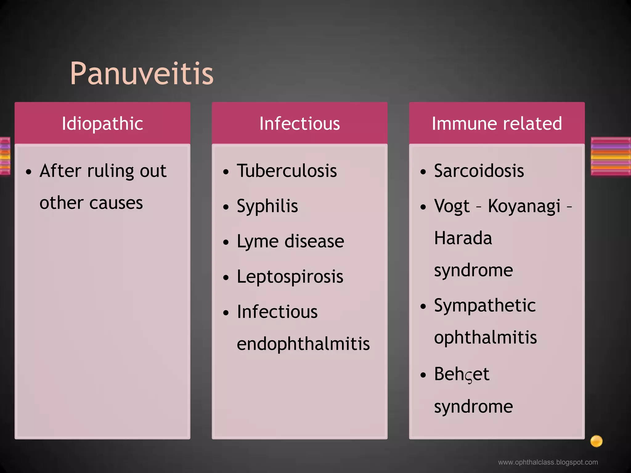
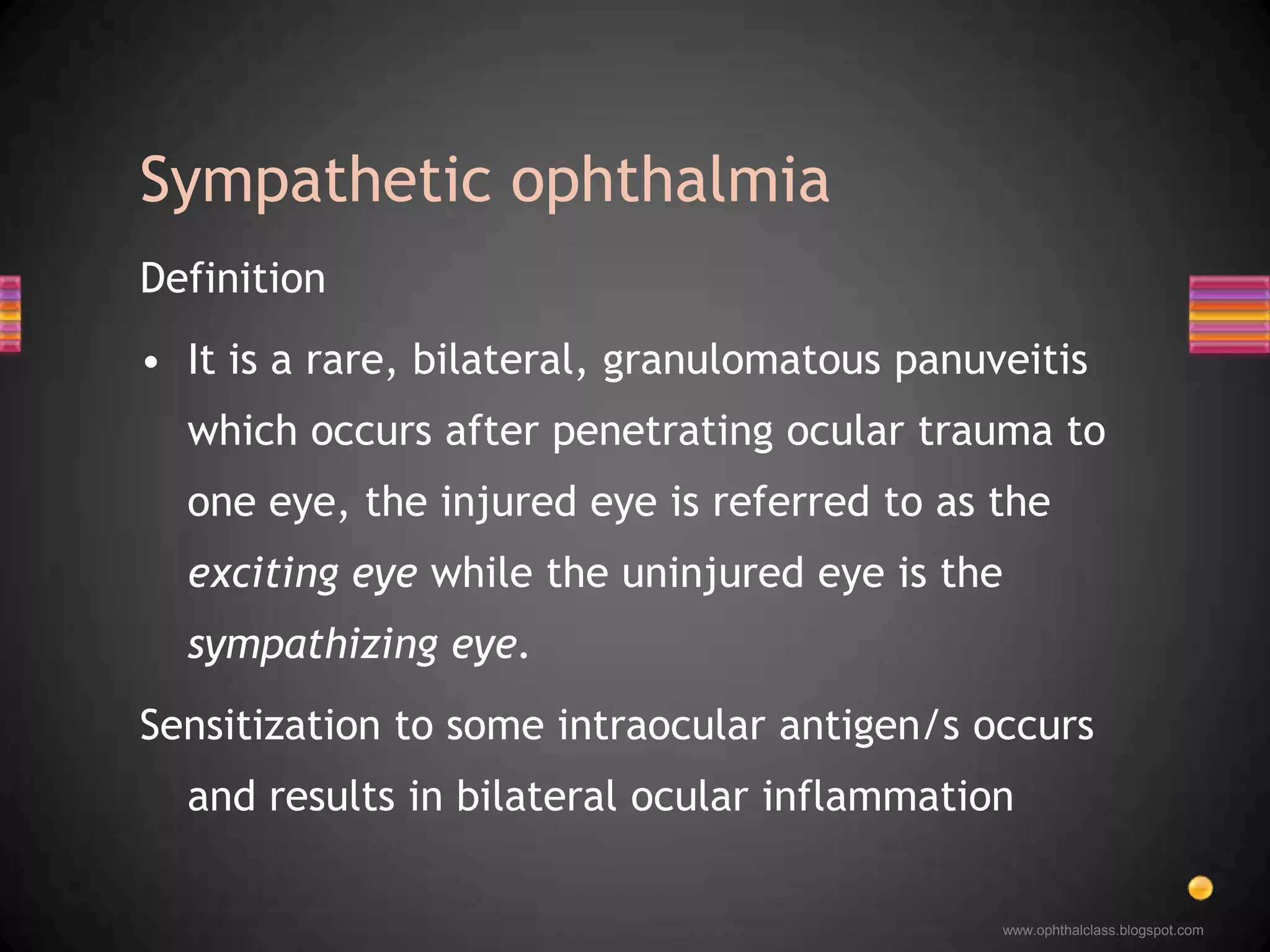
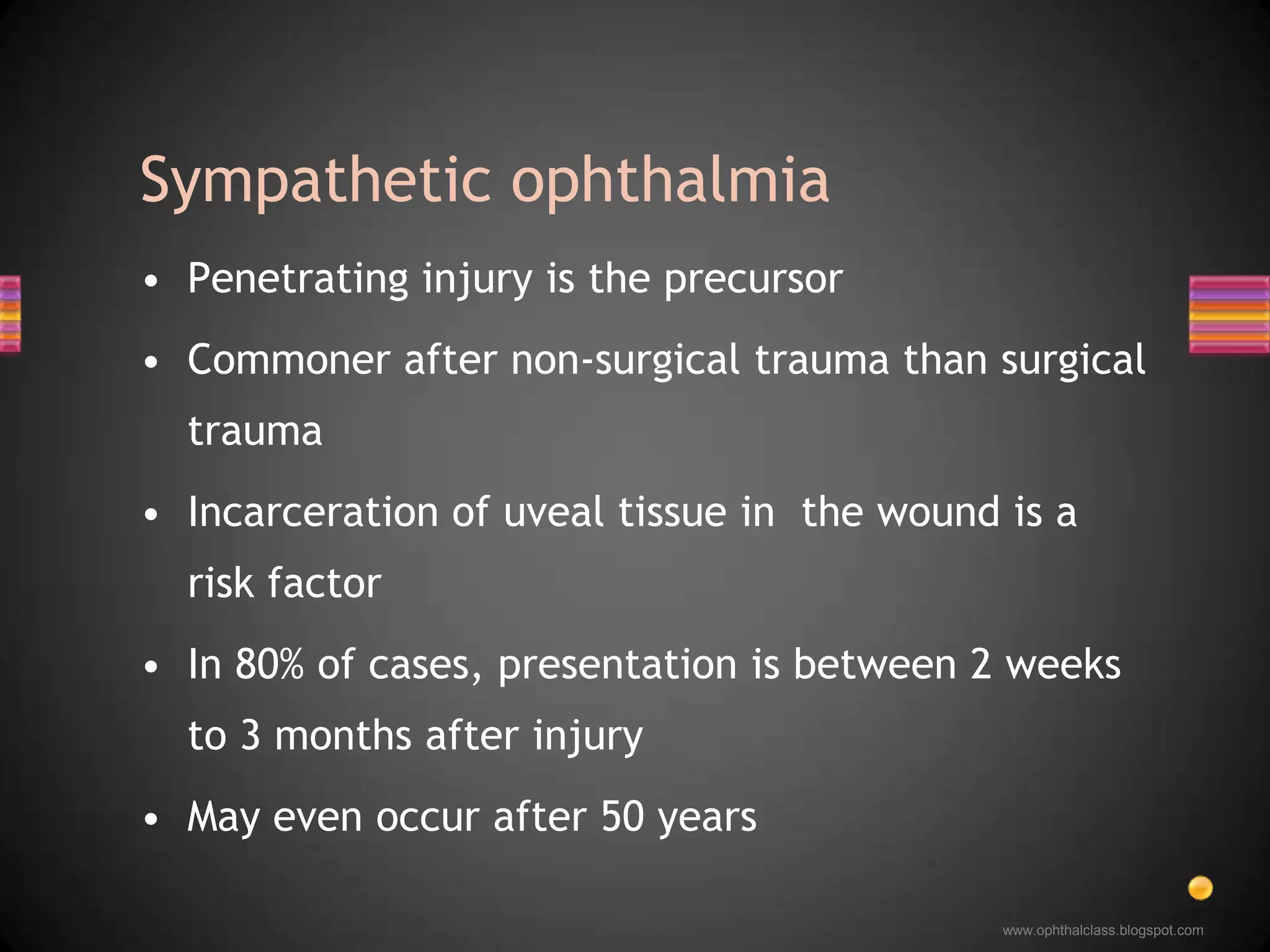
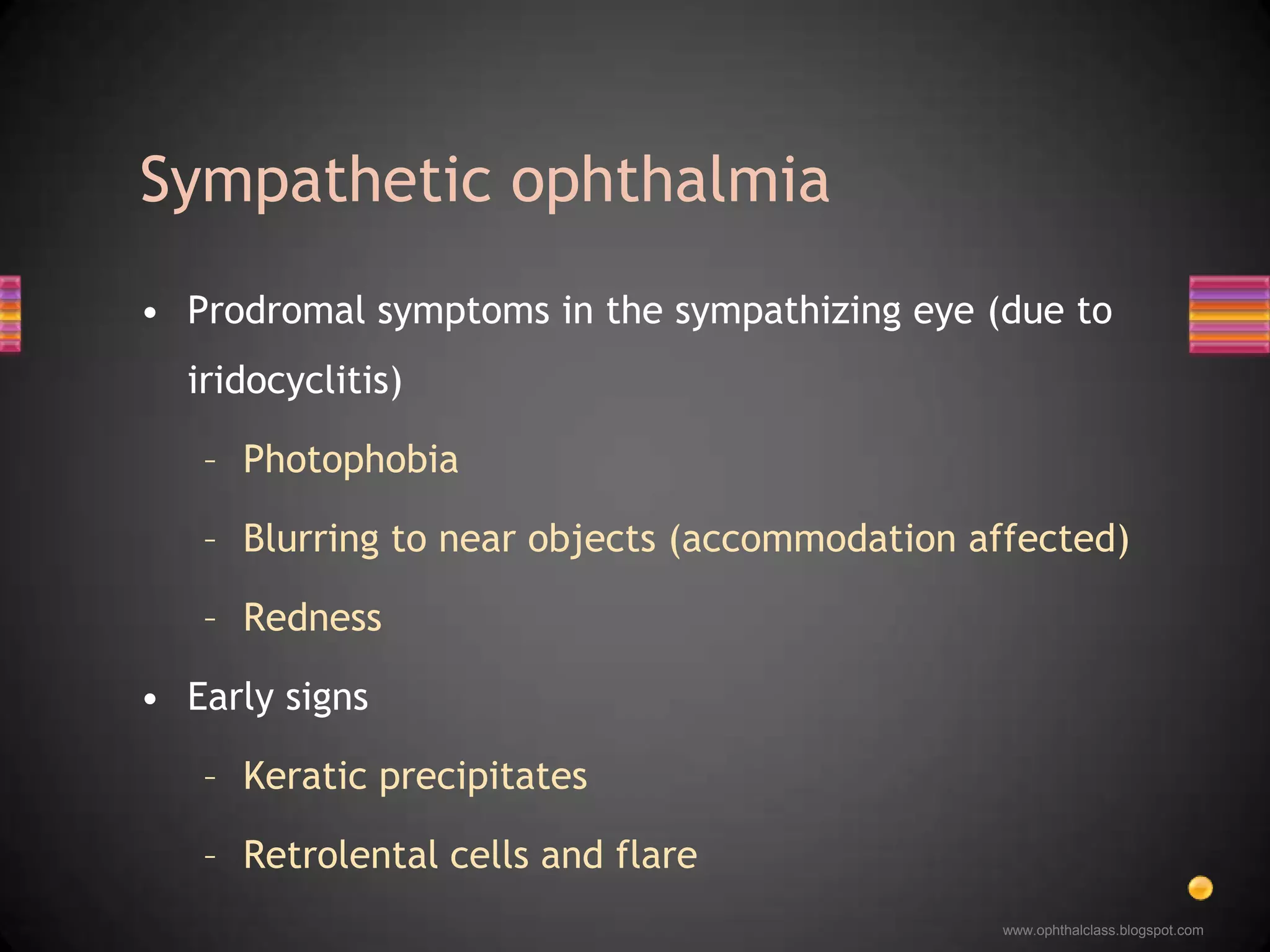
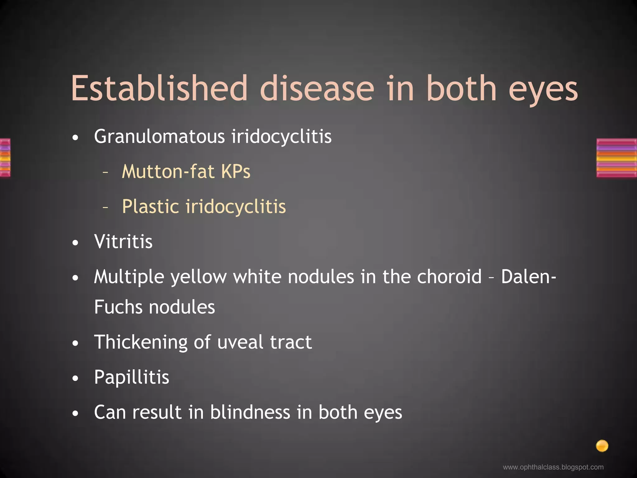
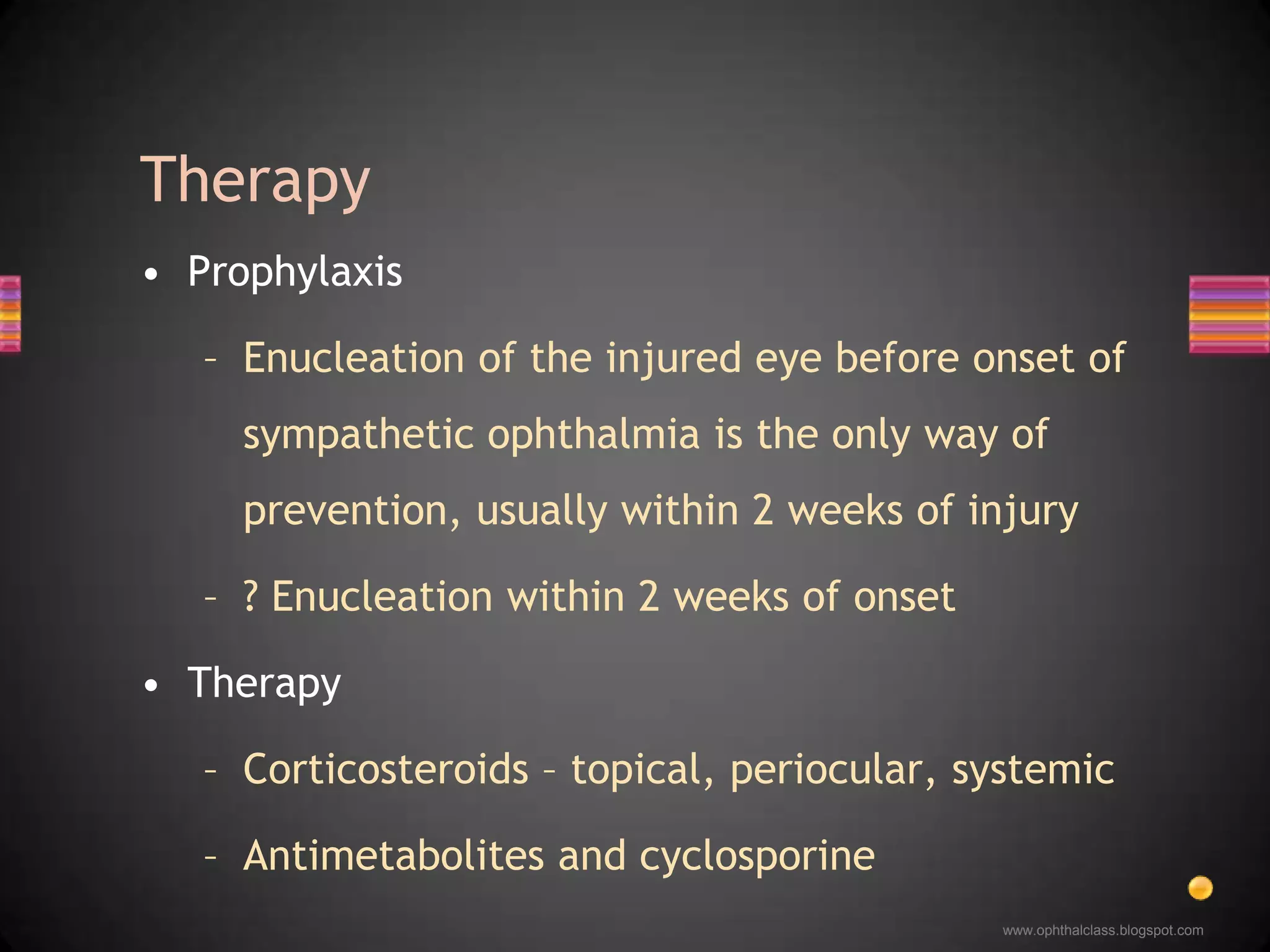
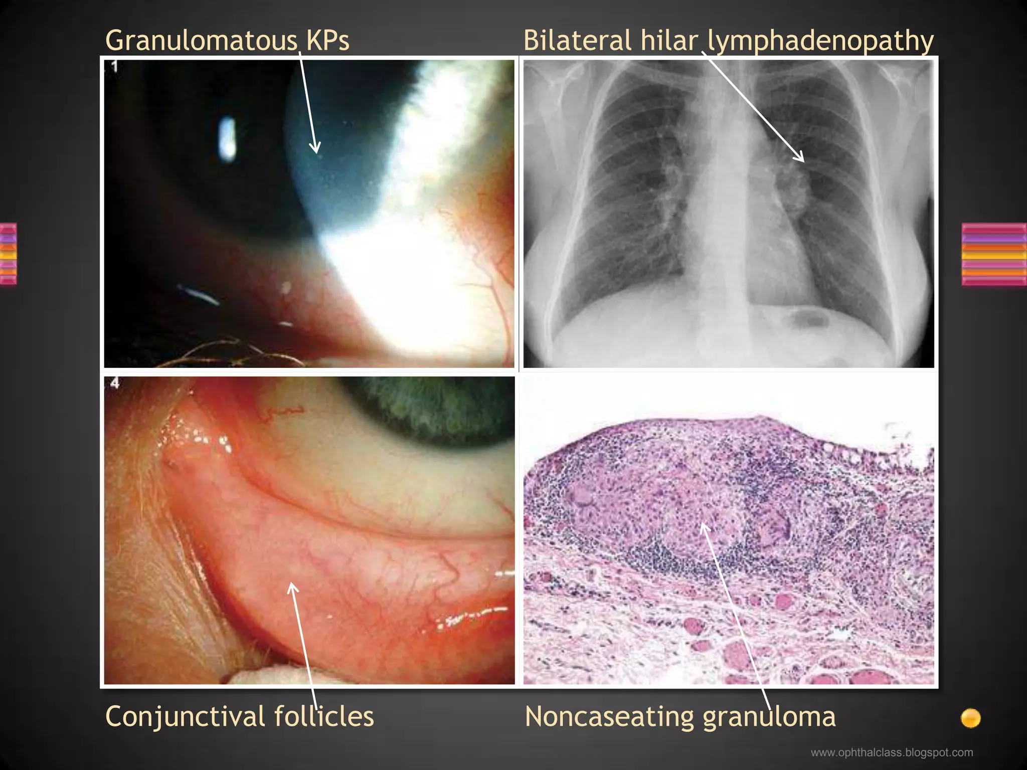
Dr. Anupama Karanthwww.ophthalclass.blogspot.comPanuveitis
Classification of uveitisAnatomic Classification of UveitisAnteriorIntermediatePosteriorPanuveitisClinical Classification of Uveitis
Anatomic classificationPanuveitisPrimary site of inflammation: anterior chamber, vitreous, retina or choroid
Clinical Classification of UveitisInfectious Bacterial / Viral / Fungal  / Parasitic  / Others Non-infectiousKnown/No known systemic association MasqueradeNeoplastic / Non-neoplastic
Panuveitis
DefinitionIt is a rare, bilateral, granulomatous panuveitis which occurs after penetrating ocular trauma to one eye, the injured eye is referred to as the exciting eye while the uninjured eye is the sympathizing eye.Sensitization to some intraocular antigen/s occurs and results in bilateral ocular inflammationSympathetic ophthalmia
Penetrating injury is the precursorCommoner after non-surgical trauma than surgical traumaIncarceration of uveal tissue in  the wound is a risk factorIn 80% of cases, presentation is between 2 weeks to 3 months after injuryMay even occur after 50 yearsSympathetic ophthalmia
Prodromal symptoms in the sympathizing eye (due to iridocyclitis)PhotophobiaBlurring to near objects (accommodation affected)Redness Early signsKeratic precipitates Retrolental cells and flareSympathetic ophthalmia
Granulomatous iridocyclitisMutton-fat KPsPlastic iridocyclitisVitritis Multiple yellow white nodules in the choroid – Dalen-Fuchs nodulesThickening of uveal tractPapillitisCan result in blindness in both eyesEstablished disease in both eyes
ProphylaxisEnucleation of the injured eye before onset of sympathetic ophthalmia is the only way of prevention, usually within 2 weeks of injury? Enucleation within 2 weeks of onsetTherapyCorticosteroids – topical, periocular, systemicAntimetabolites and cyclosporineTherapy
A few other common causes of panuveitis….
10 year history of recurrent bilateral granulomatous uveitis with waxing and waning exudative retinal detachments, on steroids and immunosuppressives…www.ophthalclass.blogspot.comhttp://www.aao.org/publications/eyenet/200804/am_rounds.cfm
 Koeppe and Busacca nodules in the iris Koeppe and Busacca nodules in the iris
 Extensive pigment alterations in the fundus Koeppe and Busacca nodules in the iris
 Extensive pigment alterations in the fundus
Dalen-Fuchs like peripheral nodules Koeppe and Busacca nodules in the iris
 Extensive pigment alterations in the fundus
Dalen-Fuchs like peripheral nodules
Subretinal fibrosisBilateral granulomatous iridocyclitisVariable vitritis, exudative retinal detachmentsCommoner in pigmented racesAssociated extraocular features important in diagnosisCSF pleocytosis, neck stiffness, seizures, paralysis (CNS)Vitiligo, alopecia, poliosis (skin)Hearing loss and tinnitus (ear)Vogt-Koyanagi-Harada disease
DiagnosisClassical clinical picture but rule out SOCSF lymphocytosisManagementVigorous use of steroids- local, periocular and systemicImmunosuppressivesVogt-Koyanagi-Harada disease
A 50 year old diabetic with blurry vision…http://www.aao.org/publications/eyenet/200710/am_rounds.cfm
Peripapillary atrophy
Peripapillary atrophyAreas of RPE atrophy with underlying large choroidal vessels visible
Peripapillary atrophyAreas of RPE atrophy with underlying large choroidal vessels visible Pigments in a bony spicule pattern adjacent to vessels
Hereditary Retinitis pigmentosaSequelae of auto-immune diseaseToxic etiologiesChloroquine, thioridazineSequelae of infectious diseaseTuberculosis Syphilis Lyme diseaseD/D Pigmentary retinopathies
RPR reactive and positive FTA-ABS…… syphilis
Secondary syphilisGranulomatous iridocyclitisIris roseola, iris papulosa, iris nodosaFocal/multifocal choroiditisRetinitis, perivasculitisPapillitis, neuroretinitisSyphilis – the great masquerader
ManagementOcular involvement to be treated as neurosyphilisCSF evaluation should be done in any syphilitic uveitisPenicillin G 2-5 million units IV 4th hourly – 2 weeksSteroids only after effective antibiotic therapySyphilis – the great masquerader
A 35 year old male patient with blurry vision…
Hypopyon uveitis, nongranulomatous, vitritis
Core lab tests…Mantoux highly positive, Chest x-ray suggestive….Tuberculosis
Chronic iridocyclitisGranulomatous / nongranulomatousChoroiditisFocal / multifocal / choroidal tuberclesRetinal vasculitis, vitritis, papillitisDifficulty - in establishing the diagnosis and ensuring treatment complianceTuberculosis
A 46 year old lady with floaters…http://www.aao.org/publications/eyenet/200801/am_rounds.cfm
Granulomatous KPs, aqueous flare and cells
InfectionsTuberculosisSyphilisLyme diseaseNon-infectiousSarcoidosis VKH syndromeD/D granulomatous iridocyclitis
Chest x-ray, serum ACE…… elevated ACE, hilar lymphadenopathySarcoidosis
Granulomatous KPsBilateral hilar lymphadenopathyConjunctival folliclesNoncaseatinggranuloma
Multisystem granulomatous diseaseOcular findingsAcute / chronic granulomatous iridocyclitisVitreous snowballsRetinal periphlebitis, candle wax drippingsChoroidal small or large granulomasSarcoidosis
Suspect in any uveitisChest x-ray or CT, serum ACE and lysozymeBiopsy from skin / conjunctiva / lacrimal glandManagementCorticosteroids – topical, periocular and systemicImmunosuppressives may be requiredSarcoidosis

Panuveitis