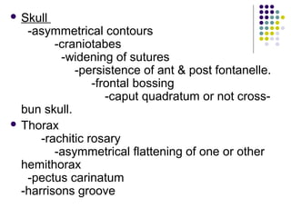
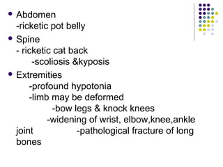
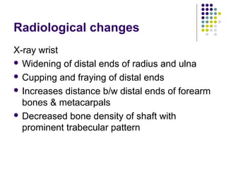
Vitamin D
Soumya Ranjan Parida
Basic B.Sc. Nursing 4th
year
Sum Nursing College
Rickets
 It is a syndrome of Vit-D deficiency
 It is characterised by various skeletal
abnormalities
 It is a disease of growing bones.
Vitamin D
Physiology
 Several compounds have vit-D activity but important
is vit-D3 (cholecalciferol)
 Exposure of skin to ultraviolet rays causes conversion
of 7-dehydrocholesterol to previt-D3
 Pre vit-D3 is converted by a nonezymatic process to
vit-D3
 vit-D3 is converted to 25-hydroxycholecalciferol in the
liver
 This 25-hydroxycholecalciferol is finally converted in
active form 1,25-dihydroxycholecalciferol in the kidney
& this step is controlled by parathormone
Functions of Vit-D
 Absorption of calcium from the gut.
 Deposition of cal phosphate in the prepratory
zone of calcification.
 Reabsorption of bones with excretion of
phosphate from the kidney along with
parathormone.
Biochemical changes
 In vitamin D deficiency, calcium levels in the blood
decrease because vitamin D is necessary for
absorption of this mineral.
 The level of parathyroid hormone, which increases
the calcium level in the blood, may increase as the
body tries to compensate for the vitamin D
deficiency.(feedback mechanism)
 Because not enough calcium and phosphate are
available to maintain healthy bones, vitamin D
deficiency may result in a bone softenning called
rickets in children or osteomalacia in adults.
Vitamin D deficiency can be caused by
 Lack of vitamin D in the diet
 By inadequate exposure to sunlight.
 A pregnant woman with vitamin D deficiency may develop
osteomalacia, and the newborn has a high risk of
developing rickets.
 People with kidney or liver disorders may be unable to
convert vitamin D into a usable form, they are at risk of
osteomalacia.
 Malabsorption disorders
 The use of certain anticonvulsants increase the risk of
vitamin D deficiency.
 Several rare hereditary forms of rickets develop because
the body cannot process (metabolize) vitamin D normally.
Clinical Features
Depends on :
 Age of child
 Severity of disease
 Underlying cause of deficiency
 Skull
-asymmetrical contours
-craniotabes
-widening of sutures
-persistence of ant & post fontanelle.
-frontal bossing
-caput quadratum or not cross-
bun skull.
 Thorax
-rachitic rosary
-asymmetrical flattening of one or other
hemithorax
-pectus carinatum
-harrisons groove
 Abdomen
-ricketic pot belly
 Spine
- ricketic cat back
-scoliosis &kyposis
 Extremities
-profound hypotonia
-limb may be deformed
-bow legs & knock knees
-widening of wrist, elbow,knee,ankle
joint -pathological fracture of long
bones
Radiological changes
X-ray wrist
 Widening of distal ends of radius and ulna
 Cupping and fraying of distal ends
 Increases distance b/w distal ends of forearm
bones & metacarpals
 Decreased bone density of shaft with
prominent trabecular pattern
Diagnosis
 The diagnosis of rickets or osteomalacia is
based on symptoms.
 Radiological- the appearance of bones on x-
rays
 Biochemical
Treatment
 involves taking daily vitamin D and calcium
supplements by mouth.
-Oral low dose -1500-5000 IU/d 6-12 weeks
f/b 400 IU/d
-Massive dose 6 lac IU oral or injection &
repeat in 3-4 weeks
 People with a chronic liver or kidney disorder
may require special formulations of vitamin D
supplements
VITAMIN D EXCESS
 Early symptoms are loss of appetite, nausea,
and vomiting, followed by excessive thirst,
weakness, nervousness, and high blood
pressure.
 Because the calcium level is high, calcium
may be deposited throughout the body,
particularly in the kidneys, blood vessels,
lungs, and heart.
 The kidneys may be permanently damaged
and malfunction
 Vitamin D excess is usually diagnosed when blood
tests detect a high calcium level in a person who
takes high doses of vitamin D.
 The diagnosis is confirmed by measuring the level
of vitamin D in the blood.
 Treatment consists of discontinuing vitamin D
supplements, following a low-calcium diet for a while
to offset the effects of a high calcium level in the
body, and taking drugs to suppress the release of
calcium from the bones.
Refractory rickets
 Develop rickets inspite of intake of vit-D
 Do not show evidence of healng rickets after
2 weeks of parentral therapy
 Causes
-Renal causes
-Malabsorbtion
-Hepatic dysfunction
-Miscellanous causes
Differential diagnosis
 Craniotabes-hydrocephalus
 Osteogenesis imperfecta
 Rosary-scurvy,chondrodystrophies
Complications
 Respiratory infections
 Pulmonary atelectesis
 Chronic GIT disturbances
 Anemia
Prevention
 Exposure to sunlight
 Oral administration of vit-D

Vitamin d

  • 1.
    Vitamin D Soumya RanjanParida Basic B.Sc. Nursing 4th year Sum Nursing College
  • 2.
    Rickets  It isa syndrome of Vit-D deficiency  It is characterised by various skeletal abnormalities  It is a disease of growing bones.
  • 3.
    Vitamin D Physiology  Severalcompounds have vit-D activity but important is vit-D3 (cholecalciferol)  Exposure of skin to ultraviolet rays causes conversion of 7-dehydrocholesterol to previt-D3  Pre vit-D3 is converted by a nonezymatic process to vit-D3  vit-D3 is converted to 25-hydroxycholecalciferol in the liver  This 25-hydroxycholecalciferol is finally converted in active form 1,25-dihydroxycholecalciferol in the kidney & this step is controlled by parathormone
  • 4.
    Functions of Vit-D Absorption of calcium from the gut.  Deposition of cal phosphate in the prepratory zone of calcification.  Reabsorption of bones with excretion of phosphate from the kidney along with parathormone.
  • 5.
    Biochemical changes  Invitamin D deficiency, calcium levels in the blood decrease because vitamin D is necessary for absorption of this mineral.  The level of parathyroid hormone, which increases the calcium level in the blood, may increase as the body tries to compensate for the vitamin D deficiency.(feedback mechanism)  Because not enough calcium and phosphate are available to maintain healthy bones, vitamin D deficiency may result in a bone softenning called rickets in children or osteomalacia in adults.
  • 6.
    Vitamin D deficiencycan be caused by  Lack of vitamin D in the diet  By inadequate exposure to sunlight.  A pregnant woman with vitamin D deficiency may develop osteomalacia, and the newborn has a high risk of developing rickets.  People with kidney or liver disorders may be unable to convert vitamin D into a usable form, they are at risk of osteomalacia.  Malabsorption disorders  The use of certain anticonvulsants increase the risk of vitamin D deficiency.  Several rare hereditary forms of rickets develop because the body cannot process (metabolize) vitamin D normally.
  • 7.
    Clinical Features Depends on:  Age of child  Severity of disease  Underlying cause of deficiency
  • 8.
     Skull -asymmetrical contours -craniotabes -wideningof sutures -persistence of ant & post fontanelle. -frontal bossing -caput quadratum or not cross- bun skull.  Thorax -rachitic rosary -asymmetrical flattening of one or other hemithorax -pectus carinatum -harrisons groove
  • 9.
     Abdomen -ricketic potbelly  Spine - ricketic cat back -scoliosis &kyposis  Extremities -profound hypotonia -limb may be deformed -bow legs & knock knees -widening of wrist, elbow,knee,ankle joint -pathological fracture of long bones
  • 10.
    Radiological changes X-ray wrist Widening of distal ends of radius and ulna  Cupping and fraying of distal ends  Increases distance b/w distal ends of forearm bones & metacarpals  Decreased bone density of shaft with prominent trabecular pattern
  • 11.
    Diagnosis  The diagnosisof rickets or osteomalacia is based on symptoms.  Radiological- the appearance of bones on x- rays  Biochemical
  • 12.
    Treatment  involves takingdaily vitamin D and calcium supplements by mouth. -Oral low dose -1500-5000 IU/d 6-12 weeks f/b 400 IU/d -Massive dose 6 lac IU oral or injection & repeat in 3-4 weeks  People with a chronic liver or kidney disorder may require special formulations of vitamin D supplements
  • 13.
    VITAMIN D EXCESS Early symptoms are loss of appetite, nausea, and vomiting, followed by excessive thirst, weakness, nervousness, and high blood pressure.  Because the calcium level is high, calcium may be deposited throughout the body, particularly in the kidneys, blood vessels, lungs, and heart.  The kidneys may be permanently damaged and malfunction
  • 14.
     Vitamin Dexcess is usually diagnosed when blood tests detect a high calcium level in a person who takes high doses of vitamin D.  The diagnosis is confirmed by measuring the level of vitamin D in the blood.  Treatment consists of discontinuing vitamin D supplements, following a low-calcium diet for a while to offset the effects of a high calcium level in the body, and taking drugs to suppress the release of calcium from the bones.
  • 15.
    Refractory rickets  Developrickets inspite of intake of vit-D  Do not show evidence of healng rickets after 2 weeks of parentral therapy  Causes -Renal causes -Malabsorbtion -Hepatic dysfunction -Miscellanous causes
  • 16.
    Differential diagnosis  Craniotabes-hydrocephalus Osteogenesis imperfecta  Rosary-scurvy,chondrodystrophies
  • 17.
    Complications  Respiratory infections Pulmonary atelectesis  Chronic GIT disturbances  Anemia
  • 18.
    Prevention  Exposure tosunlight  Oral administration of vit-D