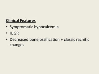
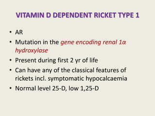
VITAMIN D
RICKETS
DR KAUSIK SUR
D.C.H,DNB
ASSISTANT PROFESSOR
DEPARTMENT OF PEDIATRICS
VIVEKANANDA INSTITUTE OF MEDICAL SCIENCES
RAMAKRISHNA MISSION SEVA PRATISTHAN
KOLKATA,INDIA
SOURCE
• Fish liver oil
• Fatty fish
• Egg yolk
• Fortified foods- formula and milk (400 IU/L)
MECHANISM OF ACTION
INTESTINAL MECHANISM
• 1,25(OH)2D3 increases the efficacy of small
intestine to absorb dietary calcium
Vitamin D receptors are most abundant in duodenum, followed by jejunum
and ileum
RENAL REABSORPTION
• 1,25(OH)2D3 increases distal tubular
reabsorption of calcium
EFFECT ON BONE
• Constant turnover (dynamic equil.) of the ions
of bone crystal between those in the
bloodstream and those stored in the bone
itself- an important feature both in formation
and repair
• Vitamin D plays an important role in the
regulation of osteoblast and osteoclast activity
and in the control of bone matrix protein
synthesis
CAUSES OF RICKETS
VITAMIN D DISORDERS
• Nutritional Vitamin D
deficiency
• Congenital Vitamin D
deficiency
• Secondary Vitamin D
deficiency
Malabsorption
Increased degradation
Decreased Liver 25-hydroxylase
• Vitamin D dependent ricket
Type 1
• Vitamin D dependent ricket
Type 2
• Chronic Renal Failure
CALCIUM DEFICIENCY
• Low intake
Diet
Premature Infant
• Malabsorption
Primary Disease
Dietary inhibitors of calcium
absorption
PHOSPHORUS DEFICIENCY
Inadequate intake
Premature infants
Aluminium containing antacids
RENAL LOSSES
• X- linked hypophosphatemic
ricket
• AD hypophosphatemic
ricket
• Hereditary
hypophosphatemic ricket
with hypercalcuria
• Overproduction of
phosphatonin
Tumour induced rickets
Mccune albright syndrome
Epidermal nevus syndrome
Neurofibromatosis
• Fanconi syndrome
• Dent Disease
DISTAL RENAL TUB. ACIDOSIS
CLINICAL FEATURES OF RICKET
GENERAL
• FTT
• Listlessness
• Protruding Abdomen,UMBILICAL HERNIA
hypotonia of abdominal wall muscles
• Muscle Weakness(specially proximal)
• Fractures
HEAD
• CRANIOTABES softening of cranial bones
• Frontal Bossing
• Delayed Fontanelle Closure
• Delayed Dentition, early numerous caries, enamel
hypoplasia- mostly deciduous teeth are concerned
• Craniosynostosis
CHEST
• RACHITIC ROSARY
Widening of costochondral junction
• Harrison Groove pulling of softened ribs by
the diaphragm during inspiration
Muscle traction on the softened rib cage
• Pectus carinatum
• Thoracic asymmetry
• Widening of thoracic bone
• Respiratory Infections
• Atelectasis impairment of air movement
BACK
Deformities of spine and pelvis are very unusual today-
described in severe long atanding rickets
• Scoliosis
• Kyphosis
• Lordosis
EXTREMITIES
• Enlargement of wrists and ankles
Growth plate widening
• Valgus or varus deformities
•WINDSWEPT DEFORMITY
combination of varus deformity of 1 leg with
valgus deformity of other leg
•Anterior bowing of tibia and femur
•Coxa Vara
•Leg pain
HYPOCALCEMIC SYMPTOMS
• Tetany
• Seizures
• Stridor due to laryngeal spasm
VARIATION IN CLINICAL PRESENTATION
BASED ON ETIOLOGY
• XLHypophosphatemic rickets- Changes in
lower extremity is dominant feature
• symptoms secendary to hypocalcemia occour
only in Ricket associated with decreased
serum calcium
INVESTIGATIONS
RADIOLOGY
• Changes are most easily visualized on PA view of wrist-
although characteristic racitic changes are seen at
other growth plates
• Alterations of the epiphyseal regions of the long bones-
most characteristic
• Accumulation of uncalcified cartilage
Widening of the radiolucent space between end of bone
shafts (metaphyseal lines) and epiphysis
• Decreased calcification
thickening of growth plate
• Edge of metaphysis loses its sharp border
FRAYING
• Edge of metaphysis changes from convex or flat
surface to a more concave surface
CUPPING (most easily seen at distal ends of radius, ulna and
fibula)
• Widening of Metaphyseal end of bone SPLAYING
• Metaphyseal lines spread laterally forming
CORTICAL SPURS
• Widening of distal ends of metaphysis
thickened wrist and ankles, rachitic rosary
• Changes of diaphysis – appear a few weeks
later
Coarse trabeculation
generalized rarefaction
Cortical thinning
Subperiosteal erosion
• Insufficient mineralization centres of
ossification are pale, irregular
• Shafts of long bones- diminished density with
thinning of cortices
• Severe rickets in young infant- shadowy shaft,
almost complete disappearance of cortices
• Older children with long standing rickets-
apparently paradoxical thickening of cortices-
from superimposition of layers of partially
mineralized osteoid
Decreased density of
Ribs
Scapula
Pelvis
skull
VITAMIN D DEFICIENCY
ETIOLOGY
• Most commonly occur in infancy due to poor intake and
inadequate cutaneous synthesis
• Formula fed infants- receive adequate vit D even without
cutaneous synthesis
• Breast fed infants rely on cutaneous synthesis or vitamin
d supplements
Cutaneous synthesis is limited due to
 Ineffectiveness of winter sun
 Avoidance of sunlight
 Decreased cutaneous synthesis due to increased skin
pigmentation
LABORATORY FINDINGS
• Hypocalcemia – variable finding due to action of
↑PTH
• Hypophosphatemia- due to PTH induced renal
loss of phosphate and decreased intestinal
absorption
• 1,25D level-N,↑,↓- secondary to up regulation
of renal 1 α hydroxylase due to
hypophosphatemia and hyperparathyroidism
• Some patients have Metabolic acidosis
secondary to PTH induced renal bicarbonate
wasting
• Generalized aminoaciduria
MANAGEMENT OF VITAMIN D
DEFICIENCY
• Should receive Vitamin D and adequate
nutritional intake of calcium and phosphorus
• STROSS THERAPY- 3-6 Lakh IU of Vitamin D
oral/IM as 2-4 doses over 1 day
0R
Daily High dose Vitamin D 2000-5000 IU/day
over 4-6 weeks
Daily Vitamin D intake of 400 IU/day
• Symptomatic Hypocalcemia- I V calcium
followed by oral calcium supplements- can be
tapered over 2-6 weeks in children who
receive adequate dietary calcium
PREVENTION OF VITAMIN D
DEFICIENCY
Universal administration of a daily
multivitamin containing 200-400 IU of
Vitamin D to children who are breast fed
For older children, the diet should be
reviewed to ensure that there is a
source of Vitamin D
THERAPEUTIC POTENTIALS OF
1,25(OH)2D3 ANALOGUES
• 25(OH)D, 1 α HYDROXYLASE and VDR are present in
 Keratinocytes
 Monocytes
 Bone
 Placenta
• Capable of regulating cell differentiation and proliferation of
both normal and malignant cells
new therapeutic modality for immune modulation incl.
 treatment of autoimmune disease or prevention of graft
rejection
 Inhibition of cell proliferation( e.g.) psoriasis
 Induction of cell differentiation(cancer)
BONE DISORDERS
• Vitamin D analogue 2b(3hydroxy proxy)- 1,252D
(ED71)- high calcemic activity and strong binding to
DBP BEING TESTED IN CLINICAL OSTEOPORESIS
TRIALS
• Several other analogues like 1α(OH) D2 and 19-nor
1,25(OH)2-D are being evaluated
• 1α(OH) D3 and 1,25(OH)2-D are widely used for
prevention and cure of renal osteodystrophy
CANCER
• Several 1,25(OH)2-D analogues have been developed
with potent anti proliferative and pro differentiating
effects on cancer cells in vitro and reduced effects on
bone metabolism
• These are potent inhibitors of cancer cell growth,
angiogenesis, metastasis with reduced calcemic
activity
Promising for their potential use in cancer treatment
SKIN
• Epidermal keratinocytes are chr. By
 Capacity to produce vitamin D3
 Presence of VDR
 Expression of 25(OH)D 1α Hydroxylase and CYP27 and 25-hydroxylase activity
Irradiated keratinocytes can fulfill their own Vitamin D
requirements and may serve as a defence mechanism against the
harmful effects of vitamin D
• Increased secretion of extracellular matrix molecules such as
fibronectin and osteopontin in vitamin D treated keratinocytes
Beneficial for wound healing
• Hyper proliferative epidermis is psoriasis responds to vitamin D
derivatives with growth arrest
HAIR
Topical vitamin D compounds alleviate or even
induce resistance against chemotherapy
dependant alopecia
IMMUNITY
• Vitamin D receptors are present in cells of the
immune system like
 antigen presenting cells(macrophages and dendritic cells)
 Activated T-lymphocytes
• Vitamin D acts as an immunomodulator
CONGENITAL VITAMIN D DEFICIENCY
• Occur when there is severe maternal vitamin
D deficiency during pregnancy
Risk factors-
 Poor dietary intake of Vitamin D
 Lack of adequate sun exposure
 Closely spaced pregnancies
Clinical Features
• Symptomatic hypocalcemia
• IUGR
• Decreased bone ossification + classic rachitic
changes
Treatment
• Vitamin D supplementation
• Adequate intake of calcium and phosphorus
• Use of prenatal vitamin D
SECENDARY VITAMIN D DEFICIENNCY
ETIOLOGY
• Liver and GI diseases incl.
1. Cholestatic Liver Disease
2. defect in bile acid metabolism
3. cystic fibrosis and other causes of pancreatic dysfunction
• Celiac disease
• Chrons disease
• Intestinal Lymphangiectasia
• Intestinal resection
• Medications- anticonvulsants (phenobarbitone,
phenytoin, isoniazid, rifampicin)
TREATMENT
Vitamin D deficiency due to malabsorption- high
dose of Vitamin D
25-D 25-50 µg/day or 5-7 µg/kg/day
superior to D3
dose adjusted based on serum 25D
or
1,25-D better absorbed in presence of fat malabsorption
or
PARENTERAL VIT. D
VITAMIN D DEPENDENT RICKET TYPE 1
• AR
• Mutation in the gene encoding renal 1α
hydroxylase
• Present during first 2 yr of life
• Can have any of the classical features of
rickets incl. symptomatic hypocalcaemia
• Normal level 25-D, low 1,25-D
TREATMENT
• Long term treatment with 1,25-D(calcitriol)
initial dose 0.25-2 µg/day
lower doses used once ricket has healed
• Adequate intake of calcium
Dose of calcitriol is adjusted to maintain a low normal serum ca,
normal serum P, high normal serun PTH
Low normal ca and high normal PTH avoids excessive dose of
calcitriol(causes hypercalcuria, nephrocalcinosis)
Monitoring-periodic addessment of urinary calcium excretion
Target <4mg/kg/day
VITAMIN D DEPENDENT RICKET TYPE 2
• AR
• Mutation in the gene encoding the vitamin D
receptor prevention of normal
physiologic response to 1,25 D
• Level of 1,25-D extremely elevated
• 50-70% have Alopecia (tend to be associated with
more severe form of the disease)
• Epidermal cysts- less common
TREATMENT
Some patients specially those without alopecia
responds to extremely high dose of vitamin
D2, 25-D or 1,25-D ( due to partially functional vitamin
d receptor)
All pts should be given a 3-6 months trial of high
dose Vitamin D and oral calcium
initial dose of 1,25-D 2µg/day(some require 50-60 µg/day)
Calcium 1000-3000mg/day
Pts not responding to high dose Vitamin D – long
term I V calcium, with possible transition to very
high dose oral calcium supplement
CHRONIC RENAL FAILURE
• Decreased activity of 1 α hydroxylase in the
kidney
• Decresaed renal excretion of phosphate
hyperphosphatemia
• Direct effect of CRF on growth hormone axis
FTT and growth retardation may be
accentuated
TREATMENT
Calcitriol (act without 1 α hydroxylase )
Normalization of serum phosphorus level by
o Dietary phosphorus restriction
o Oral phosphate binders
Correction of chronic metabolic alkalosis by
alkali
CALCIUM DEFICIENCY
• Early weaning( breast milk and formula are excellent source of calcium)
• Diet with low calcium content( <200 mg/day)
• Diet with high phytate, oxalate, phosphate( due to reliance
on green leafy vegetables decreased absorption of dietary calcium
• Children with unconventional diet (children with milk allergy)
• Transition from formula or breast milk to juice, soda,
calcium poor soy milk without alternative source of
dietary calcium
• I V nutrition without adequate calcium
• Calcium malabsorption- celiac disease, inteatinal
abetalipoproteinemia, small bowel resection
CLINICAL MANIFESTATION
• Classical s/s of rickets
• Presentation may occur during
infancy/childhood although some cases are
diagnosed at teen age
• Occur later than nutritional vitamin D
deficiency
DIAGNOSIS
• ↑alkaline phosphatase, PTH, 1,25-D
• calcium ↓ or normal
• ↓ urinary calcium
• ↓ serum P level( renal wasting from
secendary hyperparathyroidism)
TREATMENT
• Adequate calcium- as dietary supplement
350-1000 mg/day of elemental calcium
• Vitamin D supplementation- if there is
concurrent vit. D def.
PREVENTION
• Discouraging early cessation of breast feeding
• Increasing dietary sources of calcium
PHOSPHORUS DEFICIENCY
INADEQUATE INTAKE
• Decreased P absorption-
 celiac disease
 Cystic fibrosis
 Cholestatic liver disease
• Isolated P malabsorption- long term use of P
containing antacids
X LINKED HYPOPHOSPHATEMIC
RICKETS
• Most common genetic disorder causing ricket
due to hypophosphatemia
• Defective gene is on x- chromosome, but
female carriers are affected(x linked
dominant)
PATHOPHYSIOLOGY
• Defective gene is called PHEX because it is a PHosphate regulating
gene with homology to Endopeptidases on the x-chromosome
Product of this gene appears to have either a direct or an indirect role in
inactivating a phosphatonin – FGF23 may be the target phosphatonin
Absence of PHEX decreased degradation of phosphatonin
1.increased phosphate excretion
2. decreased production of 1,23-D
CLINICAL FEATURES
Abnormalities of lower extremities and poor
growth are dominant feature
Delayed dentition
Tooth abscess
Hypophosphatemia
LABORATORY FINDINGS
• High renal excretion of phosphate
• Hypophosphatemia
• Increased alkaline phosphatase
• Normal PTH and serum calcium
TREATMENT
A combination of Oral Phosphorus and 1,25-D
Phosphorus- 1-3 gm of elemental P divided into
4-5 doses
Calcitrol - 30-70 ng/kg/day in 2 div. doses
AUTOSOMAL DOMINANT
HYPOPHOSPHATEMIC RICKETS
• Less common than XLH
• Variable age of onset
• Mutation in the gene encoding FGF-23
Degradation of FGF-23 by proteases is prevented
Increased phosphatonin level( phosphatonin decreases renal
tubular reabsorption of phosphate)
Hypophosphatemia
Inhibition of 1 αhydroxylase in kidney
decreased ,25D synthesis
LABORATORY FINDINGS
Hypophosphatemia
↑alkaline phosphatase
↓ or inappropriately normal 1,25D level
TREATMENT
Similar to XLH
HEREDITARY HYPOPHOSPHATEMIC
RICKETS WITH HYPERCALCURIA
• Primary problem- renal phosphate leak
hypophosphatemia
stimulation of production of 1,25D
high level of 1,25 D increases intestinal absorption of
calcium
PTH suppression
hypercalcuria
CLINICAL MANIFESTATIONS
Dominant symptoms are
• rachitic leg abnormalities
• Muscle weakness
• Bone pain
Patients may have short stature with a
disproportionate decrease in lenth of lower
extr.
LABORATORY FINDINGS
Hypophosphatemia
Renal phosphate wasting
↑serum alkaline phosphatase
↑1,25D level
TREATMENT
Oral phosphate replacement
1-2.5gm of elemental phosphorus in 5 divided
oral doses

vitamind-100330005200-phpapp02.pdf

  • 1.
    VITAMIN D RICKETS DR KAUSIKSUR D.C.H,DNB ASSISTANT PROFESSOR DEPARTMENT OF PEDIATRICS VIVEKANANDA INSTITUTE OF MEDICAL SCIENCES RAMAKRISHNA MISSION SEVA PRATISTHAN KOLKATA,INDIA
  • 5.
    SOURCE • Fish liveroil • Fatty fish • Egg yolk • Fortified foods- formula and milk (400 IU/L)
  • 8.
  • 10.
    INTESTINAL MECHANISM • 1,25(OH)2D3increases the efficacy of small intestine to absorb dietary calcium Vitamin D receptors are most abundant in duodenum, followed by jejunum and ileum
  • 11.
    RENAL REABSORPTION • 1,25(OH)2D3increases distal tubular reabsorption of calcium
  • 12.
    EFFECT ON BONE •Constant turnover (dynamic equil.) of the ions of bone crystal between those in the bloodstream and those stored in the bone itself- an important feature both in formation and repair • Vitamin D plays an important role in the regulation of osteoblast and osteoclast activity and in the control of bone matrix protein synthesis
  • 13.
    CAUSES OF RICKETS VITAMIND DISORDERS • Nutritional Vitamin D deficiency • Congenital Vitamin D deficiency • Secondary Vitamin D deficiency Malabsorption Increased degradation Decreased Liver 25-hydroxylase • Vitamin D dependent ricket Type 1 • Vitamin D dependent ricket Type 2 • Chronic Renal Failure CALCIUM DEFICIENCY • Low intake Diet Premature Infant • Malabsorption Primary Disease Dietary inhibitors of calcium absorption PHOSPHORUS DEFICIENCY Inadequate intake Premature infants Aluminium containing antacids
  • 14.
    RENAL LOSSES • X-linked hypophosphatemic ricket • AD hypophosphatemic ricket • Hereditary hypophosphatemic ricket with hypercalcuria • Overproduction of phosphatonin Tumour induced rickets Mccune albright syndrome Epidermal nevus syndrome Neurofibromatosis • Fanconi syndrome • Dent Disease DISTAL RENAL TUB. ACIDOSIS
  • 15.
    CLINICAL FEATURES OFRICKET GENERAL • FTT • Listlessness • Protruding Abdomen,UMBILICAL HERNIA hypotonia of abdominal wall muscles • Muscle Weakness(specially proximal) • Fractures
  • 16.
    HEAD • CRANIOTABES softeningof cranial bones • Frontal Bossing • Delayed Fontanelle Closure • Delayed Dentition, early numerous caries, enamel hypoplasia- mostly deciduous teeth are concerned • Craniosynostosis
  • 17.
    CHEST • RACHITIC ROSARY Wideningof costochondral junction • Harrison Groove pulling of softened ribs by the diaphragm during inspiration Muscle traction on the softened rib cage • Pectus carinatum • Thoracic asymmetry • Widening of thoracic bone • Respiratory Infections • Atelectasis impairment of air movement
  • 18.
    BACK Deformities of spineand pelvis are very unusual today- described in severe long atanding rickets • Scoliosis • Kyphosis • Lordosis
  • 19.
    EXTREMITIES • Enlargement ofwrists and ankles Growth plate widening • Valgus or varus deformities
  • 20.
    •WINDSWEPT DEFORMITY combination ofvarus deformity of 1 leg with valgus deformity of other leg •Anterior bowing of tibia and femur •Coxa Vara •Leg pain
  • 21.
    HYPOCALCEMIC SYMPTOMS • Tetany •Seizures • Stridor due to laryngeal spasm
  • 22.
    VARIATION IN CLINICALPRESENTATION BASED ON ETIOLOGY • XLHypophosphatemic rickets- Changes in lower extremity is dominant feature • symptoms secendary to hypocalcemia occour only in Ricket associated with decreased serum calcium
  • 23.
    INVESTIGATIONS RADIOLOGY • Changes aremost easily visualized on PA view of wrist- although characteristic racitic changes are seen at other growth plates • Alterations of the epiphyseal regions of the long bones- most characteristic • Accumulation of uncalcified cartilage Widening of the radiolucent space between end of bone shafts (metaphyseal lines) and epiphysis
  • 24.
  • 26.
    • Edge ofmetaphysis loses its sharp border FRAYING • Edge of metaphysis changes from convex or flat surface to a more concave surface CUPPING (most easily seen at distal ends of radius, ulna and fibula) • Widening of Metaphyseal end of bone SPLAYING • Metaphyseal lines spread laterally forming CORTICAL SPURS • Widening of distal ends of metaphysis thickened wrist and ankles, rachitic rosary
  • 27.
    • Changes ofdiaphysis – appear a few weeks later Coarse trabeculation generalized rarefaction Cortical thinning Subperiosteal erosion
  • 28.
    • Insufficient mineralizationcentres of ossification are pale, irregular • Shafts of long bones- diminished density with thinning of cortices • Severe rickets in young infant- shadowy shaft, almost complete disappearance of cortices • Older children with long standing rickets- apparently paradoxical thickening of cortices- from superimposition of layers of partially mineralized osteoid
  • 29.
  • 30.
    VITAMIN D DEFICIENCY ETIOLOGY •Most commonly occur in infancy due to poor intake and inadequate cutaneous synthesis • Formula fed infants- receive adequate vit D even without cutaneous synthesis • Breast fed infants rely on cutaneous synthesis or vitamin d supplements Cutaneous synthesis is limited due to  Ineffectiveness of winter sun  Avoidance of sunlight  Decreased cutaneous synthesis due to increased skin pigmentation
  • 31.
    LABORATORY FINDINGS • Hypocalcemia– variable finding due to action of ↑PTH • Hypophosphatemia- due to PTH induced renal loss of phosphate and decreased intestinal absorption • 1,25D level-N,↑,↓- secondary to up regulation of renal 1 α hydroxylase due to hypophosphatemia and hyperparathyroidism • Some patients have Metabolic acidosis secondary to PTH induced renal bicarbonate wasting • Generalized aminoaciduria
  • 32.
    MANAGEMENT OF VITAMIND DEFICIENCY • Should receive Vitamin D and adequate nutritional intake of calcium and phosphorus • STROSS THERAPY- 3-6 Lakh IU of Vitamin D oral/IM as 2-4 doses over 1 day 0R Daily High dose Vitamin D 2000-5000 IU/day over 4-6 weeks Daily Vitamin D intake of 400 IU/day
  • 33.
    • Symptomatic Hypocalcemia-I V calcium followed by oral calcium supplements- can be tapered over 2-6 weeks in children who receive adequate dietary calcium
  • 34.
    PREVENTION OF VITAMIND DEFICIENCY Universal administration of a daily multivitamin containing 200-400 IU of Vitamin D to children who are breast fed For older children, the diet should be reviewed to ensure that there is a source of Vitamin D
  • 35.
    THERAPEUTIC POTENTIALS OF 1,25(OH)2D3ANALOGUES • 25(OH)D, 1 α HYDROXYLASE and VDR are present in  Keratinocytes  Monocytes  Bone  Placenta • Capable of regulating cell differentiation and proliferation of both normal and malignant cells new therapeutic modality for immune modulation incl.  treatment of autoimmune disease or prevention of graft rejection  Inhibition of cell proliferation( e.g.) psoriasis  Induction of cell differentiation(cancer)
  • 36.
    BONE DISORDERS • VitaminD analogue 2b(3hydroxy proxy)- 1,252D (ED71)- high calcemic activity and strong binding to DBP BEING TESTED IN CLINICAL OSTEOPORESIS TRIALS • Several other analogues like 1α(OH) D2 and 19-nor 1,25(OH)2-D are being evaluated • 1α(OH) D3 and 1,25(OH)2-D are widely used for prevention and cure of renal osteodystrophy
  • 37.
    CANCER • Several 1,25(OH)2-Danalogues have been developed with potent anti proliferative and pro differentiating effects on cancer cells in vitro and reduced effects on bone metabolism • These are potent inhibitors of cancer cell growth, angiogenesis, metastasis with reduced calcemic activity Promising for their potential use in cancer treatment
  • 38.
    SKIN • Epidermal keratinocytesare chr. By  Capacity to produce vitamin D3  Presence of VDR  Expression of 25(OH)D 1α Hydroxylase and CYP27 and 25-hydroxylase activity Irradiated keratinocytes can fulfill their own Vitamin D requirements and may serve as a defence mechanism against the harmful effects of vitamin D • Increased secretion of extracellular matrix molecules such as fibronectin and osteopontin in vitamin D treated keratinocytes Beneficial for wound healing • Hyper proliferative epidermis is psoriasis responds to vitamin D derivatives with growth arrest
  • 39.
    HAIR Topical vitamin Dcompounds alleviate or even induce resistance against chemotherapy dependant alopecia
  • 40.
    IMMUNITY • Vitamin Dreceptors are present in cells of the immune system like  antigen presenting cells(macrophages and dendritic cells)  Activated T-lymphocytes • Vitamin D acts as an immunomodulator
  • 41.
    CONGENITAL VITAMIN DDEFICIENCY • Occur when there is severe maternal vitamin D deficiency during pregnancy Risk factors-  Poor dietary intake of Vitamin D  Lack of adequate sun exposure  Closely spaced pregnancies
  • 42.
    Clinical Features • Symptomatichypocalcemia • IUGR • Decreased bone ossification + classic rachitic changes
  • 43.
    Treatment • Vitamin Dsupplementation • Adequate intake of calcium and phosphorus • Use of prenatal vitamin D
  • 44.
    SECENDARY VITAMIN DDEFICIENNCY ETIOLOGY • Liver and GI diseases incl. 1. Cholestatic Liver Disease 2. defect in bile acid metabolism 3. cystic fibrosis and other causes of pancreatic dysfunction • Celiac disease • Chrons disease • Intestinal Lymphangiectasia • Intestinal resection • Medications- anticonvulsants (phenobarbitone, phenytoin, isoniazid, rifampicin)
  • 45.
    TREATMENT Vitamin D deficiencydue to malabsorption- high dose of Vitamin D 25-D 25-50 µg/day or 5-7 µg/kg/day superior to D3 dose adjusted based on serum 25D or 1,25-D better absorbed in presence of fat malabsorption or PARENTERAL VIT. D
  • 46.
    VITAMIN D DEPENDENTRICKET TYPE 1 • AR • Mutation in the gene encoding renal 1α hydroxylase • Present during first 2 yr of life • Can have any of the classical features of rickets incl. symptomatic hypocalcaemia • Normal level 25-D, low 1,25-D
  • 47.
    TREATMENT • Long termtreatment with 1,25-D(calcitriol) initial dose 0.25-2 µg/day lower doses used once ricket has healed • Adequate intake of calcium Dose of calcitriol is adjusted to maintain a low normal serum ca, normal serum P, high normal serun PTH Low normal ca and high normal PTH avoids excessive dose of calcitriol(causes hypercalcuria, nephrocalcinosis) Monitoring-periodic addessment of urinary calcium excretion Target <4mg/kg/day
  • 48.
    VITAMIN D DEPENDENTRICKET TYPE 2 • AR • Mutation in the gene encoding the vitamin D receptor prevention of normal physiologic response to 1,25 D • Level of 1,25-D extremely elevated • 50-70% have Alopecia (tend to be associated with more severe form of the disease) • Epidermal cysts- less common
  • 49.
    TREATMENT Some patients speciallythose without alopecia responds to extremely high dose of vitamin D2, 25-D or 1,25-D ( due to partially functional vitamin d receptor) All pts should be given a 3-6 months trial of high dose Vitamin D and oral calcium initial dose of 1,25-D 2µg/day(some require 50-60 µg/day) Calcium 1000-3000mg/day Pts not responding to high dose Vitamin D – long term I V calcium, with possible transition to very high dose oral calcium supplement
  • 50.
    CHRONIC RENAL FAILURE •Decreased activity of 1 α hydroxylase in the kidney • Decresaed renal excretion of phosphate hyperphosphatemia • Direct effect of CRF on growth hormone axis FTT and growth retardation may be accentuated
  • 51.
    TREATMENT Calcitriol (act without1 α hydroxylase ) Normalization of serum phosphorus level by o Dietary phosphorus restriction o Oral phosphate binders Correction of chronic metabolic alkalosis by alkali
  • 52.
    CALCIUM DEFICIENCY • Earlyweaning( breast milk and formula are excellent source of calcium) • Diet with low calcium content( <200 mg/day) • Diet with high phytate, oxalate, phosphate( due to reliance on green leafy vegetables decreased absorption of dietary calcium • Children with unconventional diet (children with milk allergy) • Transition from formula or breast milk to juice, soda, calcium poor soy milk without alternative source of dietary calcium • I V nutrition without adequate calcium • Calcium malabsorption- celiac disease, inteatinal abetalipoproteinemia, small bowel resection
  • 54.
    CLINICAL MANIFESTATION • Classicals/s of rickets • Presentation may occur during infancy/childhood although some cases are diagnosed at teen age • Occur later than nutritional vitamin D deficiency
  • 55.
    DIAGNOSIS • ↑alkaline phosphatase,PTH, 1,25-D • calcium ↓ or normal • ↓ urinary calcium • ↓ serum P level( renal wasting from secendary hyperparathyroidism)
  • 56.
    TREATMENT • Adequate calcium-as dietary supplement 350-1000 mg/day of elemental calcium • Vitamin D supplementation- if there is concurrent vit. D def.
  • 57.
    PREVENTION • Discouraging earlycessation of breast feeding • Increasing dietary sources of calcium
  • 58.
    PHOSPHORUS DEFICIENCY INADEQUATE INTAKE •Decreased P absorption-  celiac disease  Cystic fibrosis  Cholestatic liver disease • Isolated P malabsorption- long term use of P containing antacids
  • 59.
    X LINKED HYPOPHOSPHATEMIC RICKETS •Most common genetic disorder causing ricket due to hypophosphatemia • Defective gene is on x- chromosome, but female carriers are affected(x linked dominant)
  • 60.
    PATHOPHYSIOLOGY • Defective geneis called PHEX because it is a PHosphate regulating gene with homology to Endopeptidases on the x-chromosome Product of this gene appears to have either a direct or an indirect role in inactivating a phosphatonin – FGF23 may be the target phosphatonin Absence of PHEX decreased degradation of phosphatonin 1.increased phosphate excretion 2. decreased production of 1,23-D
  • 61.
    CLINICAL FEATURES Abnormalities oflower extremities and poor growth are dominant feature Delayed dentition Tooth abscess Hypophosphatemia
  • 63.
    LABORATORY FINDINGS • Highrenal excretion of phosphate • Hypophosphatemia • Increased alkaline phosphatase • Normal PTH and serum calcium
  • 64.
    TREATMENT A combination ofOral Phosphorus and 1,25-D Phosphorus- 1-3 gm of elemental P divided into 4-5 doses Calcitrol - 30-70 ng/kg/day in 2 div. doses
  • 65.
    AUTOSOMAL DOMINANT HYPOPHOSPHATEMIC RICKETS •Less common than XLH • Variable age of onset • Mutation in the gene encoding FGF-23 Degradation of FGF-23 by proteases is prevented Increased phosphatonin level( phosphatonin decreases renal tubular reabsorption of phosphate) Hypophosphatemia Inhibition of 1 αhydroxylase in kidney decreased ,25D synthesis
  • 66.
    LABORATORY FINDINGS Hypophosphatemia ↑alkaline phosphatase ↓or inappropriately normal 1,25D level TREATMENT Similar to XLH
  • 67.
    HEREDITARY HYPOPHOSPHATEMIC RICKETS WITHHYPERCALCURIA • Primary problem- renal phosphate leak hypophosphatemia stimulation of production of 1,25D high level of 1,25 D increases intestinal absorption of calcium PTH suppression hypercalcuria
  • 68.
    CLINICAL MANIFESTATIONS Dominant symptomsare • rachitic leg abnormalities • Muscle weakness • Bone pain Patients may have short stature with a disproportionate decrease in lenth of lower extr.
  • 69.
    LABORATORY FINDINGS Hypophosphatemia Renal phosphatewasting ↑serum alkaline phosphatase ↑1,25D level
  • 70.
    TREATMENT Oral phosphate replacement 1-2.5gmof elemental phosphorus in 5 divided oral doses