This document discusses rickets, a disease of growing bone caused by unmineralized bone matrix. It causes include vitamin D deficiency, calcium deficiency, phosphorus deficiency, and renal losses. Symptoms include softening of the skull, chest wall abnormalities, limb deformities, and spinal curvature. Treatment involves vitamin D, calcium, and phosphorus supplementation. Refractory rickets can be caused by defects in vitamin D metabolism or low phosphate disorders. Congenital and secondary vitamin D deficiencies as well as genetic disorders affecting vitamin D metabolism can also cause refractory rickets.


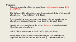
![Treatment
• Focuses on providing adequate calcium supplemention
700 mg/day[age 1-3 yr]
1,000 mg/day [4-8 yr]
1,300 mg/day [9-18 yr]
• Vitamin D supplementation is necessary if there is concurrent
vitamin D deficiency
Prevention
• Discouraging early cessation of breastfeeding
• Increasing dietary sources of calcium](https://image.slidesharecdn.com/rickets-230804103241-887f3367/85/RICKETS-pptx-38-320.jpg)