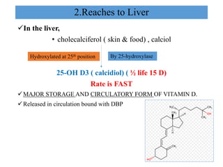
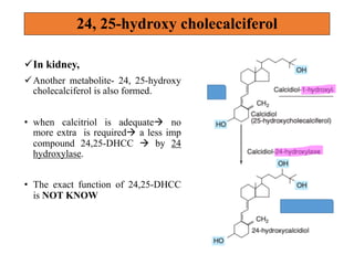
Vitamin ‘D’
Learning objectives
• Introduction
• Absorption/ transport and storage
• Formation in body
• Action/ function
• Regulation
• Deficiency
• hypervitaminosis
• Fat soluble vit.
• Resembles sterols in structure and fxn like hormoneà CHOLESTEROL.
üDietary source
• Ergocalciferol, aka vit D2(plant)
• Cholecalciferol, aka vit D3( animal)
üIn our body:
• BUT, vit D is also synthesized in on exposure to sunlight à sunshine
vitamin
• So, NOT STRICTLY VITAMIN bcozà synthesized in the skin; against
the criteria of vitamin.
Introduction
üDietary sources
• Fish, fish liver oils, egg yolk etc.
• fortified foods (milk, butter, other dairy products etc.)
üRecommended dietary allowance (RDA)
ü 400 IU or 10 mg of cholecalciferol daily. BUT,
ü In countries with good sunlight (like India) : 200 IU (or 5 mg of
cholecalciferol)
ü🌞🌝
• Dietary vit D absorbed ( bile salt)à fat soluble.
• In intestinal mucosal cellà incorporated in chylomicronsà enters in
circulation via lymph.
• In circulationà bind with vit D binding protein ( DBP)à COMPLEX
• Further, Vit D3 synthesized in skinà also binds with DBP.
• Taken up by different tissueà liver & adipose tissue stores it.
• > 90% is bound by DBP, < 1% IS FREE.
Absorption, transport and storage
👉 👉Formation
of
Vitamin D
In our body 🌝🌞
1. starts in Skin ( dermis & epidermis)
• 7-Dehydrocholesterol (intermediate of cholesterol met, skin),
• On exposure to UV light, à previtamin D à by breaking the bond betn
9 and 10 positionà B ring is opened.
• In a period of Hours à cholecalciferol ( calciol)à by photochemical
process.
• Melanin competes 7-Dehydrocholesterol in absorbing UV rays ( black
skin à less synthesis)
2.Reaches to Liver
üIn the liver,
• cholecalciferol ( skin & food) , calciol
25-OH D3 ( calcidiol) ( ½ life 15 D)
Rate is FAST
üMAJOR STORAGE AND CIRCULATORY FORM OF VITAMIN D.
üReleased in circulation bound with DBP
Hydroxylated at 25th position By 25-hydroxylase
3. Finally reaches to kidney
üIn the kidney
• Calcidiol (25-OH D) à 1-hydroxylation à to form
1,25-dihydroxyvitamin D (calcitriol) by 1-⍺ hydroxylase (Slow,rate
limiting.)
• Biologically most active vitamin D. (1000 times more potent>
calcidiol)
• ½ life : 12-14 hrs.
üBoth hydroxylase ( kidney & liver):for the hydroxylation process
• cytochrome P450,
• NADPH and
• molecular oxygen.
24, 25-hydroxy cholecalciferol
üIn kidney,
üAnother metabolite- 24, 25-hydroxy
cholecalciferol is also formed.
• when calcitriol is adequateà no
more extra is requiredà a less imp
compound 24,25-DHCC à by 24
hydroxylase.
• The exact function of 24,25-DHCC
is NOT KNOW
Functions
üCalcitriol ( 1,25-DHCC) is the biologically most active.
• Principal fxn is to maintain plasma calcium level.
qCalcitriol acts on 3 different organs.
1. intestine
2. kidney
3. bone
To Maintain plasma calcium.
( normal =9 -11 mg/dl).
• In intestine calcitriol binds with cytosolic receptor à form calcitriol
receptor complex.
• Complex reaches to nucleus à interacts with DNAà synthesis of
calcium binding protein called calbindin.
• ↑↑ calcium absorption
• ↑↑ phosphate absorption
1)Action on the intestine.
calbindin.
2. Action on bone: Diverse
üMobilizes bone mineral
• In the osteoblast of bone à ↑↑es ca++ uptake à deposited as calcium
phosphateà bone formation.
üAt the same time,
• Vitamin D is also believed to promote bone resorption & calcium
mobilization to raise the levels of Ca and P in blood in association with
PTH.
• bone resorption by ↑↑ osteoclastic activity.
• So, overall bone remodeling.
3.	Action	on	the	kidney
In one diagram: action of calcitriol.
Regulation of vit D
üRegulated by
1) PTHà direct
2) PHOSPHATEà direct
3) CALCIUMà indirect
4) 1,25 (OH)2 D ( calcitriol)à self regulated by feedback
mechanism
PTH &
hypophosphatemia
Hypocalcemia
↑↑ activity of 1-⍺ hydroxylaseà
↑↑ synthesis of calcitriol.
DIRECT
At first, it ↑↑es PTH levelà then
only ↑↑ activity of 1-⍺
hydroxylaseà↑↑ synthesis of
calcitriol.
INDIRECT
Deficiency
• Relatively less common, since can be synthesized in body.
üBUT,
• Insufficient exposure to sunlight+ low vit D dietà deficiency
• Occurs in
• Strict vegetarians
• Chronic alcoholics
• Liver or kidney disease
• Fat malabsorption syndrome
• Rickets in children
• Osteomalacia in adults
• Osteoporosis in old people
Renal	rickets	(	how	kidney	causes	rickets	?)
(renal	osteodystrophy)
↓↓ed 1-⍺ hydroxylase
Hypervitaminosis D- toxicity
• Among the vitamins, vit D is the most toxic in overdoses (10-
100 times RDA).
üLeads to
• Demineralization of bone ( resorption)+ increased calcium absorp
from intestineà hypercalcemia
• Prolonged hypercalcemiaà deposition in soft tissue like kidney &
arteriesà calcinosis
• Stones in kidney ( calculi) & rise in BP.
üIn hypervitaminosis: toxicity
• ↑↑ed calcitriolà↑↑es bone resorption BM stem cell à form more
osteoclast cell & ↑↑ osteoclastic activity.
TASK FOR YOU….. 📝
JUSTIFY- VIT D is also hormone.

Vit D pdf

  • 1.
  • 2.
    Learning objectives • Introduction •Absorption/ transport and storage • Formation in body • Action/ function • Regulation • Deficiency • hypervitaminosis
  • 3.
    • Fat solublevit. • Resembles sterols in structure and fxn like hormoneà CHOLESTEROL. üDietary source • Ergocalciferol, aka vit D2(plant) • Cholecalciferol, aka vit D3( animal) üIn our body: • BUT, vit D is also synthesized in on exposure to sunlight à sunshine vitamin • So, NOT STRICTLY VITAMIN bcozà synthesized in the skin; against the criteria of vitamin. Introduction
  • 4.
    üDietary sources • Fish,fish liver oils, egg yolk etc. • fortified foods (milk, butter, other dairy products etc.) üRecommended dietary allowance (RDA) ü 400 IU or 10 mg of cholecalciferol daily. BUT, ü In countries with good sunlight (like India) : 200 IU (or 5 mg of cholecalciferol) ü🌞🌝
  • 5.
    • Dietary vitD absorbed ( bile salt)à fat soluble. • In intestinal mucosal cellà incorporated in chylomicronsà enters in circulation via lymph. • In circulationà bind with vit D binding protein ( DBP)à COMPLEX • Further, Vit D3 synthesized in skinà also binds with DBP. • Taken up by different tissueà liver & adipose tissue stores it. • > 90% is bound by DBP, < 1% IS FREE. Absorption, transport and storage
  • 6.
  • 7.
    1. starts inSkin ( dermis & epidermis) • 7-Dehydrocholesterol (intermediate of cholesterol met, skin), • On exposure to UV light, à previtamin D à by breaking the bond betn 9 and 10 positionà B ring is opened. • In a period of Hours à cholecalciferol ( calciol)à by photochemical process. • Melanin competes 7-Dehydrocholesterol in absorbing UV rays ( black skin à less synthesis)
  • 8.
    2.Reaches to Liver üInthe liver, • cholecalciferol ( skin & food) , calciol 25-OH D3 ( calcidiol) ( ½ life 15 D) Rate is FAST üMAJOR STORAGE AND CIRCULATORY FORM OF VITAMIN D. üReleased in circulation bound with DBP Hydroxylated at 25th position By 25-hydroxylase
  • 9.
    3. Finally reachesto kidney üIn the kidney • Calcidiol (25-OH D) à 1-hydroxylation à to form 1,25-dihydroxyvitamin D (calcitriol) by 1-⍺ hydroxylase (Slow,rate limiting.) • Biologically most active vitamin D. (1000 times more potent> calcidiol) • ½ life : 12-14 hrs. üBoth hydroxylase ( kidney & liver):for the hydroxylation process • cytochrome P450, • NADPH and • molecular oxygen.
  • 10.
    24, 25-hydroxy cholecalciferol üInkidney, üAnother metabolite- 24, 25-hydroxy cholecalciferol is also formed. • when calcitriol is adequateà no more extra is requiredà a less imp compound 24,25-DHCC à by 24 hydroxylase. • The exact function of 24,25-DHCC is NOT KNOW
  • 12.
    Functions üCalcitriol ( 1,25-DHCC)is the biologically most active. • Principal fxn is to maintain plasma calcium level. qCalcitriol acts on 3 different organs. 1. intestine 2. kidney 3. bone To Maintain plasma calcium. ( normal =9 -11 mg/dl).
  • 13.
    • In intestinecalcitriol binds with cytosolic receptor à form calcitriol receptor complex. • Complex reaches to nucleus à interacts with DNAà synthesis of calcium binding protein called calbindin. • ↑↑ calcium absorption • ↑↑ phosphate absorption 1)Action on the intestine. calbindin.
  • 14.
    2. Action onbone: Diverse üMobilizes bone mineral • In the osteoblast of bone à ↑↑es ca++ uptake à deposited as calcium phosphateà bone formation. üAt the same time, • Vitamin D is also believed to promote bone resorption & calcium mobilization to raise the levels of Ca and P in blood in association with PTH. • bone resorption by ↑↑ osteoclastic activity. • So, overall bone remodeling.
  • 15.
  • 16.
    In one diagram:action of calcitriol.
  • 17.
    Regulation of vitD üRegulated by 1) PTHà direct 2) PHOSPHATEà direct 3) CALCIUMà indirect 4) 1,25 (OH)2 D ( calcitriol)à self regulated by feedback mechanism PTH & hypophosphatemia Hypocalcemia ↑↑ activity of 1-⍺ hydroxylaseà ↑↑ synthesis of calcitriol. DIRECT At first, it ↑↑es PTH levelà then only ↑↑ activity of 1-⍺ hydroxylaseà↑↑ synthesis of calcitriol. INDIRECT
  • 18.
    Deficiency • Relatively lesscommon, since can be synthesized in body. üBUT, • Insufficient exposure to sunlight+ low vit D dietà deficiency • Occurs in • Strict vegetarians • Chronic alcoholics • Liver or kidney disease • Fat malabsorption syndrome • Rickets in children • Osteomalacia in adults • Osteoporosis in old people
  • 19.
  • 20.
    Hypervitaminosis D- toxicity •Among the vitamins, vit D is the most toxic in overdoses (10- 100 times RDA). üLeads to • Demineralization of bone ( resorption)+ increased calcium absorp from intestineà hypercalcemia • Prolonged hypercalcemiaà deposition in soft tissue like kidney & arteriesà calcinosis • Stones in kidney ( calculi) & rise in BP. üIn hypervitaminosis: toxicity • ↑↑ed calcitriolà↑↑es bone resorption BM stem cell à form more osteoclast cell & ↑↑ osteoclastic activity.
  • 21.
    TASK FOR YOU…..📝 JUSTIFY- VIT D is also hormone.