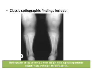
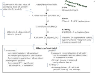
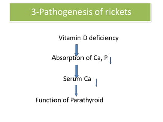
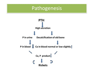
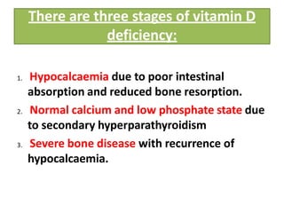
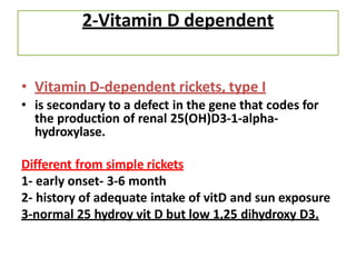
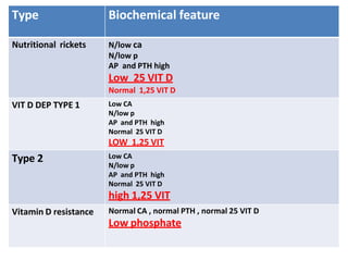
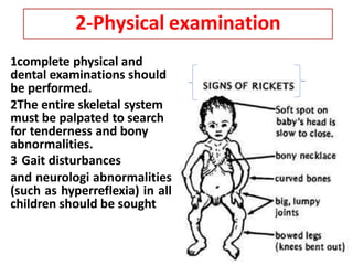
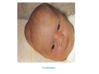
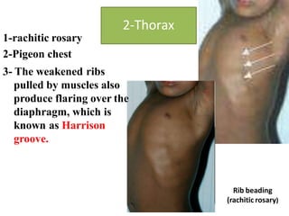
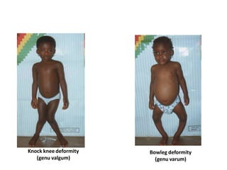
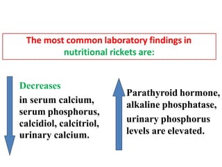
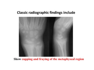
Approach to a child
with Rickets
Definition of Rickets. .
Types of rickets and how to differentiate
between them biochemically
Causes of rickets
Vitamin D metabolism.
Outline
What is Rickets?
•Rickets is a disease of growing bones due to
defective mineralization at growth plates in
growing children.
• (Osteomalacia is the same condition in adults)
• Adequate calcium and phosphate levels
are required for bone mineralization and vitamin
D is critical for calcium homeostasis
What are the different types of
rickets?
a)Vitamin D deficiency- “classical rickets”
caused by low endogenous vitamin D
b) Vitamin D dependent-
• type 1 is due to 1 alpha hydroxylase deficiency
• type 2 is due to a mutation in the vitamin D
receptor
c) Vitamin D resistant- defect in tubular
reabsorption of phosphate
1- Nutritional
• Nutritional rickets results from inadequate
sunlight exposure or inadequate intake of
dietary vitamin D, calcium, or phosphorus.
• Mostly onset is at the end of the first or during
the 2nd year.
In children, vitamin D deficiency is the most
common cause of rickets
Vitamin D Sources
• Sun light exposure
• Diet ( liver and egg yolks )
Cholecalciferol (vitamin D-3) is formed in the skin from
7-dihydrotachysterol. This steroid undergoes hydroxylation in 2 steps.
Pathophysiology - Metabolism of vitamin D
• The first hydroxylation occurs at position 25 in the liver, producing
calcidiol (25-hydroxycholecalciferol), which circulates in the plasma as
the most abundant of the vitamin D metabolites
• is a good indicator of overall vitamin D status.
Cont.........
• The second hydroxylation step occurs in the kidney at the 1 position,
where it undergoes hydroxylation to the active metabolite calcitriol
(1,25-dihydroxycholecalciferol - DHC). This cholecalciferol is not a
vitamin, but a hormone.
3-Pathogenesis of rickets
Vitamin D deficiency
Absorption of Ca, P
Serum Ca
Function of Parathyroid
Pathogenesis
PTH
High secretion
P in urine Decalcification of old bone
P in blood Ca in blood normal or low slightly
Ca, P product
Rickets
There are three stages of vitamin D
deficiency:
1. Hypocalcaemia due to poor intestinal
absorption and reduced bone resorption.
2. Normal calcium and low phosphate state due
to secondary hyperparathyroidism
3. Severe bone disease with recurrence of
hypocalcaemia.
• What are the causes of vitamin D
deficiency?
1poor exposure to sunlight
2- dark skin ( black children)
3-. Improper feeding:
1) Inadequate intake of Vitamin D
• Breast milk
• Cow’s milk
0-10IU/100ml
0.3-4IU/100ml.
2) Improper Ca and P ratio
Etiology
3. Fast growth, increased requirement (relative
deficiency)
4. Diseases ( malabsorption)
 Liver diseases, renal diseases
Gastrointestinal diseases (Celiac disease, pancreatitis )
Cystic fibrosis
Etiology
• Medications
1) Antacids
phosphate
reduce absorption of calcium and
2) Anticonvulsants ( phenytoin,phenobarbitone )
Lead to increase VIT D catabolism and inhibit ca
absorption
1) Corticosteroids
2) Loop diuretics
Etiology
2-Vitamin D dependent
• Vitamin D-dependent rickets, type I
• is secondary to a defect in the gene that codes for
the production of renal 25(OH)D3-1-alpha-
hydroxylase.
Different from simple rickets
1- early onset- 3-6 month
2- history of adequate intake of vitD and sun exposure
3-normal 25 hydroy vit D but low 1,25 dihydroxy D3.
Vitamin D-dependent rickets, type II
1. is a rare autosomal disorder caused by
mutations in the vitamin D receptor.
2. elevated levels of circulating calcitriol
differentiate this type from type I.
3. Generalized alopecia occur in 50% of the
cases .
3-Vitamin D resistant
1familial hypophosphatemic rickets
(X linked dominant.)
2 renal wasting of phosphorus at the
proximal tubule level results in
hypophosphatemia.
Cont…
• Clinical features.
1 Early age of onset and severe
deformities
2 short stature and sever dental
caries
3 Normal ca, PTH, no aminoaciduria .
Type Biochemical feature
Nutritional rickets N/low ca
N/low p
AP and PTH high
Low 25 VIT D
Normal 1,25 VIT D
VIT D DEP TYPE 1 Low CA
N/low p
AP and PTH high
Normal 25 VIT D
LOW 1,25 VIT
Type 2 Low CA
N/low p
AP and PTH high
Normal 25 VIT D
high 1,25 VIT
Vitamin D resistance Normal CA , normal PTH , normal 25 VIT D
Low phosphate
1complete physical and
dental examinations should
be performed.
2The entire skeletal system
must be palpated to search
for tenderness and bony
abnormalities.
3 Gait disturbances
and neurologi abnormalities
(such as hyperreflexia) in all
children should be sought
2-Physical examination
1-head
• 1-Craniotabes manifests
early in infants
• 2-frontal bossing and
delays the closure of the
anterior fontanelle.
• 3-Increased incidence of
cavities in the teeth
(dental caries)
Frontal bossing
Craniotabes
1-rachitic rosary
2-Pigeon chest
3- The weakened ribs
pulled by muscles also
produce flaring over the
diaphragm, which is
known as Harrison
groove.
Rib beading
(rachitic rosary)
2-Thorax
Chest deformity
Funnel chest – pectus excavatum
Pigeon chest
• 1-Bowlegs and
knock-knee
• 2-enlarged wrist
and ankle(double
malleoli)
• Lax ligament-
hypotonia
3-extremities
Knock knee deformity
(genu valgum)
Bowleg deformity
(genu varum)
3-Investigations
1-Biochemical investigations
1 serum levels of calcium (total and ionized
with serum albumin),
2 phosphorus,
3alkaline phosphatase (ALP)
4-parathyroid hormone
5 calcidiol
6 urine studies include urinalysis and levels of
urinary calcium and phosphorus.
Decreases
in serum calcium,
serum phosphorus,
calcidiol, calcitriol,
urinary calcium.
The most common laboratory findings in
nutritional rickets are:
Parathyroid hormone,
alkaline phosphatase,
urinary phosphorus
levels are elevated.
2- Radiological investigation
widening of the distal epyphysis
fraying and widening of the metaphysis
angular deformities of the arm and leg
bones.
Classic radiographic findings include
Show cupping and fraying of the metaphyseal region
• Classic radiographic findings include:
Radiographs of the knee of a 3-year-old girl with hypophosphatemia
depict severe fraying of the metaphysis.
4-Treatment
1. Special therapy: Vitamin D therapy
• A. General method: Vitamin D 2000-4000 IU/day
for 2-4 weeks, then change to
preventive dosage – 400 IU.
• B. A single large dose: For severe case, or Rickets with
complication, or those who can’t bear oral therapy.
Vitamin D3 200000 – 300000 IU, im,
preventive dosage will be used after 2-3 months.
Cont…
2-. Calcium supplementation: Dosage: 1-3
g/day
• only used for special cases, such as baby fed
mainly with cereal or infants under 3 months
of age and those who have already
developed tetany.
3-. Plastic therapy:
In children with bone deformities after 4
years old plastic surgery may be useful.
rickets.pptx

rickets.pptx

  • 1.
    Approach to achild with Rickets
  • 2.
    Definition of Rickets.. Types of rickets and how to differentiate between them biochemically Causes of rickets Vitamin D metabolism. Outline
  • 3.
    What is Rickets? •Ricketsis a disease of growing bones due to defective mineralization at growth plates in growing children. • (Osteomalacia is the same condition in adults) • Adequate calcium and phosphate levels are required for bone mineralization and vitamin D is critical for calcium homeostasis
  • 4.
    What are thedifferent types of rickets? a)Vitamin D deficiency- “classical rickets” caused by low endogenous vitamin D b) Vitamin D dependent- • type 1 is due to 1 alpha hydroxylase deficiency • type 2 is due to a mutation in the vitamin D receptor c) Vitamin D resistant- defect in tubular reabsorption of phosphate
  • 5.
    1- Nutritional • Nutritionalrickets results from inadequate sunlight exposure or inadequate intake of dietary vitamin D, calcium, or phosphorus. • Mostly onset is at the end of the first or during the 2nd year. In children, vitamin D deficiency is the most common cause of rickets
  • 6.
    Vitamin D Sources •Sun light exposure • Diet ( liver and egg yolks )
  • 7.
    Cholecalciferol (vitamin D-3)is formed in the skin from 7-dihydrotachysterol. This steroid undergoes hydroxylation in 2 steps. Pathophysiology - Metabolism of vitamin D • The first hydroxylation occurs at position 25 in the liver, producing calcidiol (25-hydroxycholecalciferol), which circulates in the plasma as the most abundant of the vitamin D metabolites • is a good indicator of overall vitamin D status.
  • 8.
    Cont......... • The secondhydroxylation step occurs in the kidney at the 1 position, where it undergoes hydroxylation to the active metabolite calcitriol (1,25-dihydroxycholecalciferol - DHC). This cholecalciferol is not a vitamin, but a hormone.
  • 10.
    3-Pathogenesis of rickets VitaminD deficiency Absorption of Ca, P Serum Ca Function of Parathyroid
  • 11.
    Pathogenesis PTH High secretion P inurine Decalcification of old bone P in blood Ca in blood normal or low slightly Ca, P product Rickets
  • 12.
    There are threestages of vitamin D deficiency: 1. Hypocalcaemia due to poor intestinal absorption and reduced bone resorption. 2. Normal calcium and low phosphate state due to secondary hyperparathyroidism 3. Severe bone disease with recurrence of hypocalcaemia.
  • 13.
    • What arethe causes of vitamin D deficiency?
  • 14.
    1poor exposure tosunlight 2- dark skin ( black children) 3-. Improper feeding: 1) Inadequate intake of Vitamin D • Breast milk • Cow’s milk 0-10IU/100ml 0.3-4IU/100ml. 2) Improper Ca and P ratio Etiology
  • 15.
    3. Fast growth,increased requirement (relative deficiency) 4. Diseases ( malabsorption)  Liver diseases, renal diseases Gastrointestinal diseases (Celiac disease, pancreatitis ) Cystic fibrosis Etiology
  • 16.
    • Medications 1) Antacids phosphate reduceabsorption of calcium and 2) Anticonvulsants ( phenytoin,phenobarbitone ) Lead to increase VIT D catabolism and inhibit ca absorption 1) Corticosteroids 2) Loop diuretics Etiology
  • 17.
    2-Vitamin D dependent •Vitamin D-dependent rickets, type I • is secondary to a defect in the gene that codes for the production of renal 25(OH)D3-1-alpha- hydroxylase. Different from simple rickets 1- early onset- 3-6 month 2- history of adequate intake of vitD and sun exposure 3-normal 25 hydroy vit D but low 1,25 dihydroxy D3.
  • 18.
    Vitamin D-dependent rickets,type II 1. is a rare autosomal disorder caused by mutations in the vitamin D receptor. 2. elevated levels of circulating calcitriol differentiate this type from type I. 3. Generalized alopecia occur in 50% of the cases .
  • 19.
    3-Vitamin D resistant 1familialhypophosphatemic rickets (X linked dominant.) 2 renal wasting of phosphorus at the proximal tubule level results in hypophosphatemia.
  • 20.
    Cont… • Clinical features. 1Early age of onset and severe deformities 2 short stature and sever dental caries 3 Normal ca, PTH, no aminoaciduria .
  • 21.
    Type Biochemical feature Nutritionalrickets N/low ca N/low p AP and PTH high Low 25 VIT D Normal 1,25 VIT D VIT D DEP TYPE 1 Low CA N/low p AP and PTH high Normal 25 VIT D LOW 1,25 VIT Type 2 Low CA N/low p AP and PTH high Normal 25 VIT D high 1,25 VIT Vitamin D resistance Normal CA , normal PTH , normal 25 VIT D Low phosphate
  • 22.
    1complete physical and dentalexaminations should be performed. 2The entire skeletal system must be palpated to search for tenderness and bony abnormalities. 3 Gait disturbances and neurologi abnormalities (such as hyperreflexia) in all children should be sought 2-Physical examination
  • 23.
    1-head • 1-Craniotabes manifests earlyin infants • 2-frontal bossing and delays the closure of the anterior fontanelle. • 3-Increased incidence of cavities in the teeth (dental caries) Frontal bossing
  • 24.
  • 25.
    1-rachitic rosary 2-Pigeon chest 3-The weakened ribs pulled by muscles also produce flaring over the diaphragm, which is known as Harrison groove. Rib beading (rachitic rosary) 2-Thorax
  • 26.
    Chest deformity Funnel chest– pectus excavatum Pigeon chest
  • 27.
    • 1-Bowlegs and knock-knee •2-enlarged wrist and ankle(double malleoli) • Lax ligament- hypotonia 3-extremities
  • 28.
    Knock knee deformity (genuvalgum) Bowleg deformity (genu varum)
  • 30.
    3-Investigations 1-Biochemical investigations 1 serumlevels of calcium (total and ionized with serum albumin), 2 phosphorus, 3alkaline phosphatase (ALP) 4-parathyroid hormone 5 calcidiol 6 urine studies include urinalysis and levels of urinary calcium and phosphorus.
  • 31.
    Decreases in serum calcium, serumphosphorus, calcidiol, calcitriol, urinary calcium. The most common laboratory findings in nutritional rickets are: Parathyroid hormone, alkaline phosphatase, urinary phosphorus levels are elevated.
  • 32.
    2- Radiological investigation wideningof the distal epyphysis fraying and widening of the metaphysis angular deformities of the arm and leg bones.
  • 33.
    Classic radiographic findingsinclude Show cupping and fraying of the metaphyseal region
  • 34.
    • Classic radiographicfindings include: Radiographs of the knee of a 3-year-old girl with hypophosphatemia depict severe fraying of the metaphysis.
  • 35.
    4-Treatment 1. Special therapy:Vitamin D therapy • A. General method: Vitamin D 2000-4000 IU/day for 2-4 weeks, then change to preventive dosage – 400 IU. • B. A single large dose: For severe case, or Rickets with complication, or those who can’t bear oral therapy. Vitamin D3 200000 – 300000 IU, im, preventive dosage will be used after 2-3 months.
  • 36.
    Cont… 2-. Calcium supplementation:Dosage: 1-3 g/day • only used for special cases, such as baby fed mainly with cereal or infants under 3 months of age and those who have already developed tetany. 3-. Plastic therapy: In children with bone deformities after 4 years old plastic surgery may be useful.