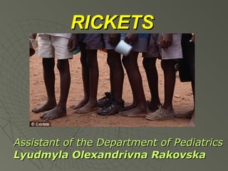
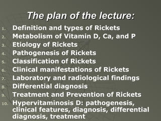
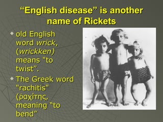
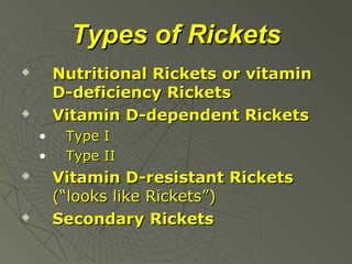
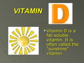
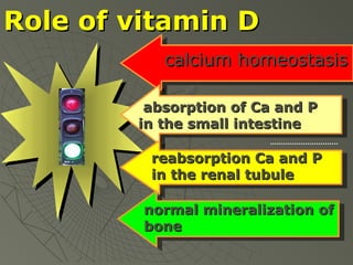
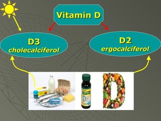
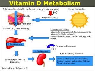
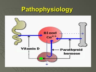
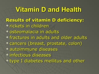
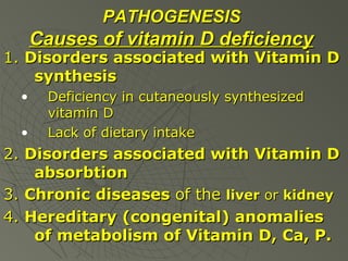
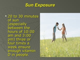
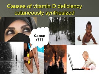
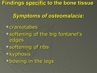
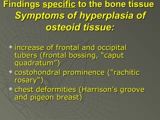
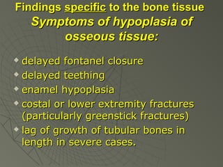
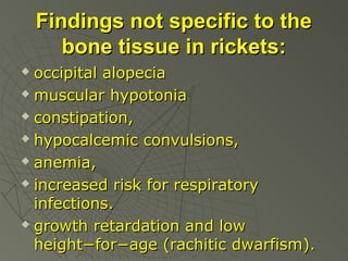
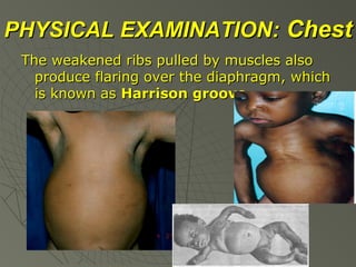
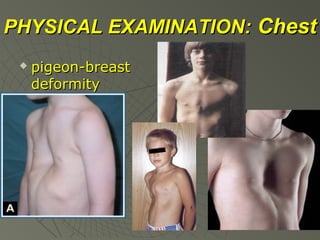
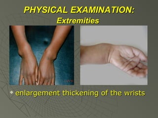
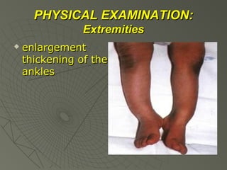
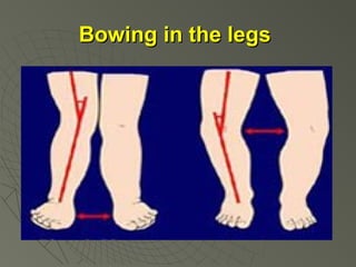
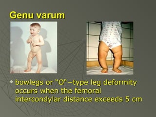
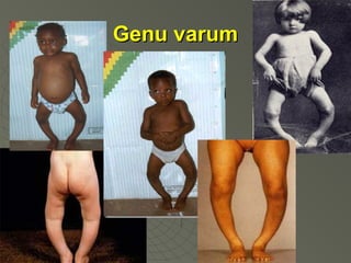
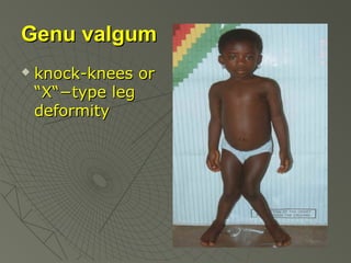
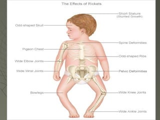
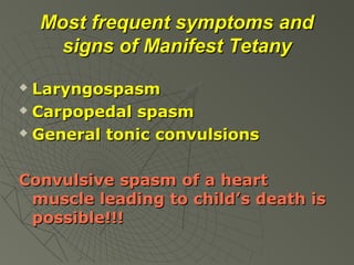
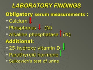
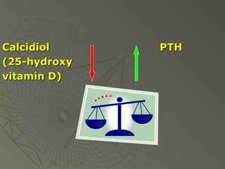
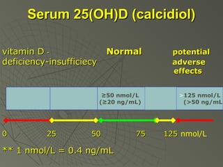
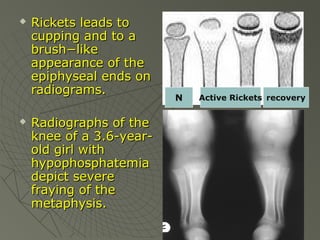
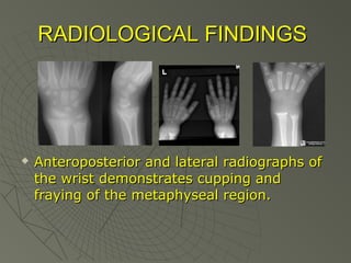
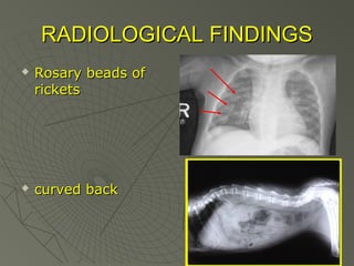
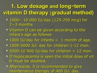
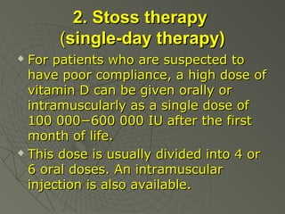
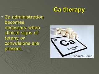
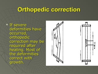
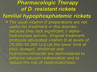
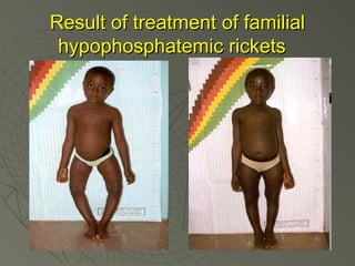
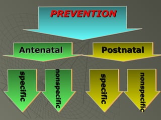
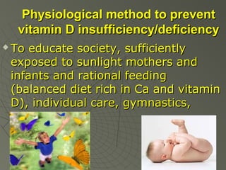
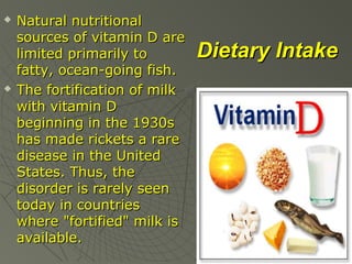
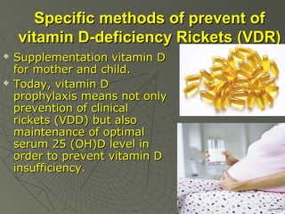
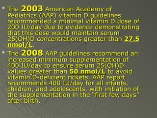
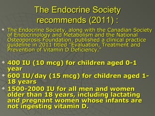
This document provides an overview of rickets presented by Lyudmyla Olexandrivna Rakovska from the Department of Pediatrics at V.N. Karazin Kharkiv National University. The 10-point plan discusses the definition, etiology, pathogenesis, classification, clinical manifestations, laboratory/radiological findings, differential diagnosis, treatment and prevention of rickets. Key points include that rickets is caused by vitamin D deficiency impairing bone mineralization, especially in children under 2 years old. Symptoms involve soft, deformed bones and skeletal abnormalities. Treatment focuses on vitamin D supplementation through either low daily doses or high single doses.
Skeletal Imaging of Nutritional Disorders in Children_Crimson PublishersCrimsonpublishersNTNF
Imaging of skeleton plays a major role in the diagnosis of nutritional disorders in children. The disorders can be1) Deficiency and2) Toxic (Overdose). The common deficiency disorders include rickets, scurvy, osteoporosis and anemias. Conventional radiology is adequate in the diagnosis, except in the grading of diagnosis of degrees of osteoporosis. The differential diagnosis is also discussed.
Tom Selleck Health: A Comprehensive Look at the Iconic Actor’s Wellness Journeygreendigital
Tom Selleck, an enduring figure in Hollywood. has captivated audiences for decades with his rugged charm, iconic moustache. and memorable roles in television and film. From his breakout role as Thomas Magnum in Magnum P.I. to his current portrayal of Frank Reagan in Blue Bloods. Selleck's career has spanned over 50 years. But beyond his professional achievements. fans have often been curious about Tom Selleck Health. especially as he has aged in the public eye.
Follow us on: Pinterest
Introduction
Many have been interested in Tom Selleck health. not only because of his enduring presence on screen but also because of the challenges. and lifestyle choices he has faced and made over the years. This article delves into the various aspects of Tom Selleck health. exploring his fitness regimen, diet, mental health. and the challenges he has encountered as he ages. We'll look at how he maintains his well-being. the health issues he has faced, and his approach to ageing .
Early Life and Career
Childhood and Athletic Beginnings
Tom Selleck was born on January 29, 1945, in Detroit, Michigan, and grew up in Sherman Oaks, California. From an early age, he was involved in sports, particularly basketball. which played a significant role in his physical development. His athletic pursuits continued into college. where he attended the University of Southern California (USC) on a basketball scholarship. This early involvement in sports laid a strong foundation for his physical health and disciplined lifestyle.
Transition to Acting
Selleck's transition from an athlete to an actor came with its physical demands. His first significant role in "Magnum P.I." required him to perform various stunts and maintain a fit appearance. This role, which he played from 1980 to 1988. necessitated a rigorous fitness routine to meet the show's demands. setting the stage for his long-term commitment to health and wellness.
Fitness Regimen
Workout Routine
Tom Selleck health and fitness regimen has evolved. adapting to his changing roles and age. During his "Magnum, P.I." days. Selleck's workouts were intense and focused on building and maintaining muscle mass. His routine included weightlifting, cardiovascular exercises. and specific training for the stunts he performed on the show.
Selleck adjusted his fitness routine as he aged to suit his body's needs. Today, his workouts focus on maintaining flexibility, strength, and cardiovascular health. He incorporates low-impact exercises such as swimming, walking, and light weightlifting. This balanced approach helps him stay fit without putting undue strain on his joints and muscles.
Importance of Flexibility and Mobility
In recent years, Selleck has emphasized the importance of flexibility and mobility in his fitness regimen. Understanding the natural decline in muscle mass and joint flexibility with age. he includes stretching and yoga in his routine. These practices help prevent injuries, improve posture, and maintain mobilit
micro teaching on communication m.sc nursing.pdfAnurag Sharma
Microteaching is a unique model of practice teaching. It is a viable instrument for the. desired change in the teaching behavior or the behavior potential which, in specified types of real. classroom situations, tends to facilitate the achievement of specified types of objectives.
These simplified slides by Dr. Sidra Arshad present an overview of the non-respiratory functions of the respiratory tract.
Learning objectives:
1. Enlist the non-respiratory functions of the respiratory tract
2. Briefly explain how these functions are carried out
3. Discuss the significance of dead space
4. Differentiate between minute ventilation and alveolar ventilation
5. Describe the cough and sneeze reflexes
Study Resources:
1. Chapter 39, Guyton and Hall Textbook of Medical Physiology, 14th edition
2. Chapter 34, Ganong’s Review of Medical Physiology, 26th edition
3. Chapter 17, Human Physiology by Lauralee Sherwood, 9th edition
4. Non-respiratory functions of the lungs https://academic.oup.com/bjaed/article/13/3/98/278874
MANAGEMENT OF ATRIOVENTRICULAR CONDUCTION BLOCK.pdfJim Jacob Roy
Cardiac conduction defects can occur due to various causes.
Atrioventricular conduction blocks ( AV blocks ) are classified into 3 types.
This document describes the acute management of AV block.
Basavarajeeyam is an important text for ayurvedic physician belonging to andhra pradehs. It is a popular compendium in various parts of our country as well as in andhra pradesh. The content of the text was presented in sanskrit and telugu language (Bilingual). One of the most famous book in ayurvedic pharmaceutics and therapeutics. This book contains 25 chapters called as prakaranas. Many rasaoushadis were explained, pioneer of dhatu druti, nadi pareeksha, mutra pareeksha etc. Belongs to the period of 15-16 century. New diseases like upadamsha, phiranga rogas are explained.
Ozempic: Preoperative Management of Patients on GLP-1 Receptor Agonists Saeid Safari
Preoperative Management of Patients on GLP-1 Receptor Agonists like Ozempic and Semiglutide
ASA GUIDELINE
NYSORA Guideline
2 Case Reports of Gastric Ultrasound
NVBDCP.pptx Nation vector borne disease control programSapna Thakur
NVBDCP was launched in 2003-2004 . Vector-Borne Disease: Disease that results from an infection transmitted to humans and other animals by blood-feeding arthropods, such as mosquitoes, ticks, and fleas. Examples of vector-borne diseases include Dengue fever, West Nile Virus, Lyme disease, and malaria.
3. The plan of the lecture:
1.
2.
3.
4.
5.
6.
7.
8.
9.
10.
Definition and types of Rickets
Metabolism of Vitamin D, Ca, and P
Etiology of Rickets
Pathogenesis of Rickets
Classification of Rickets
Clinical manifestations of Rickets
Laboratory and radiological findings
Differential diagnosis
Treatment and Prevention of Rickets
Hypervitaminosis D: pathogenesis,
clinical features, diagnosis, differential
diagnosis, treatment
4. Rickets (Rachitis)
is a metabolic disease of
growing bone that is unique
to children (especially of first
2 years) and adolescents. It
is caused by a failure of
osteoid to calcify in a growing
person.
5. Rickets
characterized by a failure of
bone tissue to be properly
mineralized, especially by
imperfect calcification, typically
resulting in soft bones and
skeletal deformities.
6. “English disease” is another
name of Rickets
old English
word wrick,
(wrickken)
means "to
twist”.
The Greek word
"rachitis"
(ῥαχίτης,
meaning “to
bend”
7. Types of Rickets
Nutritional Rickets or vitamin
D-deficiency Rickets
Vitamin D-dependent Rickets
•
•
Type I
Type II
Vitamin D-resistant Rickets
(“looks like Rickets”)
Secondary Rickets
10. Role of vitamin D
calcium homeostasis
absorption of Ca and P
in the small intestine
reabsorption Ca and P
in the renal tubule
normal mineralization of
bone
14. Vitamin D and Health
Results of vitamin D deficiency:
rickets in children
osteomalacia in adults
fractures in adults and older adults
cancers (breast, prostate, colon)
autoimmune diseases
infectious diseases
type I diabetes mellitus and other
15. PATHOGENESIS
Causes of vitamin D deficiency
1. Disorders associated with Vitamin D
synthesis
•
•
Deficiency in cutaneously synthesized
vitamin D
Lack of dietary intake
2. Disorders associated with Vitamin D
absorbtion
3. Chronic diseases of the liver or kidney
4. Hereditary (congenital) anomalies
of metabolism of Vitamin D, Ca, P.
16. Sun Exposure
20 to 30 minutes
of sun
(especially
between the
hours of 10:00
am and 3:00
pm) three or
four times a
week ensure
enough vitamin
D in people.
20. Medications are associated
with vitamin D deficiency
Steroids
Antiepileptic (anticonvulsants) drugs:
Phenobarbital and Phenytoin
(brand name Dilantin®)
Orlistat (brand names Xenical® and
alliTM) and the cholesterol-lowering
drug Cholestyramine (brand names
Questran®, LoCholest®, and
Prevalite®)
22. Hereditary (congenital)
anomalies of metabolism of
Vitamin D, Ca and P
Vit D-dependent rickets of I type
(pseudovitamin D-deficiency rickets) - defect
in the gene coding of renal 1-alpha-hydroxylase.
Autosomal recessive desease.
Vit D-dependent rickets of II type
(hereditary 1-alfa, 25-dihydroxyvitamin Dresistent rickets) - mutation exists in the
vitamin D receptors (VDR). Autosomal recessive
desease.
Vit D-resistent rickets (familial
hypophosphatemic rickets) - mutations of the
phosphate-regulating gene on the X chromosome
23. Classification of Rickets
Degree
Period of disease
Initial
Height (manifest)
Mild
(“Florid rickets”)
(I)
Moderate Reconvalescenction
(reparation)
(II)
Residual symptoms
Severe
(permanent
(III)
changes)
Course
Acute
Subacute
Recurring
24. CLINICAL MANIFESTATIONS
I. Findings specific to the bone
tissue in rickets
Symptoms of osteomalacia:
Symptoms of hyperplasia of
osteoid tissue
Symptoms of hypoplasia of
osseous tissue
II. Findings not specific to the
bone tissue in rickets
25. Findings specific to the bone tissue
Symptoms of osteomalacia:
сraniotabes
softening of the big fontanel's
edges
softening of ribs
kyphosis
bowing in the legs
26. Findings specific to the bone tissue
Symptoms of hyperplasia of
osteoid tissue:
increase of frontal and occipital
tubers (frontal bossing, “caput
quadratum”)
costohondral prominence ("rachitic
rosary")
chest deformities (Harrison’s groove
and pigeon breast)
27. Findings specific to the bone tissue
Symptoms of hypoplasia of
osseous tissue:
delayed fontanel closure
delayed teething
enamel hypoplasia
costal or lower extremity fractures
(particularly greenstick fractures)
lag of growth of tubular bones in
length in severe cases.
28. Findings not specific to the
bone tissue in rickets:
occipital alopecia
muscular hypotonia
constipation,
hypocalcemic convulsions,
anemia,
increased risk for respiratory
infections.
growth retardation and low
height−for−age (rachitic dwarfism).
29.
In acute course of rickets symptoms of
osteomalacia prevail, in subacute –
symptoms of hyperplasia of osteoid tissue.
Skeletal deformities become obvious in
stage II and worsen in stage III.
Initial period is starting from 2-3 month of
life, lasts from 2-3 weeks to 2-3 months.
In breast-fed infants whose mothers have
osteomalacia, rickets may develop before
2 mo.
Florid rickets appears toward the end of
the 1st and during the 2nd year of life.
Later in childhood, manifest vitamin D
deficient rickets is rare.
30. PHYSICAL EXAMINATION: Head
Craniotabes (areas
of thinning and
softening of bones of
the skull) manifests
early in infants older
than 2−3 months.
It detected by pressing
firmly over the
occiput or posterior
parietal bones. A
ping-pong-ball
sensation will be felt.
43. PHYSICAL EXAMINATION:
non specific symptoms
Muscular hypotonia
Generalized muscular hypotonia
"floppy baby syndrome" or "slinky
baby" (such that the baby is floppy
or slinky-like)
44. PHYSICAL EXAMINATION:
non specific symptoms
Neurologic abnormalities
observed in all cases and revealed at
initial period of disease as irritability,
interrupted sleep, sweating.
45. PHYSICAL EXAMINATION:
non specific symptoms
Occipital alopecia
Increased sweating, particularly
around the head, may also be
present as non specific symptom. It
leads to occipital alopecia and occurs
in 30%.
46.
rachitic rosary (62.1%),
craniotabes (49%), occipital
alopecia (31.4%) and
enlargement of the wrists
(27.1%) were the four most
common physical examination
findings for the age group 0−6
months
47. TETANY (SPASMOPHILIA)
disease of infants, resulting from
disturbances of Ca metabolism and
characterized by the development of
tonetic and tonoclonic convulsions.
48. Most frequent symptoms and
signs of latent Tetany
Chvostek’s sign;
Trousseau’s symptom;
Maslov’s symptom;
Erb’s symptom;
Lust’s sign
49. Most frequent symptoms and
signs of Manifest Tetany
Laryngospasm
Carpopedal spasm
General tonic convulsions
Convulsive spasm of a heart
muscle leading to child’s death is
possible!!!
50.
51.
In a patient suspected to have
rickets based on clinical findings,
the diagnosis is confirmed by
biochemical and radiological
findings.
52. LABORATORY FINDINGS
Obligatory serum measurements :
Calcium
Phosphorus
(N)
Alkaline phosphatase
(N)
Additional:
25-hydroxy vitamin D
Parathyroid hormone
Sulkovich’s test of urine
55. Sulkovich’s test
This test gives approximate
presentation about Ca content in
blood from is presents in urine.
Assessment of result:
“+” – normal level
“++, ++++” – hypercalcaemia and
hypercalciuria
58.
Rickets leads to
cupping and to a
brush−like
appearance of the
epiphyseal ends on
radiograms.
Radiographs of the
knee of a 3.6-yearold girl with
hypophosphatemia
depict severe
fraying of the
metaphysis.
N
Active Rickets recovery
61. DIFFERENTIAL DIAGNOSIS
Vitamin
Vitamin
Vitamin
D ̶ deficient rickets
D ̶ dependent rickets (types I and II)
D-resistent Rickets (“looks like Rickets”):
• Hereditary X-linked hypophosphatemic rickets with
hypocalciuria
• Familial hypophosphatrmic
• Phosphat-diabetes
• Achondrodroplasia
• Fanconi syndrome (De Toni-Debre-Fanconi desease) (types
I and II)
Pseudohypoparathyroidism
Renal tubular acidosis
Cystinosis
Tyrosinemia
Secondary Rickets (renal, gastrointestinal, tumorassociated, medications, malabsortion syndromes et
all)
62. Familial
hypophosphatemic
Low stature in the
family, dental
deformities,
orthopaedic
abnormalities, and
consanguineous
marriage indicated
for hereditary
hypophosphatemic
rickets
65. Differential diagnosis tetany
includes hyperthermic convulsions,
complicated pneumonia, meningitis,
viral diseases. In hypoparathyroidism
which is very rare in babies,
hypocalcemia and
hyperphosphatemia are usual.
Eclamptic form is necessary to
differentiate from epilepsy.
67. Ergocalciferol is a form of vitamin
D, also called vitamin D2.
It is marketed under various names
including Deltalin (Eli Lilly and Company),
Drisdol (Sanofi-Synthelabo) and Calcidol
(Patrin Pharma).
69. 1. Low dosage and long−term
vitamin D therapy (gradual method)
1000− 10 000 IU/day (125-250 mcg) for
2−3 months
Vitamin D can be given according to the
infant’s age as follows:
1000 IU/day for infants < 1 month of age,
1000-5000 IU/ day for children 1-12 mon.
5000-10 000 IU/day for children > 12 mon.
If hypocalcemia is seen the initial dose of vit
D must be doubled.
Afterwards, it is recommended to give
maintenance therapy of 400 IU/ day.
70.
Because this method requires daily
treatment, success depends on
compliance.
Levels of Ca and P are normalized in 6−10
days by this therapy, while it takes 1−2
months for PTH to reach normal levels.
Depending on the severity of the disease,
it may take 3 months for the normal
serum ALP levels to be restored and the
radiological findings of rickets to
disappear.
In this treatment model, lack of
compliance is an important cause of lack
of response
71. 2. Stoss therapy
(single-day therapy)
For patients who are suspected to
have poor compliance, a high dose of
vitamin D can be given orally or
intramuscularly as a single dose of
100 000−600 000 IU after the first
month of life.
This dose is usually divided into 4 or
6 oral doses. An intramuscular
injection is also available.
72. Stoss therapy
(single-day therapy)
Administration of 150 000−300 000 units
of vitamin D and 600 000 units (15,000
mcg) in severe rickets is an effective and
safe method of treatment.
Vitamin D (cholecalciferol) is well stored
in the body and is gradually released over
many weeks. This treatment evokes a
rapid clinical response, resulting in
biochemical recovery in a few days and
radiological recovery in 10−15 days.
73.
The single-day therapy avoids
problems with compliance and may
be helpful in differentiating
nutritional rickets from familial
hypophosphatemia rickets (FHR). In
nutritional rickets, the phosphorus
level rises in 96 hours and
radiographic healing is visible in 6-7
days. Neither happens with FHR.
75. Emergency care of tetany
10% Ca gluconate (1−2 ml/kg, or 0,3-0,5
mgkg 3 times a day) IV and slowly while
monitoring ECG (bradicardia, arrhythmia).
In convulsions it is necessary to inject i.v.
seduxen (0.1 ml/kg or 0,5 mgkg of 0,5%
solution), MgSO4 (0,2-0.5 ml/kg of 25%
solution), sodium oxybutirate (0.5 ml/kg
or 50-120 mgkg of 20% solution),
oxygen inhalations.
In laryngospasm it is necessary to create a
dominant focus of excitement in the brain
(irritation of nasal mucosa, skin with a
prick, tap).
76. Ca levels should then be maintained
with oral Ca supplements.
Infants with latent tetany may
respond orally calcium gluconate
(5%) or calcium chloride (1-2%) – 1
teaspoonful x 3 times a day.
Restriction of cow milk because of
great quantity of phosphates.
After 3-4 days – vit D 2-4 000 twice
a day is prescribed.
78. Pharmacologic Therapy
of D ̶ resistant rickets
familial hypophosphatemic rickets
The usual vitamin D preparations are not
useful for treatment in this disorder,
because they lack significant 1-alphahydroxylase activity. Original treatment
protocols advocated vitamin D at levels of
25,000-50,000 U/d (at the lower limit of
toxic dosage). Amiloride and
hydrochlorothiazide are administered to
enhance calcium reabsorption and to
reduce the risk of nephrocalcinosis.
81. Physiological method to prevent
vitamin D insufficiency/deficiency
To educate society, sufficiently
exposed to sunlight mothers and
infants and rational feeding
(balanced diet rich in Ca and vitamin
D), individual care, gymnastics,
massage.
82.
Natural nutritional
sources of vitamin D are
limited primarily to
fatty, ocean-going fish.
The fortification of milk
with vitamin D
beginning in the 1930s
has made rickets a rare
disease in the United
States. Thus, the
disorder is rarely seen
today in countries
where "fortified" milk is
available.
Dietary Intake
83. Specific methods of prevent of
vitamin D-deficiency Rickets (VDR)
Supplementation vitamin D
for mother and child.
Today, vitamin D
prophylaxis means not only
prevention of clinical
rickets (VDD) but also
maintenance of optimal
serum 25 (OH)D level in
order to prevent vitamin D
insufficiency.
84.
The daily maintenance dose of
vitamin D varies by age. It has
been reported that the dose of
prophylactic vitamin D should be
between 400 and 1000 IU/day to
maintain the serum 25(OH)D at
optimum levels (50-75 nmol/L).
85.
The 2003 American Academy of
Pediatrics (AAP) vitamin D guidelines
recommended a minimal vitamin D dose of
200 IU/day due to evidence demonstrating
that this dose would maintain serum
25(OH)D concentrations greater than 27.5
nmol/L.
The 2008 AAP guidelines recommend an
increased minimum supplementation of
400 IU/day to ensure serum 25(OH)D
values greater than 50 nmol/L to avoid
vitamin D-deficient rickets. AAP report
recommends 400 IU/day for all infants,
children, and adolescents, with initiation of
the supplementation in the “first few days”
after birth.
86. The Endocrine Society
recommends (2011) :
The Endocrine Society, along with the Canadian Society
of Endocrinology and Metabolism and the National
Osteoporosis Foundation, published a clinical practice
guideline in 2011 titled "Evaluation, Treatment and
Prevention of Vitamin D Deficiency."
400 IU (10 mcg) for children aged 0-1
year
600 IU/day (15 mcg) for children aged 118 years
1500-2000 IU for all men and women
older than 18 years, including lactating
and pregnant women whose infants are
not ingesting vitamin D.
87.
Also with a perspective to prevent
early rickets, it is recommended that
vitamin D at a dose of 2000 IU/day
should be administered during the
last trimester of pregnancy to
mothers with poor exposure to
sunlight due to various reasons and
who are at high risk of Vitamin D
deficiency.
89. Prophylactic course of vitamin D
may be interrupted by UV
irradiation.
Specific prevention isn’t carried
out in the children receiving
formula feeding.
91. HYPERVITAMINOSIS D
Hypervitaminosis D is a condition
result from toxic action of vitamin D
and is characterized with intoxication
of different degree, affection of
organs and systems and
development of hypercalcemia.
96.
An excess of vitamin D causes
abnormally high blood concentrations
of calcium (hypercalcemia), which
can cause overcalcification of the
bones, soft tissues, heart and
kidneys.
97. Laboratory findings:
A serum 25(OH)D concentration >500
nmol/L (>200 ng/mL)
hypercalcemia, hypophosphatemia.
hypomagnesemia, hypokalemia,
increased levels of cholesterin ;
metabolic acidosis.
Blood: leucocytosis;
Urine: increase of calcium, Sulkowitch's
test is positive (++++);
98. Treatment
To cancel immediately vit D, and limited
quntety of cow milk (that is rich in calcium).
Intensive detoxicative therapy:
i.v. injections of albumin, 5% solution of
glucose with Ringer’s solution, cocarboxylase,
vit C, prednisolone (1-2mg/kg), vit A (5-10000
IU/day), vit E, furosemid (1 mg/kg 3 times/d),
thyreocalcitonin (75-150 U i.m. every day), 3%
solution of ammonium chloride (1 teaspoonful 3
times a day), almagel, chlolestiramine (0.5
g/kg 3 times/day),Trilon B 50 mg/kg 2-3
times/d orally, in severe cases –i.v. are used.
Editor's Notes
Primary sources and configurations of vitamin D. Adapted from Lucas et al. (2)