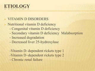
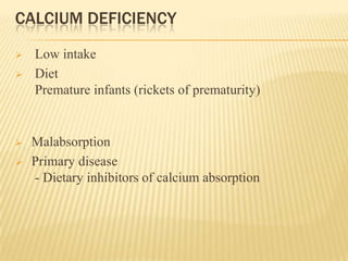
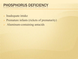
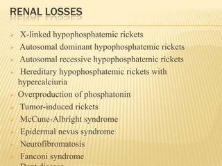
RICKETS IN CHILDREN
Sachin Soni
DNB Pediatrics
TITLE
 Vitamin D physiology
 Introduction
 Etiology
 Clinical feature
 Radiology
 Diagnosis
 Lab
 Treatment
VITAMIN D PHYSIOLOGY
 Source: -Fish, liver and oil,
- Human milk (30-40 IU/L)
- Exposure to sun light
 Vitamin D requirement:
Infants- 200IU/day (5mcg)
Children- 400IU/day (10mcg)
INTRODUCTION
Disease of growing bone due to unmineralized matrix at
the growth plates and occurs in children only before
fusion of epiphyses
ETIOLOGY
 VITAMIN D DISORDERS
 Nutritional vitamin D deficiency
- Congenital vitamin D deficiency
- Secondary vitamin D deficiency Malabsorption
- Increased degradation
- Decreased liver 25-hydroxylase
-Vitamin D–dependent rickets type 1
-Vitamin D–dependent rickets type 2
- Chronic renal failure
CALCIUM DEFICIENCY
 Low intake
 Diet
Premature infants (rickets of prematurity)
 Malabsorption
 Primary disease
- Dietary inhibitors of calcium absorption
PHOSPHORUS DEFICIENCY
 Inadequate intake
 Premature infants (rickets of prematurity)
 Aluminum-containing antacids
RENAL LOSSES
 X-linked hypophosphatemic rickets
 Autosomal dominant hypophosphatemic rickets
 Autosomal recessive hypophosphatemic rickets
 Hereditary hypophosphatemic rickets with
hypercalciuria
 Overproduction of phosphatonin
 Tumor-induced rickets
 McCune-Albright syndrome
 Epidermal nevus syndrome
 Neurofibromatosis
 Fanconi syndrome
CLINICAL FEATURES OF RICKETS
 General
- Failure to thrive
- Listlessness
- Protruding abdomen
- Muscle weakness (especially proximal)
- Fractures
HEAD
- Craniotabes
- Frontal bossing
- Delayed fontanel closure
- Delayed dentition; caries
- Craniosynostosis
 CHEST
- Rachitic rosary
- Harrison groove
- Respiratory infections and atelectasis
BACK
- Scoliosis
- Kyphosis
- Lordosis
EXTREMITIES:
- Enlargement of wrists and ankles
-Valgus or varus deformities
-Windswept deformity (combination of valgus deformity of
1leg with varus deformity of the other leg)
-Anterior bowing of the tibia and femur
-Coxa vara
-Leg pain
HYPOCALCEMIC SYMPTOMS
 Tetany
 Seizures
 Stridor due to laryngeal spasm
Deformities showing curvature of
the limbs, potbelly, and Harrison
groove.
RADIOLOGY
Wrist x-rays in a
normal child (A) and
a child with
rickets (B). Child
with rickets has
metaphyseal fraying
and cupping of the
distal radius and
ulna.
CLINICAL EVALUATION
 Dietary history
 Cutaneous synthesis
 Maternal risk
 Medication
 Malabsorption
 Renal disease
 Family history
 Physical Examination
 Lab Test
NUTRITIONAL VITAMIN D DEFICIENCY
 Vitamin D deficiency is most common cause of rickets
globally
 Most common in infancy
 Transplacental transport of vit D provide enough vit D
for first 1 to 2 months of life.
 Skin pigmentation
LABORATORY FINDINGS
Elevated: Decreased:
Alkaline phosphatase Calcium
Parathyroid hormone Phosphorus
Dihydroxyvitamin D Hydroxyvitamin D
Disorder Ca Pi PTH 25-(OH)D 1,25-(OH)2D ALK PHOS URINE Ca URINE Pi
Vitamin D
deficiency
N, ↓ ↓ ↑ ↓ ↓, N, ↑ ↑ ↓ ↑
VDDR, type 1 N, ↓ ↓ ↑ N ↓ ↑ ↓ ↑
VDDR, type 2 N, ↓ ↓ ↑ N ↑↑ ↑ ↓ ↑
Chronic renal
failure
N, ↓ ↑ ↑ N ↓ ↑ N, ↓ ↓
Dietary Pi
deficiency
N ↓ N, ↓ N ↑ ↑ ↑ ↓
XLH N ↓ N N RD ↑ ↓ ↑
ADHR N ↓ N N RD ↑ ↓ ↑
HHRH N ↓ N, ↓ N RD ↑ ↑ ↑
ARHR N ↓ N N RD ↑ ↓ ↑
Tumor-induced
rickets
N ↓ N N RD ↑ ↓ ↑
Fanconi
syndrome
N ↓ N N RD or ↑ ↑ ↓ or ↑ ↑
Dietary Ca
deficiency
N, ↓ ↓ ↑ N ↑ ↑ ↓ ↑
TREATMENT
 Stoss therapy – 300000 – 600000 IU Vitamin D oral or
IM, 2-4 doses over one day
 Alternatively high dose vit D, 2000-5000 IU/day over 4-
6 wk
 Followed by oral Vit D :
< 1 year of age - 400IU
> 1 years of age- 600IU
 Symptomatic hypocalcemia – IV calcium gluconate 100
mg/kg followed by oral calcium or calcitrol -
0.05mcg/kg/day
PROGNOSIS
 Most of children have excellent prognosis
 Severe disease causing permanent deformity and
short stature
PREVENTION
 Daily multivitamin contain- 400IU vit D for infants
while 600 IU/day for older children
SECONDARY VITAMIN D DEFICIENCY
 GI diseases - Cholestatic liver disease,
- Cystic fibrosis, pancreatic dysfunction,
- Defects in bile acid metabolism,
- Celiac disease, Crohn disease. intestinal
- lymphangiectasia
- Intestinal resection.
 Severe liver disease decreases 25-D formation due to insufficient
enzyme activity
 vitamin D deficiency due to liver disease usually requires a loss of
>90% of liver function.
 Medication- Phenobarbital or phenytoin
- isoniazid or rifampin.
TREATMENT
 high doses of vitamin D- 25-D
(25-50 g/day or 5-7g/kg/day)
 1,25-D, or with parenteral vitamin D.
 Degradation of vitamin D by the CYP system
- Acute therapy as for nutritional deficiency
followed by
 long-term administration of high doses of vitamin D
- 1,000 IU/day) as much as 4,000 IU/day
VITAMIN D–DEPENDENT RICKETS, TYPE 1
 Autosomal recessive disorder
 Mutations in the gene encoding renal 1α-
hydroxylase
 1st 2 yr of life
 Classic features symptomatic hypocalcemia
 Normal levels of 25-D
 Low or normal levels of 1,25-D
 Renal tubular dysfunction- Metabolic acidosis and
generalized aminoaciduria
 Teatment- 1,25-D (calcitriol)- 0.25-2 g/day
VITAMIN D–DEPENDENT RICKETS, TYPE 2
 Autosomal recessive disorder
 Mutations in gene encoding vitamin D receptor
 Levels of 1,25-D are extremely elevated
 Present during infancy, might not be diagnosed
until adulthood.
 50-70% of children have alopecia, range from
alopecia areata to alopecia totalis.
 Epidermal cysts are less common
THANK YOU

Rickets in children

  • 1.
    RICKETS IN CHILDREN SachinSoni DNB Pediatrics
  • 2.
    TITLE  Vitamin Dphysiology  Introduction  Etiology  Clinical feature  Radiology  Diagnosis  Lab  Treatment
  • 3.
  • 4.
     Source: -Fish,liver and oil, - Human milk (30-40 IU/L) - Exposure to sun light  Vitamin D requirement: Infants- 200IU/day (5mcg) Children- 400IU/day (10mcg)
  • 5.
    INTRODUCTION Disease of growingbone due to unmineralized matrix at the growth plates and occurs in children only before fusion of epiphyses
  • 6.
    ETIOLOGY  VITAMIN DDISORDERS  Nutritional vitamin D deficiency - Congenital vitamin D deficiency - Secondary vitamin D deficiency Malabsorption - Increased degradation - Decreased liver 25-hydroxylase -Vitamin D–dependent rickets type 1 -Vitamin D–dependent rickets type 2 - Chronic renal failure
  • 7.
    CALCIUM DEFICIENCY  Lowintake  Diet Premature infants (rickets of prematurity)  Malabsorption  Primary disease - Dietary inhibitors of calcium absorption
  • 8.
    PHOSPHORUS DEFICIENCY  Inadequateintake  Premature infants (rickets of prematurity)  Aluminum-containing antacids
  • 9.
    RENAL LOSSES  X-linkedhypophosphatemic rickets  Autosomal dominant hypophosphatemic rickets  Autosomal recessive hypophosphatemic rickets  Hereditary hypophosphatemic rickets with hypercalciuria  Overproduction of phosphatonin  Tumor-induced rickets  McCune-Albright syndrome  Epidermal nevus syndrome  Neurofibromatosis  Fanconi syndrome
  • 10.
    CLINICAL FEATURES OFRICKETS  General - Failure to thrive - Listlessness - Protruding abdomen - Muscle weakness (especially proximal) - Fractures
  • 11.
    HEAD - Craniotabes - Frontalbossing - Delayed fontanel closure - Delayed dentition; caries - Craniosynostosis  CHEST - Rachitic rosary - Harrison groove - Respiratory infections and atelectasis
  • 12.
    BACK - Scoliosis - Kyphosis -Lordosis EXTREMITIES: - Enlargement of wrists and ankles -Valgus or varus deformities -Windswept deformity (combination of valgus deformity of 1leg with varus deformity of the other leg) -Anterior bowing of the tibia and femur -Coxa vara -Leg pain
  • 13.
    HYPOCALCEMIC SYMPTOMS  Tetany Seizures  Stridor due to laryngeal spasm
  • 14.
    Deformities showing curvatureof the limbs, potbelly, and Harrison groove.
  • 15.
    RADIOLOGY Wrist x-rays ina normal child (A) and a child with rickets (B). Child with rickets has metaphyseal fraying and cupping of the distal radius and ulna.
  • 16.
    CLINICAL EVALUATION  Dietaryhistory  Cutaneous synthesis  Maternal risk  Medication  Malabsorption  Renal disease  Family history  Physical Examination  Lab Test
  • 17.
    NUTRITIONAL VITAMIN DDEFICIENCY  Vitamin D deficiency is most common cause of rickets globally  Most common in infancy  Transplacental transport of vit D provide enough vit D for first 1 to 2 months of life.  Skin pigmentation
  • 18.
    LABORATORY FINDINGS Elevated: Decreased: Alkalinephosphatase Calcium Parathyroid hormone Phosphorus Dihydroxyvitamin D Hydroxyvitamin D
  • 19.
    Disorder Ca PiPTH 25-(OH)D 1,25-(OH)2D ALK PHOS URINE Ca URINE Pi Vitamin D deficiency N, ↓ ↓ ↑ ↓ ↓, N, ↑ ↑ ↓ ↑ VDDR, type 1 N, ↓ ↓ ↑ N ↓ ↑ ↓ ↑ VDDR, type 2 N, ↓ ↓ ↑ N ↑↑ ↑ ↓ ↑ Chronic renal failure N, ↓ ↑ ↑ N ↓ ↑ N, ↓ ↓ Dietary Pi deficiency N ↓ N, ↓ N ↑ ↑ ↑ ↓ XLH N ↓ N N RD ↑ ↓ ↑ ADHR N ↓ N N RD ↑ ↓ ↑ HHRH N ↓ N, ↓ N RD ↑ ↑ ↑ ARHR N ↓ N N RD ↑ ↓ ↑ Tumor-induced rickets N ↓ N N RD ↑ ↓ ↑ Fanconi syndrome N ↓ N N RD or ↑ ↑ ↓ or ↑ ↑ Dietary Ca deficiency N, ↓ ↓ ↑ N ↑ ↑ ↓ ↑
  • 20.
    TREATMENT  Stoss therapy– 300000 – 600000 IU Vitamin D oral or IM, 2-4 doses over one day  Alternatively high dose vit D, 2000-5000 IU/day over 4- 6 wk  Followed by oral Vit D : < 1 year of age - 400IU > 1 years of age- 600IU  Symptomatic hypocalcemia – IV calcium gluconate 100 mg/kg followed by oral calcium or calcitrol - 0.05mcg/kg/day
  • 21.
    PROGNOSIS  Most ofchildren have excellent prognosis  Severe disease causing permanent deformity and short stature
  • 22.
    PREVENTION  Daily multivitamincontain- 400IU vit D for infants while 600 IU/day for older children
  • 23.
    SECONDARY VITAMIN DDEFICIENCY  GI diseases - Cholestatic liver disease, - Cystic fibrosis, pancreatic dysfunction, - Defects in bile acid metabolism, - Celiac disease, Crohn disease. intestinal - lymphangiectasia - Intestinal resection.  Severe liver disease decreases 25-D formation due to insufficient enzyme activity  vitamin D deficiency due to liver disease usually requires a loss of >90% of liver function.  Medication- Phenobarbital or phenytoin - isoniazid or rifampin.
  • 24.
    TREATMENT  high dosesof vitamin D- 25-D (25-50 g/day or 5-7g/kg/day)  1,25-D, or with parenteral vitamin D.  Degradation of vitamin D by the CYP system - Acute therapy as for nutritional deficiency followed by  long-term administration of high doses of vitamin D - 1,000 IU/day) as much as 4,000 IU/day
  • 25.
    VITAMIN D–DEPENDENT RICKETS,TYPE 1  Autosomal recessive disorder  Mutations in the gene encoding renal 1α- hydroxylase  1st 2 yr of life  Classic features symptomatic hypocalcemia  Normal levels of 25-D  Low or normal levels of 1,25-D  Renal tubular dysfunction- Metabolic acidosis and generalized aminoaciduria  Teatment- 1,25-D (calcitriol)- 0.25-2 g/day
  • 26.
    VITAMIN D–DEPENDENT RICKETS,TYPE 2  Autosomal recessive disorder  Mutations in gene encoding vitamin D receptor  Levels of 1,25-D are extremely elevated  Present during infancy, might not be diagnosed until adulthood.  50-70% of children have alopecia, range from alopecia areata to alopecia totalis.  Epidermal cysts are less common
  • 27.