Download as PDF, PPTX


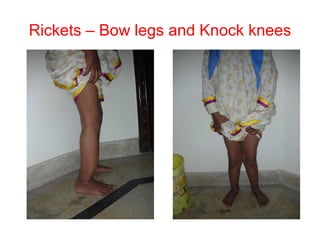


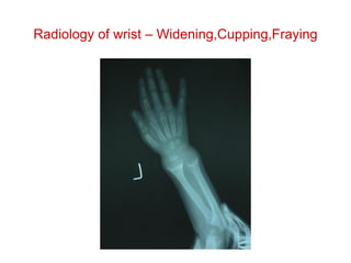


This document discusses a case of a 2-year old child who is unable to stand or walk. On examination, the child has signs consistent with rickets including an open fontanel, wide wrists, and prominent costochondral junctions. The diagnosis is nutritional rickets due to vitamin D deficiency. Treatment involves high dose vitamin D supplementation to correct the deficiency and allow for proper bone mineralization and healing of rickets. Unresponsive cases may be due to other underlying causes. Prevention involves routine vitamin D supplementation of children.


