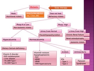
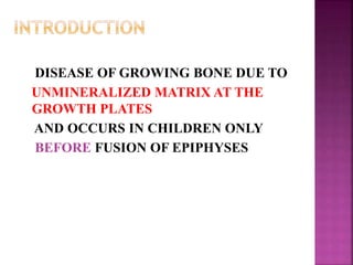
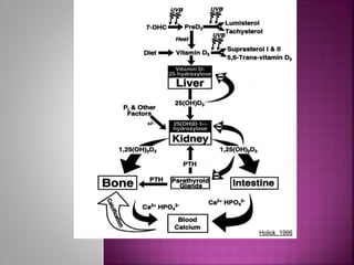
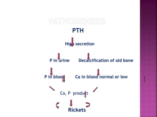
DR SREEKRISHNA R
PG RESIDENT IN PAEDIATRICS
MGM MEDICAL COLLEGE INDORE
DISEASE OF GROWING BONE DUE TO
UNMINERALIZED MATRIX AT THE
GROWTH PLATES
AND OCCURS IN CHILDREN ONLY
BEFORE FUSION OF EPIPHYSES
 CALCIUM AND PHOSPHATE
 Constitutes the crystalline component of bone
Deficiency leads to disease ( Rickets and/or osteomalacia)
 RICKETS:
 Deficient mineralization at growth plate
 OSTEOMALACIA:
 impaired mineralization of the bone matrix.
 Open plates: Occur in Osteomalacia and rickets.
 Closed plates: Happens in osteomalacia only!
What is Rickets ?
Impaired Apoptosis of Terminally Differentiated Chondrocytes in the Growth Plate
Responsible for Clinical & Radiological Signs of Rickets
What is Rickets ?
 Disease of the growing child
 Impaired mineralisation of the growth plate & osteoid
 Low serum phosphate is fundamental to pathogenesis of rickets
Normal Growth Plate Rachitic Growth Plate
Apoptosis of
Hypertrophic
Chondrocytes
caused by
PHOSPHATE ions
HYPOPHOSPHATEMIA
No Apoptosis of
Hypertrophic
Chondrocytes
VITAMIN D DISORDERS(most
common)
CALCIUM DEFICIENCY
PHOSPHORUS DEFICIENCY
RENAL LOSSES
VITAMIN D DISORDERS CALCIUM DEFICIENCY
 Nutritional
 Secondary
Malabsorption
 Vitamin D–dependent rickets
type 1
 Vitamin D–dependent rickets
type 2
 Chronic renal failure
LOW INTAKE
 Diet
 Premature infants (rickets of
prematurity
MALABSORPTION
 Primary disease
 Dietary inhibitors of calcium
absorption
PHOSPHORUS DEFICIENCY RENAL LOSSES
INADEQUATE INTAKE
 Premature infants
 Aluminum-containing
antacids
 X-linked dom hypophosphatemic
 Autosomal dominant
hypophosphatemic
 Autosomal recessive
hypophosphatemic
 Hereditary hypophosphatemic
rickets with hypercalciuria
 Overproduction of phosphatonin
 Fanconi syndrome
 Dent disease
 Distal renal tubular acidosis
GENERAL
- Failure to thrive
- Listlessness
- Protruding abdomen
- Muscle weakness (especially proximal)
- Fractures
- Craniotabes
- Frontal bossing
- Delayed fontanel closure
- Delayed dentition; caries
- Craniosynostosis
CHEST
- Rachitic rosary
- Harrison groove
- Respiratory infections and atelectasis
 Tetany
 Seizures
 Stridor due to laryngeal spasm
 Frontal bossing Rachitic rosary
Knock knee deformity
(genu valgum)
Bowleg deformity
(genu varum)
A teenage male with rickets.
Note deformities of legs (bow legs)
and compromised height.
 Fraying
 Cupping
 Widening of the distal end of the metaphysis
 Rachitic rosary
 Coarse trabeculation of the diaphysis
 Generalized rarefaction
 MOST COMMON CAUSE OF RICKETS
 PEOPLE AT RISK
 Children age 6 to 24 months old
 Dark skinned people
 Premature babies
 Exclusively breast-fed babies
 Source: -Fish, liver and oil,
- Human milk (30-40 IU/L)
- Exposure to sun light
 Vitamin D requirement:
< 1 yr- 400IU/day
>1 yr- 600IU/day
PTH
High secretion
P in urine Decalcification of old bone
P in blood Ca in blood normal or low
Ca, P product
Rickets
LABORATORY FINDINGS
 Hypocalcemia or nornmal calcium
 Hypophosphatemia
“ 25-D levels low”
 Variation in 1,25-D levels (low, normal, or high)
 1,25-D is only low when there is severe vitamin D
deficiency.
 Metabolic acidosis secondary to PTH-induced renal
bicarbonate wasting.
 Generalized aminoaciduria.
 HISTORY OF POOR VITAMIN D INTAKE.
 RISK FACTORS FOR DECREASED CUTANEOUS SYNTHESIS
 RADIOGRAPHIC CHANGES CONSISTENT WITH RICKETS
 TYPICAL LABORATORY FINDINGS.
 A NORMAL PTH LEVEL ALMOST NEVER
OCCURS WITH VITAMIN D DEFICIENCY AND
SUGGESTS A PRIMARY PHOSPHATE DISORDER.
TREATMENT
 2 strategies
 STOSS THERAPY,(300,000-600,000 IU of vitamin D
are administered orally or intramuscularly as 2-4
doses over 1 day)
 DAILY THERAPY
 2,000-5,000 IU/day over 4-6 wk
 EITHER STRATEGY SHOULD BE FOLLOWED BY DAILY VITAMIN D
INTAKE OF 400 IU/DAY IF <1 YR OLD OR 600 IU/DAY IF >1 YR
 ENSURE THAT CHILDREN RECEIVE ADEQUATE DIETARY CALCIUM
AND PHOSPHORUS
Rare occurs in severe maternal vitamin D
deficiency during pregnancy
Risk factor in mother
 POOR DIETARY INTAKE OF VITAMIN D.
 LACK OF ADEQUATE SUN EXPOSURE.
 CLOSELY SPACED PREGNANCIES.
 CLINICAL FEATURES
 symptomatic hypocalcemia, tetany
 intrauterine growth retardation
 decreased bone ossification
 classic rachitic changes.
 TREATMENT
 vitamin D supplementation
 adequate intake of calcium and phosphorus
 PREVENTION
 Use of prenatal vitamins containing vitamin D
 ETIOLOGY
 INADEQUATE ABSORPTION
 DECREASED HYDROXYLATION IN THE LIVER
 INCREASED DEGRADATION
 Occurs Secondary to liver and GI diseases
 CHOLESTATIC LIVER DISEASE,
 DEFECTS IN BILE ACID METABOLISM
 CYSTIC FIBROSIS
 PANCREATIC DYSFUNCTION,
 CELIAC DISEASE
 CROHN DISEASE.
 INTESTINAL LYMPHANGIECTASIA AND AFTER INTESTINAL RESECTION.
=Degradation of vitamin D
 Phenobarbitone and phenytoin cytochrome P450
inducers
TREATMENT
 High doses of vitamin D. 50mcg/day or 5-
7mcg/kg/day.
 Alternatively, may be treated with 1,25-D
 Stoss therapy
 Is an autosomal recessive disorder
 Etiology
 Due to mutations in the gene encoding renal 1α-
hydroxylase
 Prevents conversion of 25-D into 1,25-D
 Presentation
 during the 1st 2 yr of life
 classic features of rickets
 symptomatic hypocalcemia
 LAB FINDINGS
 Normal levels of 25-D
 low levels of 1,25-D
 high PTH
 low serum phosphorus levels
 Treatment
 long-term 1,25-D (calcitriol) with 0.25-2 g/day.
 ensure adequate intake of calcium.
 Monitor periodic urinary calcium excretion,
(<4 mg/kg/day).
 Excessive dosing can cause hypercalciuria &
nephrocalcinosis.
 Is an autosomal recessive disorder
 Etiology
 mutations in the gene encoding the vitamin D
receptor
 prevents a normal physiologic response to 1,25-D
 Most cases present during infancy.
 50-70% of children have alopecia.
 less commonly Epidermal cysts is seen
 LAB FINDINGS
 1,25-D are extremely elevated
 TREATMENT
 HIGH DOSES OF VITAMIN D2, 25-D OR 1,25-D
(2 G/DAY- AS HIGH AS 50-60 G/DAY.)
 CALCIUM 1,000-3,000 MG/DAY
 3-6 MO TRIAL OF HIGH-DOSE VITAMIN D AND ORAL
CALCIUM
 INTRAVENOUS CALCIUM WHO DO NOT RESPOND TO ORAL.
 There is decreased activity of 1α-hydroxylase in
the kidney.
 leading to diminished production of 1,25-d
 patients have “hyperphosphatemia” due to
decreased renal excretion.
 TREATMENT
 therapy with calcitriol
 leads adequate absorption of calcium
 directly suppresses the parathyroid gland.
 ETIOLOGY
 Rickets occurs secondary to inadequate dietary
calcium <200 mg/day.
 Children who receive parentral nutrition
without adequate calcium
 Early weaning from milk and milk produts
 Malabsorption of calcium can occur in
 CELIAC DISEASE,
 INTESTINAL ABETALIPOPROTEINEMIA,
 SMALL BOWEL RESECTION
Diagnosis
 Lab findings include increased levels of alkaline
phosphatase, PTH, and 1,25-D
 Calcium levels may be normal or low
 Decreased urinary excretion of calcium
 Low phosphorus levels due to renal wasting of
phosphate

 In coexisting nutritional vitamin D deficiency, have
low 25-D levels
 provide adequate calcium, dietary supplement
(doses of 700 [1-3 yr age]m/k/d
1000 (4-8)
1300(>8)
 vitamin d supplementation
 discouraging early cessation of breast-feeding.
 increasing dietary sources of calcium.
 RICKETS IN VERY LOW BIRTHWEIGHT INFANTS
 PATHOGENESIS
 80% TRANSFER OF CALCIUM AND PHOSPHORUS
OCCURS DURING THE 3RD TRIMESTER.
 PREMATURE BIRTH INTERRUPTS THIS PROCESS.
 MOST CASES OF RICKETS OCCUR IN INFANTS WITH A
BIRTHWEIGHT <1,000G
RISK FACTORS
 LOW BIRTHWEIGHT & YOUNGER GESTATIONAL AGE.
 UNSUPPLEMENTED BREAST MILK AND STANDARD INFANT
FORMULA DO NOT CONTAIN ENOUGH CALCIUM AND
PHOSPHORUS
 CHOLESTATIC JAUNDICE IN PRETERM
 PROLONGED USE OF PARENTERAL NUTRITION
 MEDICATIONS SUCH AS DIURETICS AND CORTICOSTEROIDS
 OCCURS 1-4 MO AFTER BIRTH
 INFANTS CAN HAVE NONTRAUMATIC FRACTURES OF
LEGS, ARMS, AND RIBS.
 RACHITIC RESPIRATORY DISTRESS USUALLY
DEVELOPS >5 WK AFTER BIRTH
 FRONTAL BOSSING, RACHITIC ROSARY,
CRANIOTABES, AND WIDENED WRISTS AND ANKLES.
 LONG TERM EFFECT IS ENAMEL HYPOPLASIA
 Serum phosphorus level is low
 Normal levels of 25-D
 1,25-D are high
 Hypercalciuria indicates that phosphorus is
the limiting nutrient for bone mineralization
 Alkaline phosphatase levels are often
elevated
 WEEKLY MEASUREMENTS OF CALCIUM,
PHOSPHORUS, AND ALKALINE PHOSPHATASE.
 X-RAY FOR RICKETS AT 6-8 WK OF AGE
PREVENTION
 ADEQUATE AMOUNTS OF CALCIUM, PHOSPHORUS, AND
VITAMIN D
 EARLY TRANSITION TO ENTERAL FEEDINGS
 HUMAN MILK FORTIFIED WITH CALCIUM AND
PHOSPHORUS OR PRETERM INFANT FORMULA, WITH
HIGH CONCENTRATIONS
PHOSPHORUS DEFICIENCY RENAL LOSSES
INADEQUATE INTAKE
 Premature infants
 Aluminum-containing
antacids
 X-linked dom hypophosphatemic
 Autosomal dominant
hypophosphatemic
 Autosomal recessive
hypophosphatemic
 Hereditary hypophosphatemic
rickets with hypercalciuria
 Overproduction of phosphatonin
 Fanconi syndrome
 Dent disease
 Distal renal tubular acidosis
PHOSPHATONIN
 HUMORAL MEDIATOR THAT DECREASES RENAL
TUBULAR REABSORPTION OF PHOSPHATE
 DECREASES THE ACTIVITY OF RENAL 1Α-
HYDROXYLASE
 INCREASED LEVELS OF PHOSPHATONIN CAUSE
MANY OF THE PHOSPHATE-WASTING DISEASES
X LINKED DOM
HYPOPHOSH
RICKETS
AUTSO
DOM
HYPOPH
OS
RICKETS
AUTO
RECESSI
HYPOPH
OS
RICKETS
HEREDI HYPOPHOS RICK
WITH HYPER CALCIURIA
MOST COMMON GENET
DISORDER CAUSING
HYPOPHOSPHA
LESS
COMMON
RARE RARE AUTO DOM
PHEX GENE MUTATION FG 23 GENE
MUTA
MUTA
DENTIN MATRIX
PROTEIN
MUTATION IN SOD –PHOS CO
TRANSPORTER IN PROX TUBULE
PREDOM LOWER EXTREMITY
INVOLVEMENT
MAY BE SHORT STATURE ONLY
RACHITIC LEG ABNORM
BONE PAIN,MUSLE WEAKNESS
DISPROPOR DECREASE IN LENGTH OF
LOWER EXTRE
FAMILY HISTORY OF RENAL STONE
WITH HYPERCALCIURIA
HYPO PHOSP
INCREASED ALP
LOW TO NOR 1 25
NOR PTH
SIMILAR TO
XLP
SIMILAR TO
XLP
HYPO PHOS
INCRE ALP
INCREASED 1,25 D
LOW PTH
PHOSP (1-3 G/D) DIV DOSES SIMILAR IN SIMILAR IN XLP PHOSP ()1-2.5 G) DIVIDED DOSES
 Tumor-induced osteomalacia
 McCune-Albright syndrome.
 Epidermal nevus syndrome.
 Neurofibromatosis
 Above all produses phospate deficency can
lead to rickets. But more common in adults
 IT IS SECONDARY TO GENERALIZED DYSFUNCTION OF
THE PROXIMAL RENAL TUBULE.
 THERE IS LOSS OF PHOSPHATE, AMINO ACIDS,
BICARBONATE, GLUCOSE, URATE.
 THERE IS HYPOPHOSPHATEMIA DUE TO PHOSPHATE
LOSSES, LEADS TO RICKETS
 PROXIMAL RENAL TUBULAR ACIDOSIS DUE TO
BICARBONATE LOSSES LEADS TO BONE DISSOLUTION
 FTT IS A CONSEQUENCE OF BOTH RICKETS AND RTA.
o USUALLY MANIFESTS WITH FAILURE TO THRIVE
METABOLIC ACIDOSIS WITH AN INABILITY TO ACIDIFY
URINE
HYPERCALCIURIA AND NEPHROCALCINOSIS ARE TYPICALLY
PRESENT..
RICKETS IS VARIABLE, AND IT RESPONDS TO ALKALI
THERAPY
DISORDER Ca Pi PTH 25-
OHD
1,25-
(OH)2D
ALK
PHOS
URINE
Ca
URINE
Pi
Vitamin D
deficiency
N,
↓
↓ ↑ ↓ ↓, N, ↑ ↑ ↓ ↑
VDDR, type 1 N,
↓
↓ ↑ N ↓ ↑ ↓ ↑
VDDR, type 2 N,
↓
↓ ↑ N ↑↑ ↑ ↓ ↑
Chronic Renal
Failure
N,
↓
↑ ↑ N ↓ ↑ N, ↓ ↓
Dietary Pi
deficiency
N ↓ N, ↓ N ↑ ↑ ↑ ↓
XLH N ↓ N N RD ↑ ↓ ↑
ADHR N ↓ N N RD ↑ ↓ ↑
HHRH N ↓ N, ↓ N RD ↑ ↑ ↑
Tumor-induced
rickets
N ↓ N N RD ↑ ↓ ↑
Fanconi
syndrome
N ↓ N N RD or ↑ ↑ ↓ or ↑ ↑
Dietary Ca
deficiency
N,
↓
↓ ↑ N ↑ ↑ ↓ ↑
 DIET HISTORY- VIT D DEF
 CUTANEOUS SUN EXPOSURE:CULTURE,CLOTHING
 MATERNAL RISK FACTOR FOR VIT D
 MALABSORPTION:GI SYMPT, LIVER DISEASE
 CHILD ON ANTICONVULSANT,AL ANTACID
 RENAL DISEASE:CRF,POLYURIA-FANCONI
 FAMILY HISTORY OF SHORT STATURE,BONE
DISEASE,UNEXPLAINED DEATH OF SIBLING(CYSTINOSIS,
FANCONI)
 ALOPECIA-VIT D DEPENDENT RICKETS TYPE 2
Rickets
Heals Does not heal
Nutritional rickets Refractory rickets
s.Urea,Creat-High
Azotemic rickets
sUrea,Creat-Normal
PseudoHypopathyroidism
Acidosis
RTA
- Type 1
- Type 2
VitaminD resistant
Hypophosphatemic
- XLH
- ADHR
- ARHR
- HHR Hypercalciuria
Vitamin D disorder
-VitD malabsorption
- Liver disease
- Vit.DDR Type I
- Vit.DDR Type II
Stoss therapy
Xray,Ca,Po4,Alk.P,U
rea, Creat,ABG
Phosp N or Low Phosp- High
Chronic Renal Failure
Renal osteodystrophy
Non Azotemic rickets
Hypocalcemia Normocalcemia
Dietary Calcium deficiency
Aminoaciduria
Fanconi
Syndrome
 MANY CASES OF RICKETS ARE NUTRITIONAL AND RESPOND TO VITAMIN D
THERAPY
 .
 A SINGLE DOSE OF 6 LAKH IU INTRAMUSCULAR (IM) IS THE MOST PRACTICAL WAY
TO TREAT NUTRITIONAL RICKETS ALONG WITH CALCIUM.
 IN CASE OF NONRESPONSE, THE RENAL CAUSES SHOULD BE CONSIDERED.
 A NORMAL SERUM CREATININE EXCLUDES RENAL OSTEODYSTROPHY.
 THE PRESENCE OF ACIDOSIS POINTS TOWARDS RTA.
 HYPERCALCIURIA OCCURS DUE TO BONE RESORPTION AS A RESULT OF CHRONIC
ACIDOSIS AND DECREASED RENAL TUBULAR REABSORPTION OF CALCIUM.
 FURTHER DIFFERENTIATION BETWEEN TYPES 1 AND 2 IS BY URINE PH.
 THE ABSENCE OF ACIDOSIS INDICATES EITHER HYPOPHOSPHATEMIC RICKETS OR
VDDR.
 HYPOPHOSPHATEMIC RICKETS SHOWS RENAL PHOSPHATE WASTING.
 VDDR CAN BE IDENTIFIED BY MEASURING SERUM VITAMIN D.
References
 Nelson textbook of pediatrics
 Bagga textbook pedia Nephrology
 Sperling pediatrics Endocrinology.
Rickets a brief outlook

Rickets a brief outlook

  • 1.
    DR SREEKRISHNA R PGRESIDENT IN PAEDIATRICS MGM MEDICAL COLLEGE INDORE
  • 2.
    DISEASE OF GROWINGBONE DUE TO UNMINERALIZED MATRIX AT THE GROWTH PLATES AND OCCURS IN CHILDREN ONLY BEFORE FUSION OF EPIPHYSES
  • 3.
     CALCIUM ANDPHOSPHATE  Constitutes the crystalline component of bone Deficiency leads to disease ( Rickets and/or osteomalacia)  RICKETS:  Deficient mineralization at growth plate  OSTEOMALACIA:  impaired mineralization of the bone matrix.  Open plates: Occur in Osteomalacia and rickets.  Closed plates: Happens in osteomalacia only!
  • 4.
    What is Rickets? Impaired Apoptosis of Terminally Differentiated Chondrocytes in the Growth Plate Responsible for Clinical & Radiological Signs of Rickets
  • 5.
    What is Rickets?  Disease of the growing child  Impaired mineralisation of the growth plate & osteoid  Low serum phosphate is fundamental to pathogenesis of rickets Normal Growth Plate Rachitic Growth Plate Apoptosis of Hypertrophic Chondrocytes caused by PHOSPHATE ions HYPOPHOSPHATEMIA No Apoptosis of Hypertrophic Chondrocytes
  • 6.
    VITAMIN D DISORDERS(most common) CALCIUMDEFICIENCY PHOSPHORUS DEFICIENCY RENAL LOSSES
  • 7.
    VITAMIN D DISORDERSCALCIUM DEFICIENCY  Nutritional  Secondary Malabsorption  Vitamin D–dependent rickets type 1  Vitamin D–dependent rickets type 2  Chronic renal failure LOW INTAKE  Diet  Premature infants (rickets of prematurity MALABSORPTION  Primary disease  Dietary inhibitors of calcium absorption
  • 8.
    PHOSPHORUS DEFICIENCY RENALLOSSES INADEQUATE INTAKE  Premature infants  Aluminum-containing antacids  X-linked dom hypophosphatemic  Autosomal dominant hypophosphatemic  Autosomal recessive hypophosphatemic  Hereditary hypophosphatemic rickets with hypercalciuria  Overproduction of phosphatonin  Fanconi syndrome  Dent disease  Distal renal tubular acidosis
  • 9.
    GENERAL - Failure tothrive - Listlessness - Protruding abdomen - Muscle weakness (especially proximal) - Fractures
  • 10.
    - Craniotabes - Frontalbossing - Delayed fontanel closure - Delayed dentition; caries - Craniosynostosis CHEST - Rachitic rosary - Harrison groove - Respiratory infections and atelectasis
  • 11.
     Tetany  Seizures Stridor due to laryngeal spasm
  • 13.
     Frontal bossingRachitic rosary
  • 14.
    Knock knee deformity (genuvalgum) Bowleg deformity (genu varum)
  • 15.
    A teenage malewith rickets. Note deformities of legs (bow legs) and compromised height.
  • 16.
     Fraying  Cupping Widening of the distal end of the metaphysis  Rachitic rosary  Coarse trabeculation of the diaphysis  Generalized rarefaction
  • 19.
     MOST COMMONCAUSE OF RICKETS  PEOPLE AT RISK  Children age 6 to 24 months old  Dark skinned people  Premature babies  Exclusively breast-fed babies
  • 21.
     Source: -Fish,liver and oil, - Human milk (30-40 IU/L) - Exposure to sun light  Vitamin D requirement: < 1 yr- 400IU/day >1 yr- 600IU/day
  • 23.
    PTH High secretion P inurine Decalcification of old bone P in blood Ca in blood normal or low Ca, P product Rickets
  • 24.
    LABORATORY FINDINGS  Hypocalcemiaor nornmal calcium  Hypophosphatemia “ 25-D levels low”  Variation in 1,25-D levels (low, normal, or high)  1,25-D is only low when there is severe vitamin D deficiency.  Metabolic acidosis secondary to PTH-induced renal bicarbonate wasting.  Generalized aminoaciduria.
  • 25.
     HISTORY OFPOOR VITAMIN D INTAKE.  RISK FACTORS FOR DECREASED CUTANEOUS SYNTHESIS  RADIOGRAPHIC CHANGES CONSISTENT WITH RICKETS  TYPICAL LABORATORY FINDINGS.  A NORMAL PTH LEVEL ALMOST NEVER OCCURS WITH VITAMIN D DEFICIENCY AND SUGGESTS A PRIMARY PHOSPHATE DISORDER.
  • 26.
    TREATMENT  2 strategies STOSS THERAPY,(300,000-600,000 IU of vitamin D are administered orally or intramuscularly as 2-4 doses over 1 day)  DAILY THERAPY  2,000-5,000 IU/day over 4-6 wk  EITHER STRATEGY SHOULD BE FOLLOWED BY DAILY VITAMIN D INTAKE OF 400 IU/DAY IF <1 YR OLD OR 600 IU/DAY IF >1 YR  ENSURE THAT CHILDREN RECEIVE ADEQUATE DIETARY CALCIUM AND PHOSPHORUS
  • 27.
    Rare occurs insevere maternal vitamin D deficiency during pregnancy Risk factor in mother  POOR DIETARY INTAKE OF VITAMIN D.  LACK OF ADEQUATE SUN EXPOSURE.  CLOSELY SPACED PREGNANCIES.
  • 28.
     CLINICAL FEATURES symptomatic hypocalcemia, tetany  intrauterine growth retardation  decreased bone ossification  classic rachitic changes.  TREATMENT  vitamin D supplementation  adequate intake of calcium and phosphorus  PREVENTION  Use of prenatal vitamins containing vitamin D
  • 29.
     ETIOLOGY  INADEQUATEABSORPTION  DECREASED HYDROXYLATION IN THE LIVER  INCREASED DEGRADATION  Occurs Secondary to liver and GI diseases  CHOLESTATIC LIVER DISEASE,  DEFECTS IN BILE ACID METABOLISM  CYSTIC FIBROSIS  PANCREATIC DYSFUNCTION,  CELIAC DISEASE  CROHN DISEASE.  INTESTINAL LYMPHANGIECTASIA AND AFTER INTESTINAL RESECTION.
  • 30.
    =Degradation of vitaminD  Phenobarbitone and phenytoin cytochrome P450 inducers TREATMENT  High doses of vitamin D. 50mcg/day or 5- 7mcg/kg/day.  Alternatively, may be treated with 1,25-D  Stoss therapy
  • 31.
     Is anautosomal recessive disorder  Etiology  Due to mutations in the gene encoding renal 1α- hydroxylase  Prevents conversion of 25-D into 1,25-D  Presentation  during the 1st 2 yr of life  classic features of rickets  symptomatic hypocalcemia
  • 32.
     LAB FINDINGS Normal levels of 25-D  low levels of 1,25-D  high PTH  low serum phosphorus levels  Treatment  long-term 1,25-D (calcitriol) with 0.25-2 g/day.  ensure adequate intake of calcium.  Monitor periodic urinary calcium excretion, (<4 mg/kg/day).  Excessive dosing can cause hypercalciuria & nephrocalcinosis.
  • 33.
     Is anautosomal recessive disorder  Etiology  mutations in the gene encoding the vitamin D receptor  prevents a normal physiologic response to 1,25-D  Most cases present during infancy.  50-70% of children have alopecia.  less commonly Epidermal cysts is seen
  • 34.
     LAB FINDINGS 1,25-D are extremely elevated  TREATMENT  HIGH DOSES OF VITAMIN D2, 25-D OR 1,25-D (2 G/DAY- AS HIGH AS 50-60 G/DAY.)  CALCIUM 1,000-3,000 MG/DAY  3-6 MO TRIAL OF HIGH-DOSE VITAMIN D AND ORAL CALCIUM  INTRAVENOUS CALCIUM WHO DO NOT RESPOND TO ORAL.
  • 35.
     There isdecreased activity of 1α-hydroxylase in the kidney.  leading to diminished production of 1,25-d  patients have “hyperphosphatemia” due to decreased renal excretion.  TREATMENT  therapy with calcitriol  leads adequate absorption of calcium  directly suppresses the parathyroid gland.
  • 36.
     ETIOLOGY  Ricketsoccurs secondary to inadequate dietary calcium <200 mg/day.  Children who receive parentral nutrition without adequate calcium  Early weaning from milk and milk produts  Malabsorption of calcium can occur in  CELIAC DISEASE,  INTESTINAL ABETALIPOPROTEINEMIA,  SMALL BOWEL RESECTION
  • 37.
    Diagnosis  Lab findingsinclude increased levels of alkaline phosphatase, PTH, and 1,25-D  Calcium levels may be normal or low  Decreased urinary excretion of calcium  Low phosphorus levels due to renal wasting of phosphate   In coexisting nutritional vitamin D deficiency, have low 25-D levels
  • 38.
     provide adequatecalcium, dietary supplement (doses of 700 [1-3 yr age]m/k/d 1000 (4-8) 1300(>8)  vitamin d supplementation  discouraging early cessation of breast-feeding.  increasing dietary sources of calcium.
  • 39.
     RICKETS INVERY LOW BIRTHWEIGHT INFANTS  PATHOGENESIS  80% TRANSFER OF CALCIUM AND PHOSPHORUS OCCURS DURING THE 3RD TRIMESTER.  PREMATURE BIRTH INTERRUPTS THIS PROCESS.  MOST CASES OF RICKETS OCCUR IN INFANTS WITH A BIRTHWEIGHT <1,000G
  • 40.
    RISK FACTORS  LOWBIRTHWEIGHT & YOUNGER GESTATIONAL AGE.  UNSUPPLEMENTED BREAST MILK AND STANDARD INFANT FORMULA DO NOT CONTAIN ENOUGH CALCIUM AND PHOSPHORUS  CHOLESTATIC JAUNDICE IN PRETERM  PROLONGED USE OF PARENTERAL NUTRITION  MEDICATIONS SUCH AS DIURETICS AND CORTICOSTEROIDS
  • 41.
     OCCURS 1-4MO AFTER BIRTH  INFANTS CAN HAVE NONTRAUMATIC FRACTURES OF LEGS, ARMS, AND RIBS.  RACHITIC RESPIRATORY DISTRESS USUALLY DEVELOPS >5 WK AFTER BIRTH  FRONTAL BOSSING, RACHITIC ROSARY, CRANIOTABES, AND WIDENED WRISTS AND ANKLES.  LONG TERM EFFECT IS ENAMEL HYPOPLASIA
  • 42.
     Serum phosphoruslevel is low  Normal levels of 25-D  1,25-D are high  Hypercalciuria indicates that phosphorus is the limiting nutrient for bone mineralization  Alkaline phosphatase levels are often elevated
  • 43.
     WEEKLY MEASUREMENTSOF CALCIUM, PHOSPHORUS, AND ALKALINE PHOSPHATASE.  X-RAY FOR RICKETS AT 6-8 WK OF AGE PREVENTION  ADEQUATE AMOUNTS OF CALCIUM, PHOSPHORUS, AND VITAMIN D  EARLY TRANSITION TO ENTERAL FEEDINGS  HUMAN MILK FORTIFIED WITH CALCIUM AND PHOSPHORUS OR PRETERM INFANT FORMULA, WITH HIGH CONCENTRATIONS
  • 44.
    PHOSPHORUS DEFICIENCY RENALLOSSES INADEQUATE INTAKE  Premature infants  Aluminum-containing antacids  X-linked dom hypophosphatemic  Autosomal dominant hypophosphatemic  Autosomal recessive hypophosphatemic  Hereditary hypophosphatemic rickets with hypercalciuria  Overproduction of phosphatonin  Fanconi syndrome  Dent disease  Distal renal tubular acidosis
  • 45.
    PHOSPHATONIN  HUMORAL MEDIATORTHAT DECREASES RENAL TUBULAR REABSORPTION OF PHOSPHATE  DECREASES THE ACTIVITY OF RENAL 1Α- HYDROXYLASE  INCREASED LEVELS OF PHOSPHATONIN CAUSE MANY OF THE PHOSPHATE-WASTING DISEASES
  • 47.
    X LINKED DOM HYPOPHOSH RICKETS AUTSO DOM HYPOPH OS RICKETS AUTO RECESSI HYPOPH OS RICKETS HEREDIHYPOPHOS RICK WITH HYPER CALCIURIA MOST COMMON GENET DISORDER CAUSING HYPOPHOSPHA LESS COMMON RARE RARE AUTO DOM PHEX GENE MUTATION FG 23 GENE MUTA MUTA DENTIN MATRIX PROTEIN MUTATION IN SOD –PHOS CO TRANSPORTER IN PROX TUBULE PREDOM LOWER EXTREMITY INVOLVEMENT MAY BE SHORT STATURE ONLY RACHITIC LEG ABNORM BONE PAIN,MUSLE WEAKNESS DISPROPOR DECREASE IN LENGTH OF LOWER EXTRE FAMILY HISTORY OF RENAL STONE WITH HYPERCALCIURIA HYPO PHOSP INCREASED ALP LOW TO NOR 1 25 NOR PTH SIMILAR TO XLP SIMILAR TO XLP HYPO PHOS INCRE ALP INCREASED 1,25 D LOW PTH PHOSP (1-3 G/D) DIV DOSES SIMILAR IN SIMILAR IN XLP PHOSP ()1-2.5 G) DIVIDED DOSES
  • 48.
     Tumor-induced osteomalacia McCune-Albright syndrome.  Epidermal nevus syndrome.  Neurofibromatosis  Above all produses phospate deficency can lead to rickets. But more common in adults
  • 49.
     IT ISSECONDARY TO GENERALIZED DYSFUNCTION OF THE PROXIMAL RENAL TUBULE.  THERE IS LOSS OF PHOSPHATE, AMINO ACIDS, BICARBONATE, GLUCOSE, URATE.  THERE IS HYPOPHOSPHATEMIA DUE TO PHOSPHATE LOSSES, LEADS TO RICKETS  PROXIMAL RENAL TUBULAR ACIDOSIS DUE TO BICARBONATE LOSSES LEADS TO BONE DISSOLUTION  FTT IS A CONSEQUENCE OF BOTH RICKETS AND RTA.
  • 50.
    o USUALLY MANIFESTSWITH FAILURE TO THRIVE METABOLIC ACIDOSIS WITH AN INABILITY TO ACIDIFY URINE HYPERCALCIURIA AND NEPHROCALCINOSIS ARE TYPICALLY PRESENT.. RICKETS IS VARIABLE, AND IT RESPONDS TO ALKALI THERAPY
  • 51.
    DISORDER Ca PiPTH 25- OHD 1,25- (OH)2D ALK PHOS URINE Ca URINE Pi Vitamin D deficiency N, ↓ ↓ ↑ ↓ ↓, N, ↑ ↑ ↓ ↑ VDDR, type 1 N, ↓ ↓ ↑ N ↓ ↑ ↓ ↑ VDDR, type 2 N, ↓ ↓ ↑ N ↑↑ ↑ ↓ ↑ Chronic Renal Failure N, ↓ ↑ ↑ N ↓ ↑ N, ↓ ↓ Dietary Pi deficiency N ↓ N, ↓ N ↑ ↑ ↑ ↓ XLH N ↓ N N RD ↑ ↓ ↑ ADHR N ↓ N N RD ↑ ↓ ↑ HHRH N ↓ N, ↓ N RD ↑ ↑ ↑ Tumor-induced rickets N ↓ N N RD ↑ ↓ ↑ Fanconi syndrome N ↓ N N RD or ↑ ↑ ↓ or ↑ ↑ Dietary Ca deficiency N, ↓ ↓ ↑ N ↑ ↑ ↓ ↑
  • 52.
     DIET HISTORY-VIT D DEF  CUTANEOUS SUN EXPOSURE:CULTURE,CLOTHING  MATERNAL RISK FACTOR FOR VIT D  MALABSORPTION:GI SYMPT, LIVER DISEASE  CHILD ON ANTICONVULSANT,AL ANTACID  RENAL DISEASE:CRF,POLYURIA-FANCONI  FAMILY HISTORY OF SHORT STATURE,BONE DISEASE,UNEXPLAINED DEATH OF SIBLING(CYSTINOSIS, FANCONI)  ALOPECIA-VIT D DEPENDENT RICKETS TYPE 2
  • 53.
    Rickets Heals Does notheal Nutritional rickets Refractory rickets s.Urea,Creat-High Azotemic rickets sUrea,Creat-Normal PseudoHypopathyroidism Acidosis RTA - Type 1 - Type 2 VitaminD resistant Hypophosphatemic - XLH - ADHR - ARHR - HHR Hypercalciuria Vitamin D disorder -VitD malabsorption - Liver disease - Vit.DDR Type I - Vit.DDR Type II Stoss therapy Xray,Ca,Po4,Alk.P,U rea, Creat,ABG Phosp N or Low Phosp- High Chronic Renal Failure Renal osteodystrophy Non Azotemic rickets Hypocalcemia Normocalcemia Dietary Calcium deficiency Aminoaciduria Fanconi Syndrome
  • 54.
     MANY CASESOF RICKETS ARE NUTRITIONAL AND RESPOND TO VITAMIN D THERAPY  .  A SINGLE DOSE OF 6 LAKH IU INTRAMUSCULAR (IM) IS THE MOST PRACTICAL WAY TO TREAT NUTRITIONAL RICKETS ALONG WITH CALCIUM.  IN CASE OF NONRESPONSE, THE RENAL CAUSES SHOULD BE CONSIDERED.  A NORMAL SERUM CREATININE EXCLUDES RENAL OSTEODYSTROPHY.  THE PRESENCE OF ACIDOSIS POINTS TOWARDS RTA.  HYPERCALCIURIA OCCURS DUE TO BONE RESORPTION AS A RESULT OF CHRONIC ACIDOSIS AND DECREASED RENAL TUBULAR REABSORPTION OF CALCIUM.  FURTHER DIFFERENTIATION BETWEEN TYPES 1 AND 2 IS BY URINE PH.  THE ABSENCE OF ACIDOSIS INDICATES EITHER HYPOPHOSPHATEMIC RICKETS OR VDDR.  HYPOPHOSPHATEMIC RICKETS SHOWS RENAL PHOSPHATE WASTING.  VDDR CAN BE IDENTIFIED BY MEASURING SERUM VITAMIN D.
  • 55.
    References  Nelson textbookof pediatrics  Bagga textbook pedia Nephrology  Sperling pediatrics Endocrinology.