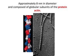
MICRO FILAMENTS


     BURGOS
• Cells are capable of remarkable
  motility. The neural crest cells in a
  vertebrate embryo leave the
  developing nervous system and
  migrate across the entire width of
  the embryo, forming such diverse
  products as the pigment cells.
Approximately 8 nm in diameter
and composed of globular subunits of the protein
                    actin.
Microfilament Assembly and Disassembly

• Before it is incorporated into a filament, an actin
  monomer binds a molecule of ATP.

• Actin is an ATPase, just as tubulin is a GTPase, and
  the role of ATP in actin assembly is similar to that of
  GTP in microtubule assembly.
• As the cell progresses into mitosis, the preprophase
  band is lost and microtubules reappear in the form
  of the mitotic spindle.
Actin assembly in vitro
• Electron micrograph of a short actin filament
  that was labeled with S1 myosin and then
  used to nucleate actin polymerization.

• The addition of actin subunits occurs much
  more rapidly at the barbed (plus) end than at
  the pointed (minus) end of the existing
  filament.
Myosin: The Molecular Motor of Actin
             Filaments
• Fluorescence micrograph showing fine
  processes (neurites) growing out from a
  microscopic fragment of mouse embryonic
  nervous tissue.

• The neurites (stained green) are growing
  outward on a glass coverslip coated with strips
  of laminin (stained red).
MUSCLE CONTRACTILITY

• Skeletal muscles derive their name from the fact
  that most of them are anchored to bones that they
  move.

• They are under voluntary control and can be
  consciously commanded to contract. Skeletal
  muscle cells have a highly unorthodox structure.
Levels of organization of a skeletal
              muscle.
• A single, cylindrically shaped muscle cell is
  typically 10 to 100 m thick, over 100 mm long,
  and contains hundreds of nuclei.

• Because of these properties, a skeletal muscle
  cell is more appropriately called a muscle
  fiber. Muscle fibers have multiple nuclei
  because each fiber is a product of the fusion
  of large numbers of mononucleated
  myoblasts (premuscle cells) in the embryo.
• Skeletal muscle cells may have the most highly
  ordered internal structure of any cell in the body.

• A longitudinal section of a muscle fiber reveals a
  cable made up of hundreds of thinner, cylindrical
  strands, called myofibrils.
• Each myofibril consists of a repeating linear
  array of contractile units, called sarcomeres.

• Each sarcomere in turn exhibits a
  characteristic banding pattern, which gives the
  muscle fiber a striped or striated appearance.
The Composition and Organization of Thick and Thin
                      Filaments

• The thin filaments of a skeletal muscle contain two
  other proteins, tropomyosin and troponin.

• Each thick filament is composed of several hundred
  myosin II molecules together with small amounts of
  other proteins. Like the filaments that form in vitro
  the polarity of the thick filaments of muscle cells is
  reversed at the center of the sarcomere.
• Tropomyosin is an elongated molecule
  (approximately 40 nm long) that fits securely
  into the grooves within the thin filament. Each
  rod-shaped tropomyosin molecule is
  associated with seven actin subunits along the
  thin filament.

• Troponin is a globular protein complex
  composed of three subunits, each having an
  important and distinct role in the overall
  function of the molecule.
The functional anatomy of a muscle fiber.

- Calcium is housed in the elaborate network of
internal membranes that make up the sarcoplasmic
reticulum (SR).When an impulse arrives by means of a
motor neuron, it is carried into the interior of the
fiber A along the membrane of the transverse tubule
to the SR. The calcium gates of the SR open, releasing
calcium into the cytosol. The binding of calcium ions
to troponin molecules of the thin filaments leads to
the events described in the following figure and the
contraction of the fiber.
NONMUSCLE MOTILITY
• The study of nonmuscle motility is more
  challenging because the critical components
  tend to be present in less ordered, more
  labile, transient arrangements. Moreover, they
  are typically restricted to a thin cortex just
  beneath the plasma membrane.
• The cortex is an active region of the cell,
  responsible for such processes as the
  ingestion of extracellular materials, the
  extension of processes during cell movement,
  and the constriction of a single animal cell into
  two cells during cell division.
• All of these processes are dependent on the
  assembly of microfilaments in the cortex.
Actin-Binding Proteins

• The organization and behavior of actin
  filaments inside cells are determined by a
  remarkable variety of actin-binding proteins
  that affect the localized assembly or
  disassembly of the actin filaments, their
  physical properties, and their interactions with
  one another and with cellular organelles.
Nucleating proteins.
• The slowest step in the formation of an actin
  filament is the first step, nucleation, which
  requires that at least two or three actin
  monomers come together in the proper
  orientation to begin formation of the polymer.
Monomer-sequestering proteins.
• Thymosins (e.g., thymosin 4) are proteins that
  bind to actin-ATP monomers (often called G-
  actin) and prevent them from polymerizing.
• Proteins with this activity are described as
  actin monomer-sequestering proteins.
End-blocking (capping) proteins.
• Proteins of this group regulate the length of
  actin filaments by binding to one or the other
  end of the filaments, forming a cap that blocks
  both loss and gain of subunits.
• If the fast-growing, barbed end of a filament is
  capped, depolymerization may proceed at the
  opposite end, resulting in the disassembly of
  the filament.
Monomer-polymerizing proteins.
• Profilin is a small protein that binds to the
  same site on an actin monomer as does
  thymosin. However, rather than inhibiting
  polymerization, profilin promotes the growth
  of actin filaments.
Actin filament-depolymerizing
                proteins.

• Members of the cofilin family of proteins
  (including cofilin, ADF, and depactin) bind to
  actin-ADP subunits present within the body
  and at the pointed end of actin filaments.
Cross-linking proteins.
• Proteins of this group are able to alter the
  three-dimensional organization of a
  population of actin filaments.
• Each of these proteins has two or more actin-
  binding sites and therefore can cross-link two
  or more separate actin filaments.
Filament-severing proteins.
• Proteins of this class have the ability to bind to
  the side of an existing filament and break it in
  two. Severing proteins (e.g., gelsolin) may also
  promote the incorporation of actin monomers
  by creating additional free barbed ends, or
  they may cap the fragments they generate.
Membrane-binding proteins.
• These activities are generally facilitated by
  linking the actin filaments to the plasma
  membrane indirectly, by means of attachment
  to a peripheral membrane protein.
Changes in Cell Shape during Embryonic Development


• Each part of the body has a characteristic shape and
  internal architecture that arises during embryonic
  development: the spinal cord is basically a hollow
  tube, the kidney consists of microscopic tubules,
  each lung is composed of microscopic air spaces,
  and so forth.
• Numerous cellular activities are necessary.
Changes in cell shape are brought about largely
by changes in the orientation of cytoskeletal
elements within the cells. One of the best
examples of this phenomenon is seen in the
early stages of the development of the nervous
system.
Micro filaments

Micro filaments

  • 1.
  • 2.
    • Cells arecapable of remarkable motility. The neural crest cells in a vertebrate embryo leave the developing nervous system and migrate across the entire width of the embryo, forming such diverse products as the pigment cells.
  • 3.
    Approximately 8 nmin diameter and composed of globular subunits of the protein actin.
  • 4.
    Microfilament Assembly andDisassembly • Before it is incorporated into a filament, an actin monomer binds a molecule of ATP. • Actin is an ATPase, just as tubulin is a GTPase, and the role of ATP in actin assembly is similar to that of GTP in microtubule assembly.
  • 5.
    • As thecell progresses into mitosis, the preprophase band is lost and microtubules reappear in the form of the mitotic spindle.
  • 6.
  • 7.
    • Electron micrographof a short actin filament that was labeled with S1 myosin and then used to nucleate actin polymerization. • The addition of actin subunits occurs much more rapidly at the barbed (plus) end than at the pointed (minus) end of the existing filament.
  • 8.
    Myosin: The MolecularMotor of Actin Filaments
  • 9.
    • Fluorescence micrographshowing fine processes (neurites) growing out from a microscopic fragment of mouse embryonic nervous tissue. • The neurites (stained green) are growing outward on a glass coverslip coated with strips of laminin (stained red).
  • 10.
    MUSCLE CONTRACTILITY • Skeletalmuscles derive their name from the fact that most of them are anchored to bones that they move. • They are under voluntary control and can be consciously commanded to contract. Skeletal muscle cells have a highly unorthodox structure.
  • 11.
    Levels of organizationof a skeletal muscle.
  • 12.
    • A single,cylindrically shaped muscle cell is typically 10 to 100 m thick, over 100 mm long, and contains hundreds of nuclei. • Because of these properties, a skeletal muscle cell is more appropriately called a muscle fiber. Muscle fibers have multiple nuclei because each fiber is a product of the fusion of large numbers of mononucleated myoblasts (premuscle cells) in the embryo.
  • 13.
    • Skeletal musclecells may have the most highly ordered internal structure of any cell in the body. • A longitudinal section of a muscle fiber reveals a cable made up of hundreds of thinner, cylindrical strands, called myofibrils.
  • 14.
    • Each myofibrilconsists of a repeating linear array of contractile units, called sarcomeres. • Each sarcomere in turn exhibits a characteristic banding pattern, which gives the muscle fiber a striped or striated appearance.
  • 15.
    The Composition andOrganization of Thick and Thin Filaments • The thin filaments of a skeletal muscle contain two other proteins, tropomyosin and troponin. • Each thick filament is composed of several hundred myosin II molecules together with small amounts of other proteins. Like the filaments that form in vitro the polarity of the thick filaments of muscle cells is reversed at the center of the sarcomere.
  • 16.
    • Tropomyosin isan elongated molecule (approximately 40 nm long) that fits securely into the grooves within the thin filament. Each rod-shaped tropomyosin molecule is associated with seven actin subunits along the thin filament. • Troponin is a globular protein complex composed of three subunits, each having an important and distinct role in the overall function of the molecule.
  • 17.
    The functional anatomyof a muscle fiber. - Calcium is housed in the elaborate network of internal membranes that make up the sarcoplasmic reticulum (SR).When an impulse arrives by means of a motor neuron, it is carried into the interior of the fiber A along the membrane of the transverse tubule to the SR. The calcium gates of the SR open, releasing calcium into the cytosol. The binding of calcium ions to troponin molecules of the thin filaments leads to the events described in the following figure and the contraction of the fiber.
  • 19.
    NONMUSCLE MOTILITY • Thestudy of nonmuscle motility is more challenging because the critical components tend to be present in less ordered, more labile, transient arrangements. Moreover, they are typically restricted to a thin cortex just beneath the plasma membrane.
  • 20.
    • The cortexis an active region of the cell, responsible for such processes as the ingestion of extracellular materials, the extension of processes during cell movement, and the constriction of a single animal cell into two cells during cell division. • All of these processes are dependent on the assembly of microfilaments in the cortex.
  • 21.
    Actin-Binding Proteins • Theorganization and behavior of actin filaments inside cells are determined by a remarkable variety of actin-binding proteins that affect the localized assembly or disassembly of the actin filaments, their physical properties, and their interactions with one another and with cellular organelles.
  • 22.
    Nucleating proteins. • Theslowest step in the formation of an actin filament is the first step, nucleation, which requires that at least two or three actin monomers come together in the proper orientation to begin formation of the polymer.
  • 23.
    Monomer-sequestering proteins. • Thymosins(e.g., thymosin 4) are proteins that bind to actin-ATP monomers (often called G- actin) and prevent them from polymerizing. • Proteins with this activity are described as actin monomer-sequestering proteins.
  • 24.
    End-blocking (capping) proteins. •Proteins of this group regulate the length of actin filaments by binding to one or the other end of the filaments, forming a cap that blocks both loss and gain of subunits. • If the fast-growing, barbed end of a filament is capped, depolymerization may proceed at the opposite end, resulting in the disassembly of the filament.
  • 25.
    Monomer-polymerizing proteins. • Profilinis a small protein that binds to the same site on an actin monomer as does thymosin. However, rather than inhibiting polymerization, profilin promotes the growth of actin filaments.
  • 26.
    Actin filament-depolymerizing proteins. • Members of the cofilin family of proteins (including cofilin, ADF, and depactin) bind to actin-ADP subunits present within the body and at the pointed end of actin filaments.
  • 27.
    Cross-linking proteins. • Proteinsof this group are able to alter the three-dimensional organization of a population of actin filaments. • Each of these proteins has two or more actin- binding sites and therefore can cross-link two or more separate actin filaments.
  • 28.
    Filament-severing proteins. • Proteinsof this class have the ability to bind to the side of an existing filament and break it in two. Severing proteins (e.g., gelsolin) may also promote the incorporation of actin monomers by creating additional free barbed ends, or they may cap the fragments they generate.
  • 29.
    Membrane-binding proteins. • Theseactivities are generally facilitated by linking the actin filaments to the plasma membrane indirectly, by means of attachment to a peripheral membrane protein.
  • 30.
    Changes in CellShape during Embryonic Development • Each part of the body has a characteristic shape and internal architecture that arises during embryonic development: the spinal cord is basically a hollow tube, the kidney consists of microscopic tubules, each lung is composed of microscopic air spaces, and so forth.
  • 31.
    • Numerous cellularactivities are necessary. Changes in cell shape are brought about largely by changes in the orientation of cytoskeletal elements within the cells. One of the best examples of this phenomenon is seen in the early stages of the development of the nervous system.