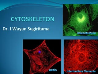
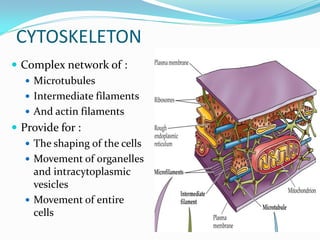
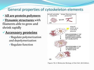
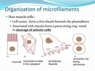
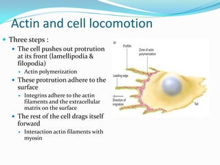
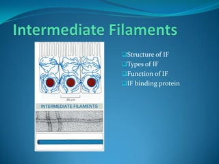
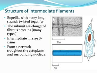
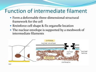
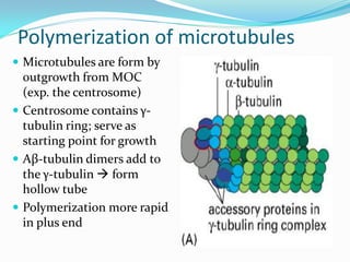
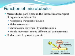
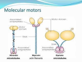
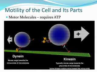
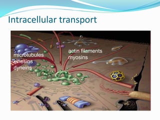
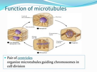
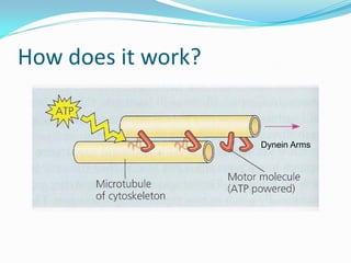
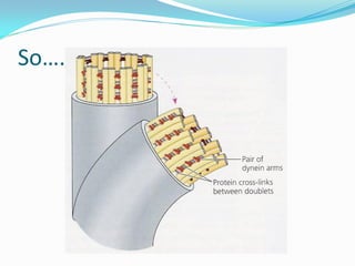
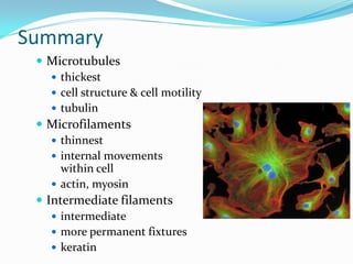
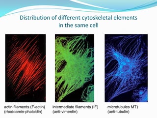
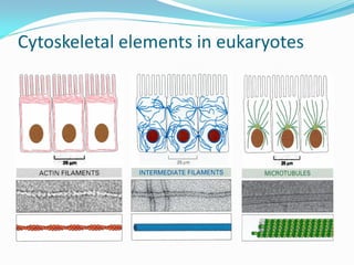
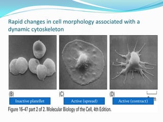
Dr. I Wayan Sugiritama
QUESTIONS:
 How cell maintain their shape ?
 How cell organize its organelles?
 How cell transport vesicles?
 How the segregation of chromosomes into daughter
  cells at mitosis ?
 How epithelial cell can withstand to the mechanical
  stress?
 How spermatozoa can reach the eggs ?
 How leucoyte can move to the extracelluler space ?
Cytoskeleton: the skeleton of a cell


                  =


                  Cells need a (cyto)skeleton to:
                  •create shape                     dynamic!
                  •change shape
                  •allow movement
CYTOSKELETON
 Complex network of :
    Microtubules
    Intermediate filaments
    And actin filaments
 Provide for :
    The shaping of the cells
    Movement of organelles
     and intracytoplasmic
     vesicles
    Movement of entire
     cells
General properties of cytoskeleton elements
All are protein polymers
Dynamic structures with
filaments able to grow and
shrink rapidly
Accessory proteins
    Regulate polymerization
    and depolymerization
    Regulate function
Structure of actin filaments
Polymerization of actin
filaments
Organization of actin filaments
Actin binding protein
Function of actin filaments
Structure of actin filaments
 Composed of two chains of globular
    subunit (G-actin), coiled each other to
    form a filamentous prot. (F-actin)
   Thinnest class of fibers (6 nm thick)
   Has stuctural polarity
   Associated with a large number actin-
    binding protein  variety of organization
    and function
   Depending on isoelectric point :
     α-actin of muscle
     β-actin & γ-actin of non muscle
Actins polymerization
 Actin filaments can grow by
  addition of actin monomer
  at either end
 When filament reach desire
  length, capping proteins
  attach to the plus end and
  terminating polymerization


                                8
Actin monomer binding proteins
 Control pool of
  unpolymerized actin
 Two proteins
   Profilin
     Inhibits addition of
      monomers to pointed
      (slowgrowing) end
   Thymosin β4
       If a filament is capped at both
        ends it is effectively stabilized
Actin binding protein
Actin bundling protein : hold   Cross-linking protein : hold
actin filaments together in     actin filaments in a gel-like
parallel bundle (microvilli)    meshwork (cell cortex)
Actin binding protein
Filament-seve ring protein :
                                    Motor protein
convert actin gel to a more fluid
state (gelsolin)
Organization of microfilaments
Microfilaments can organized in many forms :
 Skeletal muscle : paracrystalline array integrated with
  myosin filaments
Organization of microfilaments
 Non muscle cells :
   Cell cortex : form a thin sheath beneath the plasmallema
   Associated with myosin form a purse string ring result
    in cleavage of mitotic cells


                                                    contractile ring
    microvilli contractile bundles   lamellipodia   during
               in the cytoplasm      filopodia      cell division
Actin and cell locomotion
 Three steps :
    The cell pushes out protrution
     at its front (lamellipodia &
     filopodia)
        Actin polymerization
    These protrution adhere to the
     surface
        Integrins adhere to the actin
         filaments and the extracellular
         matrix on the surface
    The rest of the cell drags itself
     forward
        Interaction actin filaments with
         myosin
Structure of IF
Types of IF
Function of IF
IF binding protein
Structure of Intermediate filaments
• Ropelike with many long
  strands twisted together
• The subunit are elongated
  fibrous proteins (many
  types)
• Intermediate in size 8-
  12nm
• Form a network
  troughout the cytoplasm
  and surrounding nucleus
Polymerization of Subunit structure
•The subunit :
        •N-terminal globular head
        •C-terminal globular tail
        •Central elongated rod
        domain

•The subunit form stable dimer
•Two dimer form tetramer
•Tetramer bind to one another
and-to-end generate ropelike
Types of intermediate filaments
According to protein subunit, Intermediate filaments in the cytoplasm can be
grouped into:
Intermediate filament binding protein
 Link, stabilized and reinforced the intermediate
 filaments into three-dimensional network :
   Fillagrin : binds keratin filaments into bundles
   Synamin & Plectin : binds desmin & vimentin, links
    intermediates filaments to microtubules, actin and
    desmosome
   Plakins : maintenance of contact between keratin and
    hemidesmosomes of epithelial cells
Function of intermediate filament
   Tensile strength cells enable to withstand the
   mechanical stress (streched)


   Provide stuctural support for the cell
Function of intermediate filament
   Form a deformable three-dimensional structural
    framework for the cell
   Rreinforce cell shape & fix organelle location
   The nuclear envelope is supported by a meshwork of
    intermediate filaments
 The structure of
  microtubules
 Assembly of mirotubules
 Microtubule function
 Microtubule association with
  motor protein
 Structure and function of
  cilia and flagella
Structure of Microtubules
  Hollow tube about 25 nm in diameter
  The subunit is heterodimer α and β
   tubulin
  Polarized : having plus end & minus
   end

  Dynamic structure : grow or shrink as
  more tubulin molecules are added or
  removed
Polymerization of microtubules
 Microtubules are form by
  outgrowth from MOC
  (exp. the centrosome)
 Centrosome contains γ-
  tubulin ring; serve as
  starting point for growth
 Αβ-tubulin dimers add to
  the γ-tubulin  form
  hollow tube
 Polymerization more rapid
  in plus end
Function of microtubules
 Microtubules participate in the intracellular transport
 of organelles and vesicles
   Axoplasmic transport of neuron
   Melanin transport
   Chromosome movement by mitotic spindle
   Vesicle movement among different cell compartments
 Under control by motor protein
Molecular motors


 microtubules   actin filaments   microtubules
Motility of the Cell and Its Parts
 Motor Molecules – requires ATP
Intracellular transport

                actin filaments
 microtubules   myosins
 kinesins
 dyneins
Function of microtubules


 Pair of centrioles
  organize microtubules guiding chromosomes in
  cell division
Cilia & Flagella
 Motile processes, with higly
  organized microtubule core
 Core consist of 9 pairs of
  microtubules arround 2 central
  microtubule (axoneme)
 bending of cilia & flagella is
  driven by motor protein
  (Dynein)
 At the base is basal body, that
  control the assembly of the
  axoneme
Cilia
  Cilia = numerous & short (hair-like)
  Oar-like movement
    alternating power & recovery strokes
    generate force perpendicular to cilia’s axis
flagella
   Flagella = 1-2/cell & longer (whip-like)
      move unicellular & small multicellular organisms by
       propelling water past them
      undulatory movement , force generated parallel to
       flagellum’s axis
      cilia sweep mucus & debris from lungs
      flagellum of sperm cells
How does it work?


                    Dynein Arms
So….
Summary
  Microtubules
     thickest
     cell structure & cell motility
     tubulin
  Microfilaments
     thinnest
     internal movements
      within cell
     actin, myosin
  Intermediate filaments
     intermediate
     more permanent fixtures
     keratin
Distribution of different cytoskeletal elements
                         in the same cell


actin filaments (F-actin)   intermediate filaments (IF)   microtubules MT)
(rhodoamin-phaloidin)       (anti-vimentin)               (anti-tubulin)
Cytoskeletal elements in eukaryotes
Rapid changes in cell morphology associated with a
dynamic cytoskeleton


  Inactive platellet   Active (spread)   Active (contract)
Without the cytoskeleton ?


Wounds would never heal !
Muscle would be uselless !
Sperm never reach the egg !
cytosceleton

cytosceleton

  • 1.
    Dr. I WayanSugiritama
  • 2.
    QUESTIONS:  How cellmaintain their shape ?  How cell organize its organelles?  How cell transport vesicles?  How the segregation of chromosomes into daughter cells at mitosis ?  How epithelial cell can withstand to the mechanical stress?  How spermatozoa can reach the eggs ?  How leucoyte can move to the extracelluler space ?
  • 3.
    Cytoskeleton: the skeletonof a cell = Cells need a (cyto)skeleton to: •create shape dynamic! •change shape •allow movement
  • 4.
    CYTOSKELETON  Complex networkof :  Microtubules  Intermediate filaments  And actin filaments  Provide for :  The shaping of the cells  Movement of organelles and intracytoplasmic vesicles  Movement of entire cells
  • 5.
    General properties ofcytoskeleton elements All are protein polymers Dynamic structures with filaments able to grow and shrink rapidly Accessory proteins Regulate polymerization and depolymerization Regulate function
  • 6.
    Structure of actinfilaments Polymerization of actin filaments Organization of actin filaments Actin binding protein Function of actin filaments
  • 7.
    Structure of actinfilaments  Composed of two chains of globular subunit (G-actin), coiled each other to form a filamentous prot. (F-actin)  Thinnest class of fibers (6 nm thick)  Has stuctural polarity  Associated with a large number actin- binding protein  variety of organization and function  Depending on isoelectric point :  α-actin of muscle  β-actin & γ-actin of non muscle
  • 8.
    Actins polymerization  Actinfilaments can grow by addition of actin monomer at either end  When filament reach desire length, capping proteins attach to the plus end and terminating polymerization 8
  • 9.
    Actin monomer bindingproteins  Control pool of unpolymerized actin  Two proteins  Profilin  Inhibits addition of monomers to pointed (slowgrowing) end  Thymosin β4  If a filament is capped at both ends it is effectively stabilized
  • 10.
    Actin binding protein Actinbundling protein : hold Cross-linking protein : hold actin filaments together in actin filaments in a gel-like parallel bundle (microvilli) meshwork (cell cortex)
  • 11.
    Actin binding protein Filament-severing protein : Motor protein convert actin gel to a more fluid state (gelsolin)
  • 12.
    Organization of microfilaments Microfilamentscan organized in many forms :  Skeletal muscle : paracrystalline array integrated with myosin filaments
  • 13.
    Organization of microfilaments Non muscle cells :  Cell cortex : form a thin sheath beneath the plasmallema  Associated with myosin form a purse string ring result in cleavage of mitotic cells contractile ring microvilli contractile bundles lamellipodia during in the cytoplasm filopodia cell division
  • 14.
    Actin and celllocomotion  Three steps :  The cell pushes out protrution at its front (lamellipodia & filopodia)  Actin polymerization  These protrution adhere to the surface  Integrins adhere to the actin filaments and the extracellular matrix on the surface  The rest of the cell drags itself forward  Interaction actin filaments with myosin
  • 15.
    Structure of IF Typesof IF Function of IF IF binding protein
  • 16.
    Structure of Intermediatefilaments • Ropelike with many long strands twisted together • The subunit are elongated fibrous proteins (many types) • Intermediate in size 8- 12nm • Form a network troughout the cytoplasm and surrounding nucleus
  • 17.
    Polymerization of Subunitstructure •The subunit : •N-terminal globular head •C-terminal globular tail •Central elongated rod domain •The subunit form stable dimer •Two dimer form tetramer •Tetramer bind to one another and-to-end generate ropelike
  • 18.
    Types of intermediatefilaments According to protein subunit, Intermediate filaments in the cytoplasm can be grouped into:
  • 19.
    Intermediate filament bindingprotein  Link, stabilized and reinforced the intermediate filaments into three-dimensional network :  Fillagrin : binds keratin filaments into bundles  Synamin & Plectin : binds desmin & vimentin, links intermediates filaments to microtubules, actin and desmosome  Plakins : maintenance of contact between keratin and hemidesmosomes of epithelial cells
  • 20.
    Function of intermediatefilament  Tensile strength cells enable to withstand the mechanical stress (streched)  Provide stuctural support for the cell
  • 21.
    Function of intermediatefilament  Form a deformable three-dimensional structural framework for the cell  Rreinforce cell shape & fix organelle location  The nuclear envelope is supported by a meshwork of intermediate filaments
  • 22.
     The structureof microtubules  Assembly of mirotubules  Microtubule function  Microtubule association with motor protein  Structure and function of cilia and flagella
  • 23.
    Structure of Microtubules  Hollow tube about 25 nm in diameter  The subunit is heterodimer α and β tubulin  Polarized : having plus end & minus end  Dynamic structure : grow or shrink as more tubulin molecules are added or removed
  • 24.
    Polymerization of microtubules Microtubules are form by outgrowth from MOC (exp. the centrosome)  Centrosome contains γ- tubulin ring; serve as starting point for growth  Αβ-tubulin dimers add to the γ-tubulin  form hollow tube  Polymerization more rapid in plus end
  • 25.
    Function of microtubules Microtubules participate in the intracellular transport of organelles and vesicles  Axoplasmic transport of neuron  Melanin transport  Chromosome movement by mitotic spindle  Vesicle movement among different cell compartments  Under control by motor protein
  • 26.
    Molecular motors microtubules actin filaments microtubules
  • 27.
    Motility of theCell and Its Parts  Motor Molecules – requires ATP
  • 28.
    Intracellular transport actin filaments microtubules myosins kinesins dyneins
  • 29.
    Function of microtubules Pair of centrioles organize microtubules guiding chromosomes in cell division
  • 30.
    Cilia & Flagella Motile processes, with higly organized microtubule core  Core consist of 9 pairs of microtubules arround 2 central microtubule (axoneme)  bending of cilia & flagella is driven by motor protein (Dynein)  At the base is basal body, that control the assembly of the axoneme
  • 31.
    Cilia  Cilia= numerous & short (hair-like)  Oar-like movement  alternating power & recovery strokes  generate force perpendicular to cilia’s axis
  • 32.
    flagella Flagella = 1-2/cell & longer (whip-like)  move unicellular & small multicellular organisms by propelling water past them  undulatory movement , force generated parallel to flagellum’s axis  cilia sweep mucus & debris from lungs  flagellum of sperm cells
  • 33.
    How does itwork? Dynein Arms
  • 34.
  • 35.
    Summary  Microtubules  thickest  cell structure & cell motility  tubulin  Microfilaments  thinnest  internal movements within cell  actin, myosin  Intermediate filaments  intermediate  more permanent fixtures  keratin
  • 36.
    Distribution of differentcytoskeletal elements in the same cell actin filaments (F-actin) intermediate filaments (IF) microtubules MT) (rhodoamin-phaloidin) (anti-vimentin) (anti-tubulin)
  • 37.
  • 38.
    Rapid changes incell morphology associated with a dynamic cytoskeleton Inactive platellet Active (spread) Active (contract)
  • 39.
    Without the cytoskeleton? Wounds would never heal ! Muscle would be uselless ! Sperm never reach the egg !