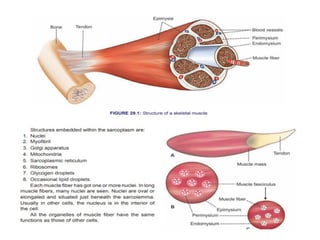
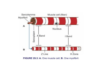
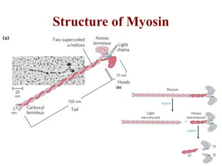
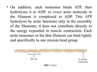
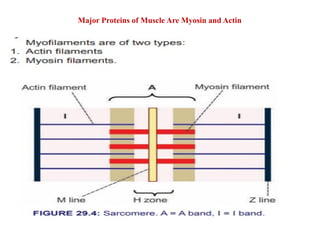
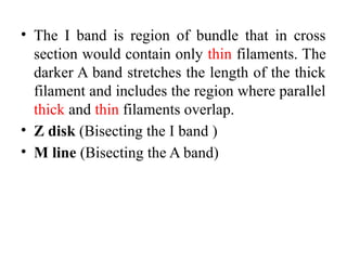
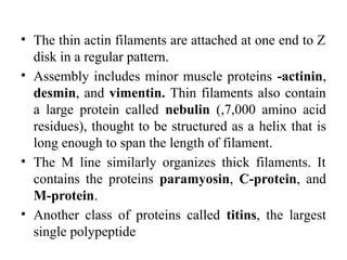
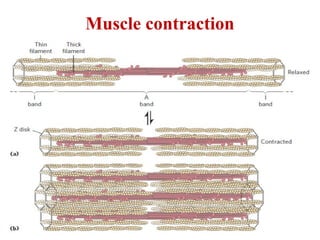
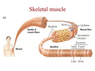
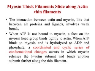
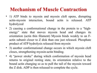
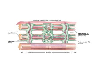
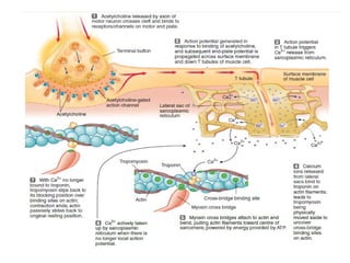
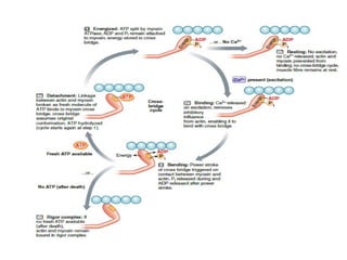
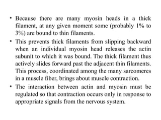
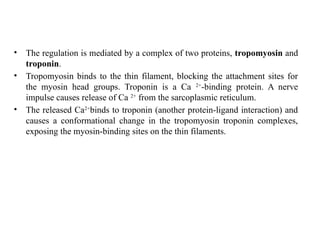
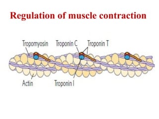
Structure and Function of Muscles
Human Physiology
Myosin
• Myosin (Mr 520,000) has six subunits: two heavy chains (each
of Mr 220,000) and four light chains (each of Mr 20,000).
• The heavy chains account for much of the overall structure. At
their carboxyl termini, they are arranged as extended helices,
wrapped around each other in a fibrous, left-handed coiled coil
similar to that of -keratin.
• At its amino terminus, each heavy chain has a large globular
domain containing a site where ATP is hydrolyzed.
• The light chains are associated with the globular domains. When
myosin is treated briefly with the protease trypsin, much of the
fibrous tail is cleaved off, dividing the protein into components
called light and heavy meromyosin.
Structure of Myosin
Myosin continue….
• In muscle cells, molecules of myosin aggregate
to form structures called thick filaments
• These rod-like structures are the core of the
contractile unit. Within a thick filament, several
hundred myosin molecules are arranged with
their fibrous “tails” associated to form a long
bipolar structure. The globular domains project
from either end of this structure, in regular
stacked arrays.
Actin
• The second major muscle protein, actin, is
abundant in almost all eukaryotic cells.
• Monomeric actin, called G-actin (globular
actin; Mr 42,000), associate to form a long
polymer called F-actin (filamentous actin). The
thin filament consists of F-actin, along with
the proteins troponin and tropomyosin.
• The filamentous parts of thin filaments
assemble as successive monomeric actin
molecules add to one end.
• On addition, each monomer binds ATP, then
hydrolyzes it to ADP, so every actin molecule in
the filament is complexed to ADP. This ATP
hydrolysis by actin functions only in the assembly
of the filaments; it does not contribute directly to
the energy expended in muscle contraction. Each
actin monomer in the thin filament can bind tightly
and specifically to one myosin head group.
Additional Proteins Ordered Thin and Thick
Filaments
• Skeletal muscle consists of parallel bundles of muscle
fibers, single fiber, very large, multinucleated cell.
• Each fiber contains about 1,000 myofibrils, 2 m in
diameter, regularly arrayed thick and thin filaments
complexed to other proteins.
• A system of flat membranous vesicles called the
sarcoplasmic reticulum surrounds each myofibril.
• Muscle fibers reveal alternating regions of high and
low electron density, called the A bands and I bands
Major Proteins of Muscle Are Myosin and Actin
• The I band is region of bundle that in cross
section would contain only thin filaments. The
darker A band stretches the length of the thick
filament and includes the region where parallel
thick and thin filaments overlap.
• Z disk (Bisecting the I band )
• M line (Bisecting the A band)
• The thin actin filaments are attached at one end to Z
disk in a regular pattern.
• Assembly includes minor muscle proteins -actinin,
desmin, and vimentin. Thin filaments also contain
a large protein called nebulin (,7,000 amino acid
residues), thought to be structured as a helix that is
long enough to span the length of filament.
• The M line similarly organizes thick filaments. It
contains the proteins paramyosin, C-protein, and
M-protein.
• Another class of proteins called titins, the largest
single polypeptide
Muscle contraction
Skeletal muscle
Myosin Thick Filaments Slide along Actin
thin filaments
• The interaction between actin and myosin, like that
between all proteins and ligands, involves weak
bonds.
• When ATP is not bound to myosin, a face on the
myosin head group binds tightly to actin. When ATP
binds to myosin and is hydrolyzed to ADP and
phosphate, a coordinated and cyclic series of
conformational changes occurs in which myosin
releases the F-actin subunit and binds another
subunit farther along the thin filament.
Mechanism of Muscle Contraction
• 1) ATP binds to myosin and myosin cleft opens, disrupting
actin-myosin interaction, bound actin is released. ATP
hydrolyzed
• 2) causing a conformational change in the protein to a “high-
energy” state that moves myosin head and changes its
orientation (actin thin filament) Myosin binds weakly to an F-
actin subunit closer to Z disk than one just released. Phosphate
product of ATP hydrolysis released from myosin
• 3) another conformational change occurs in which myosin cleft
closes, strengthening myosin-actin binding.
• 4) “power stroke” during which conformation of myosin head
returns to original resting state, its orientation relative to the
bound actin changing so as to pull the tail of the myosin toward
the Z disk. ADP is then released to complete the cycle.
• Because there are many myosin heads in a thick
filament, at any given moment some (probably 1% to
3%) are bound to thin filaments.
• This prevents thick filaments from slipping backward
when an individual myosin head releases the actin
subunit to which it was bound. The thick filament thus
actively slides forward past the adjacent thin filaments.
This process, coordinated among the many sarcomeres
in a muscle fiber, brings about muscle contraction.
• The interaction between actin and myosin must be
regulated so that contraction occurs only in response to
appropriate signals from the nervous system.
• The regulation is mediated by a complex of two proteins, tropomyosin and
troponin.
• Tropomyosin binds to the thin filament, blocking the attachment sites for
the myosin head groups. Troponin is a Ca 2+
-binding protein. A nerve
impulse causes release of Ca 2+
from the sarcoplasmic reticulum.
• The released Ca2+
binds to troponin (another protein-ligand interaction) and
causes a conformational change in the tropomyosin troponin complexes,
exposing the myosin-binding sites on the thin filaments.
Regulation of muscle contraction
References
• D. L. Nelson and M. M. Cox. 2017. Lehninger
Principles of Biochemistry. 7th
edition. Worth
Publishers, New York.
• P. C. Champe., R. A. Harvey and D. R. Ferrier.
2017. Biochemistry: Lippincott's Illustrated
Reviews. 7th
edition. Lippincott Williams and
Wilkins. U.S.A.
• Internet Source
• Question & Answer Session
• Interactive Session

Structure and Function of muscles.pptx

  • 1.
    Structure and Functionof Muscles Human Physiology
  • 7.
    Myosin • Myosin (Mr520,000) has six subunits: two heavy chains (each of Mr 220,000) and four light chains (each of Mr 20,000). • The heavy chains account for much of the overall structure. At their carboxyl termini, they are arranged as extended helices, wrapped around each other in a fibrous, left-handed coiled coil similar to that of -keratin. • At its amino terminus, each heavy chain has a large globular domain containing a site where ATP is hydrolyzed. • The light chains are associated with the globular domains. When myosin is treated briefly with the protease trypsin, much of the fibrous tail is cleaved off, dividing the protein into components called light and heavy meromyosin.
  • 8.
  • 9.
    Myosin continue…. • Inmuscle cells, molecules of myosin aggregate to form structures called thick filaments • These rod-like structures are the core of the contractile unit. Within a thick filament, several hundred myosin molecules are arranged with their fibrous “tails” associated to form a long bipolar structure. The globular domains project from either end of this structure, in regular stacked arrays.
  • 10.
    Actin • The secondmajor muscle protein, actin, is abundant in almost all eukaryotic cells. • Monomeric actin, called G-actin (globular actin; Mr 42,000), associate to form a long polymer called F-actin (filamentous actin). The thin filament consists of F-actin, along with the proteins troponin and tropomyosin. • The filamentous parts of thin filaments assemble as successive monomeric actin molecules add to one end.
  • 11.
    • On addition,each monomer binds ATP, then hydrolyzes it to ADP, so every actin molecule in the filament is complexed to ADP. This ATP hydrolysis by actin functions only in the assembly of the filaments; it does not contribute directly to the energy expended in muscle contraction. Each actin monomer in the thin filament can bind tightly and specifically to one myosin head group.
  • 12.
    Additional Proteins OrderedThin and Thick Filaments • Skeletal muscle consists of parallel bundles of muscle fibers, single fiber, very large, multinucleated cell. • Each fiber contains about 1,000 myofibrils, 2 m in diameter, regularly arrayed thick and thin filaments complexed to other proteins. • A system of flat membranous vesicles called the sarcoplasmic reticulum surrounds each myofibril. • Muscle fibers reveal alternating regions of high and low electron density, called the A bands and I bands
  • 13.
    Major Proteins ofMuscle Are Myosin and Actin
  • 14.
    • The Iband is region of bundle that in cross section would contain only thin filaments. The darker A band stretches the length of the thick filament and includes the region where parallel thick and thin filaments overlap. • Z disk (Bisecting the I band ) • M line (Bisecting the A band)
  • 15.
    • The thinactin filaments are attached at one end to Z disk in a regular pattern. • Assembly includes minor muscle proteins -actinin, desmin, and vimentin. Thin filaments also contain a large protein called nebulin (,7,000 amino acid residues), thought to be structured as a helix that is long enough to span the length of filament. • The M line similarly organizes thick filaments. It contains the proteins paramyosin, C-protein, and M-protein. • Another class of proteins called titins, the largest single polypeptide
  • 16.
  • 17.
  • 18.
    Myosin Thick FilamentsSlide along Actin thin filaments • The interaction between actin and myosin, like that between all proteins and ligands, involves weak bonds. • When ATP is not bound to myosin, a face on the myosin head group binds tightly to actin. When ATP binds to myosin and is hydrolyzed to ADP and phosphate, a coordinated and cyclic series of conformational changes occurs in which myosin releases the F-actin subunit and binds another subunit farther along the thin filament.
  • 19.
    Mechanism of MuscleContraction • 1) ATP binds to myosin and myosin cleft opens, disrupting actin-myosin interaction, bound actin is released. ATP hydrolyzed • 2) causing a conformational change in the protein to a “high- energy” state that moves myosin head and changes its orientation (actin thin filament) Myosin binds weakly to an F- actin subunit closer to Z disk than one just released. Phosphate product of ATP hydrolysis released from myosin • 3) another conformational change occurs in which myosin cleft closes, strengthening myosin-actin binding. • 4) “power stroke” during which conformation of myosin head returns to original resting state, its orientation relative to the bound actin changing so as to pull the tail of the myosin toward the Z disk. ADP is then released to complete the cycle.
  • 23.
    • Because thereare many myosin heads in a thick filament, at any given moment some (probably 1% to 3%) are bound to thin filaments. • This prevents thick filaments from slipping backward when an individual myosin head releases the actin subunit to which it was bound. The thick filament thus actively slides forward past the adjacent thin filaments. This process, coordinated among the many sarcomeres in a muscle fiber, brings about muscle contraction. • The interaction between actin and myosin must be regulated so that contraction occurs only in response to appropriate signals from the nervous system.
  • 24.
    • The regulationis mediated by a complex of two proteins, tropomyosin and troponin. • Tropomyosin binds to the thin filament, blocking the attachment sites for the myosin head groups. Troponin is a Ca 2+ -binding protein. A nerve impulse causes release of Ca 2+ from the sarcoplasmic reticulum. • The released Ca2+ binds to troponin (another protein-ligand interaction) and causes a conformational change in the tropomyosin troponin complexes, exposing the myosin-binding sites on the thin filaments.
  • 25.
  • 26.
    References • D. L.Nelson and M. M. Cox. 2017. Lehninger Principles of Biochemistry. 7th edition. Worth Publishers, New York. • P. C. Champe., R. A. Harvey and D. R. Ferrier. 2017. Biochemistry: Lippincott's Illustrated Reviews. 7th edition. Lippincott Williams and Wilkins. U.S.A. • Internet Source
  • 27.
    • Question &Answer Session • Interactive Session

Editor's Notes

  • #7 The globular domain—called myosin subfragment 1, or S1, or simply the myosin head group—is liberated from heavy meromyosin by cleavage with papain. The S1 fragment is the motor domain that makes muscle contraction possible. S1 fragments can be crystallized, and their overall structure as determined by Ivan Rayment and Hazel Holden
  • #8 Myosin. (a) Myosin has two heavy chains (in two shades of pink), the carboxyl termini forming an extended coiled coil (tail) and the amino termini having globular domains (heads). Two light chains (blue) are associated with each myosin head. (b) Cleavage with trypsin and papain separates the myosin heads (S1 fragments) from the tails.
  • #10 . In muscle, molecules of monomeric actin, called G-actin (globular actin; Mr 42,000), associate to form a long polymer called F-actin (filamentous actin). The thin filament consists of F-actin, along with the proteins troponin and tropomyosin. The filamentous parts of thin filaments assemble as successive monomeric actin molecules add to one end.
  • #12 Skeletal muscle consists of parallel bundles of muscle fibers, each fiber a single, very large, multinucleated cell, 20 to 100 m in diameter, formed from many cells fused together; a single fiber often spans the length ofthe muscle. Each fiber contains about 1,000 myofibrils, 2 m in diameter, each consisting of a vast number of regularly arrayed thick and thin filaments complexed to other proteins. A system of flat membranous vesicles called the sarcoplasmic reticulum surrounds each myofibril. Examined under the electron microscope, muscle fibers reveal alternating regions of high and low electron density, called the A bands and I bands.
  • #14 The A and I bands arise from the arrangement of thick and thin filaments, which are aligned and partially overlapping. Bisecting the I band is a thin structure called the Z disk, perpendicular to the thin filaments and serving as an anchor to which the thin filaments are attached. The A band, too, is bisected by a thin line, the M line or M disk, a region of high electron density in the middle of the thick filaments. The entire contractile unit, consisting of bundles of thick filaments interleaved at either end with bundles of thin filaments, is called the sarcomere. The arrangement of interleaved bundles allows the thick and thin filaments to slide past each other (by a mechanism discussed below), causing a progressive shortening of each sarcomere
  • #16 Muscle contraction. Thick filaments are bipolar structures created by the association of many myosin molecules. (a) Muscle contraction occurs by the sliding of the thick and thin filaments past each other so that the Z disks in neighboring I bands draw closer together. (b) The thick and thin filaments are interleaved such that each thick filament is surrounded by six thin filaments.
  • #17 Skeletal muscle. (a) Muscle fibers consist of single, elongated, multinucleated cells that arise from the fusion of many precursor cells. The fibers are made up of many myofibrils (only six are shown here for simplicity) surrounded by the membranous sarcoplasmic reticulum. The organization of thick and thin filaments in a myofibril gives it a striated appearance. When muscle contracts, the I bands narrow and the Z disks come closer together.
  • #19 Each cycle generates about 3 to 4 pN (piconewtons) of force and moves the thick filament 5 to 10 nm relative to the thin filament.
  • #25 Regulation of muscle contraction by tropomyosin and troponin. Tropomyosin and troponin are bound to F-actin in the thin filaments. In the relaxed muscle, these two proteins are arranged around the actin filaments so as to block the binding sites for myosin. Tropomyosin is a two-stranded coiled coil of helices, the same structural motif as in –keratin. It forms head-to-tail polymers twisting around the two actin chains. Troponin is attached to the actin-tropomyosin complex at regular intervals of 38.5 nm. Troponin consists of three different subunits: I, C, and T. Troponin I prevents binding of the myosin head to actin; troponin C has a binding site for Ca21; and troponin T links the entire troponin complex to tropomyosin. When the muscle receives a neural signal to initiate contraction, Ca21 is released from the sarcoplasmic reticulum (see Fig. 5–29a) and binds to troponin C. This causes a conformational change in troponin C, which alters the positions of troponin I and tropomyosin so as to relieve the inhibition by troponin I and allow muscle contraction.