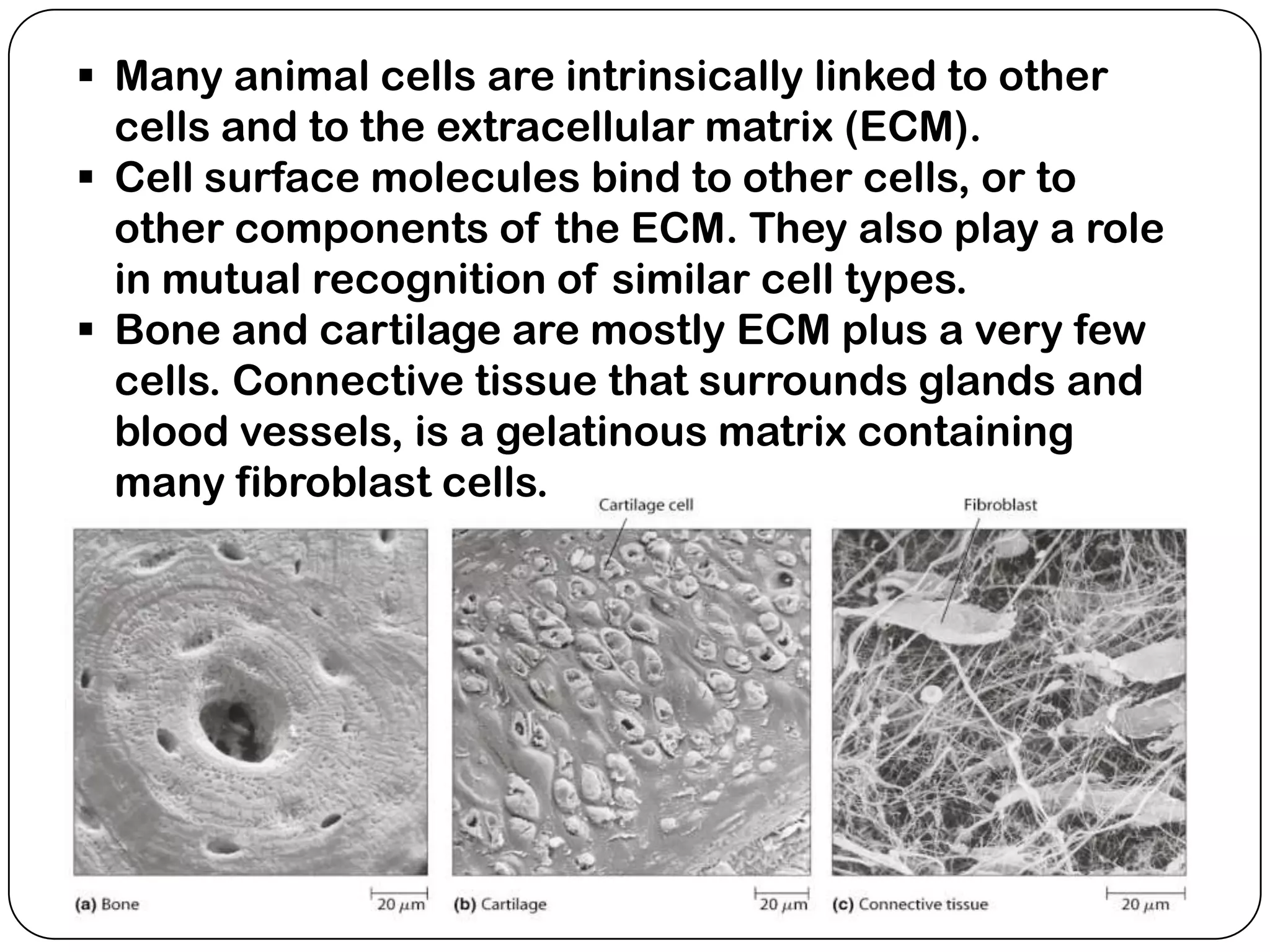
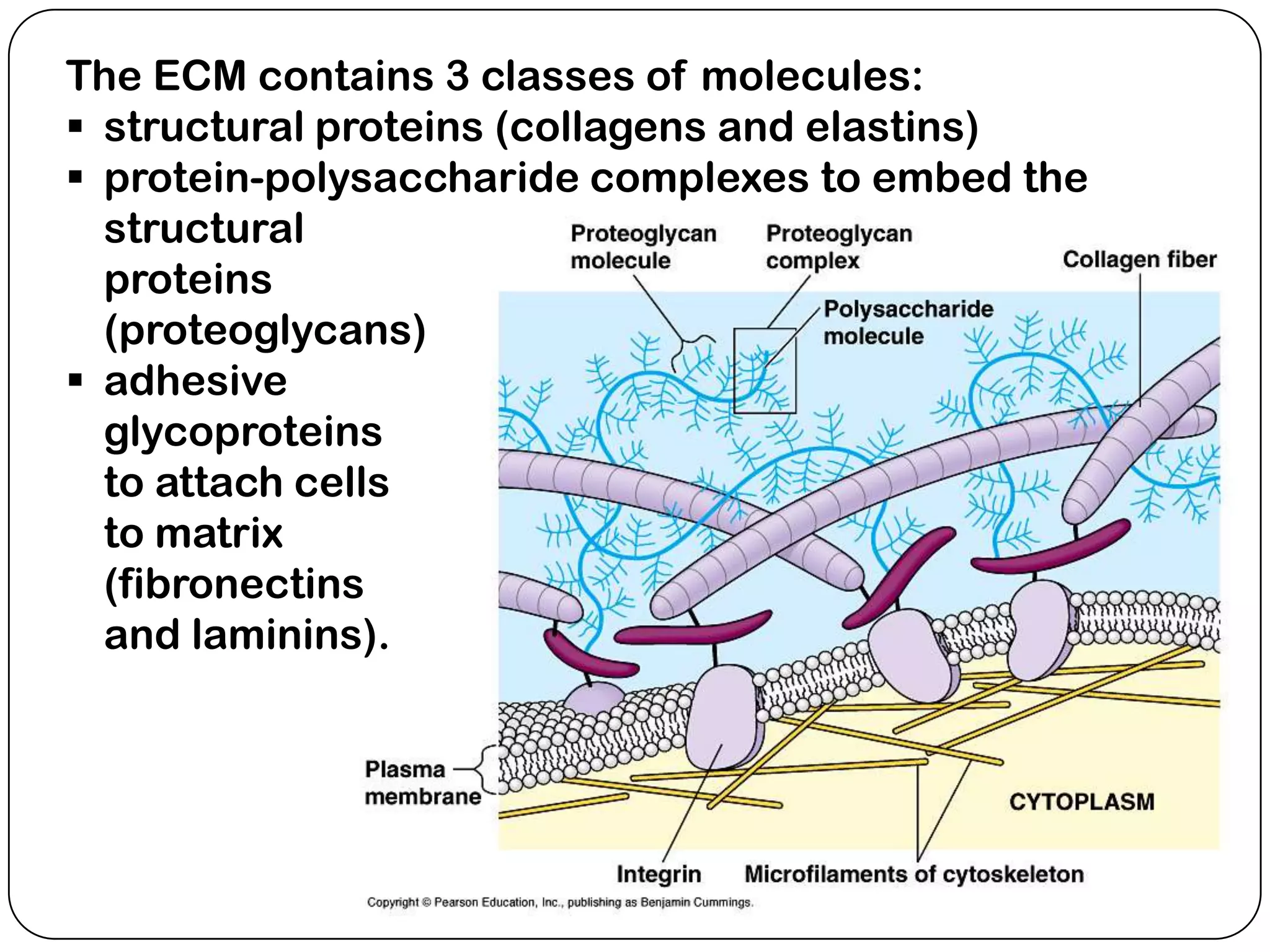
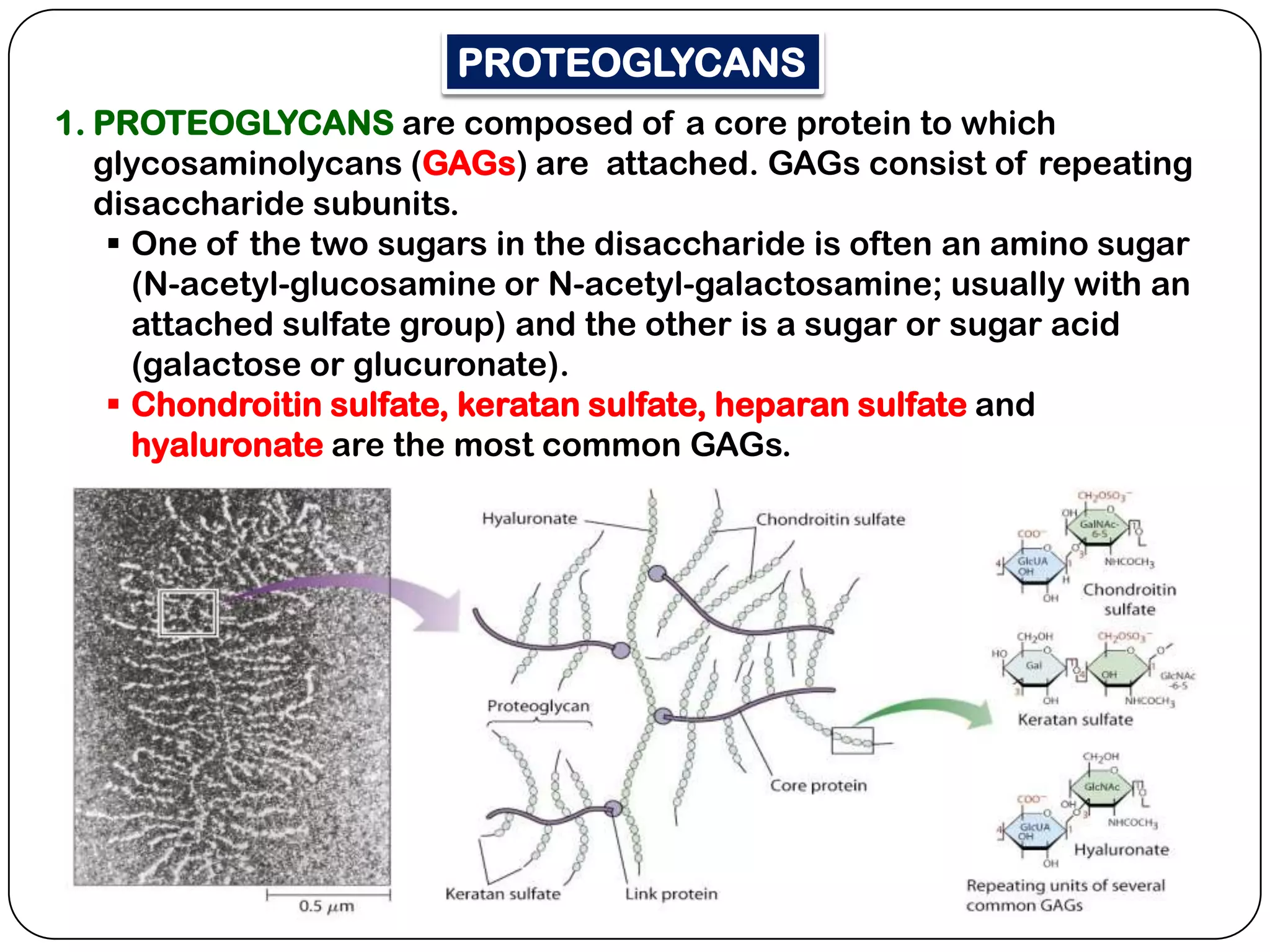
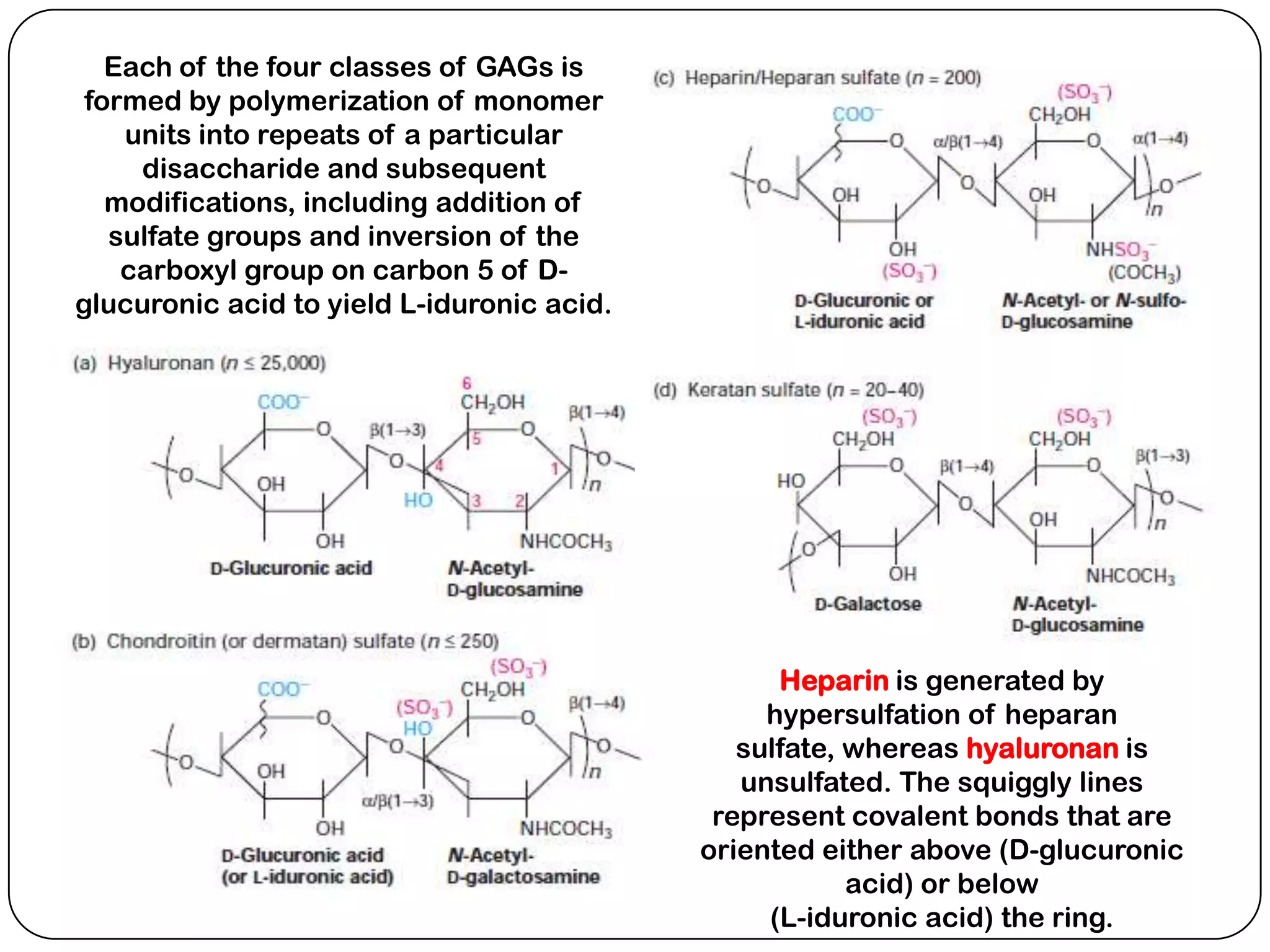
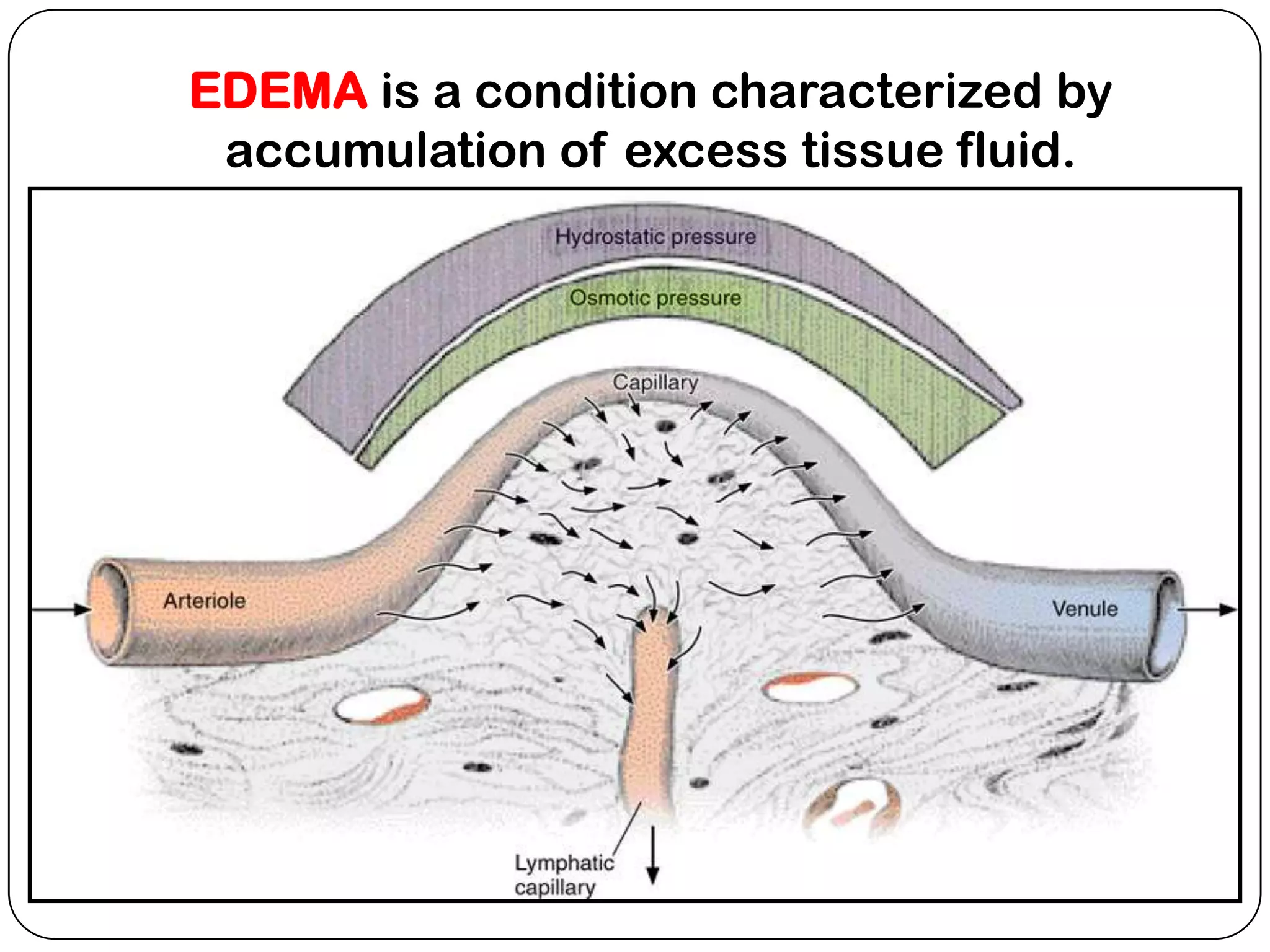
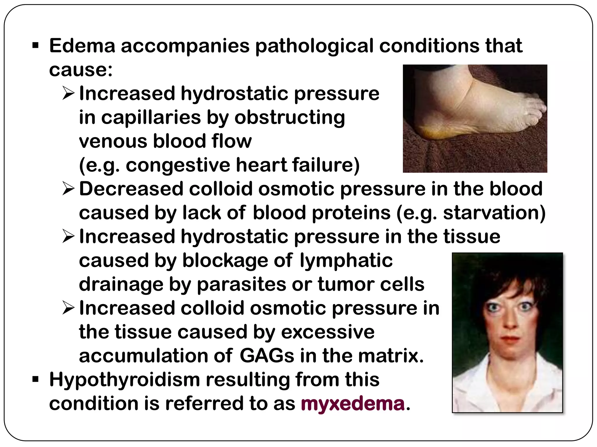
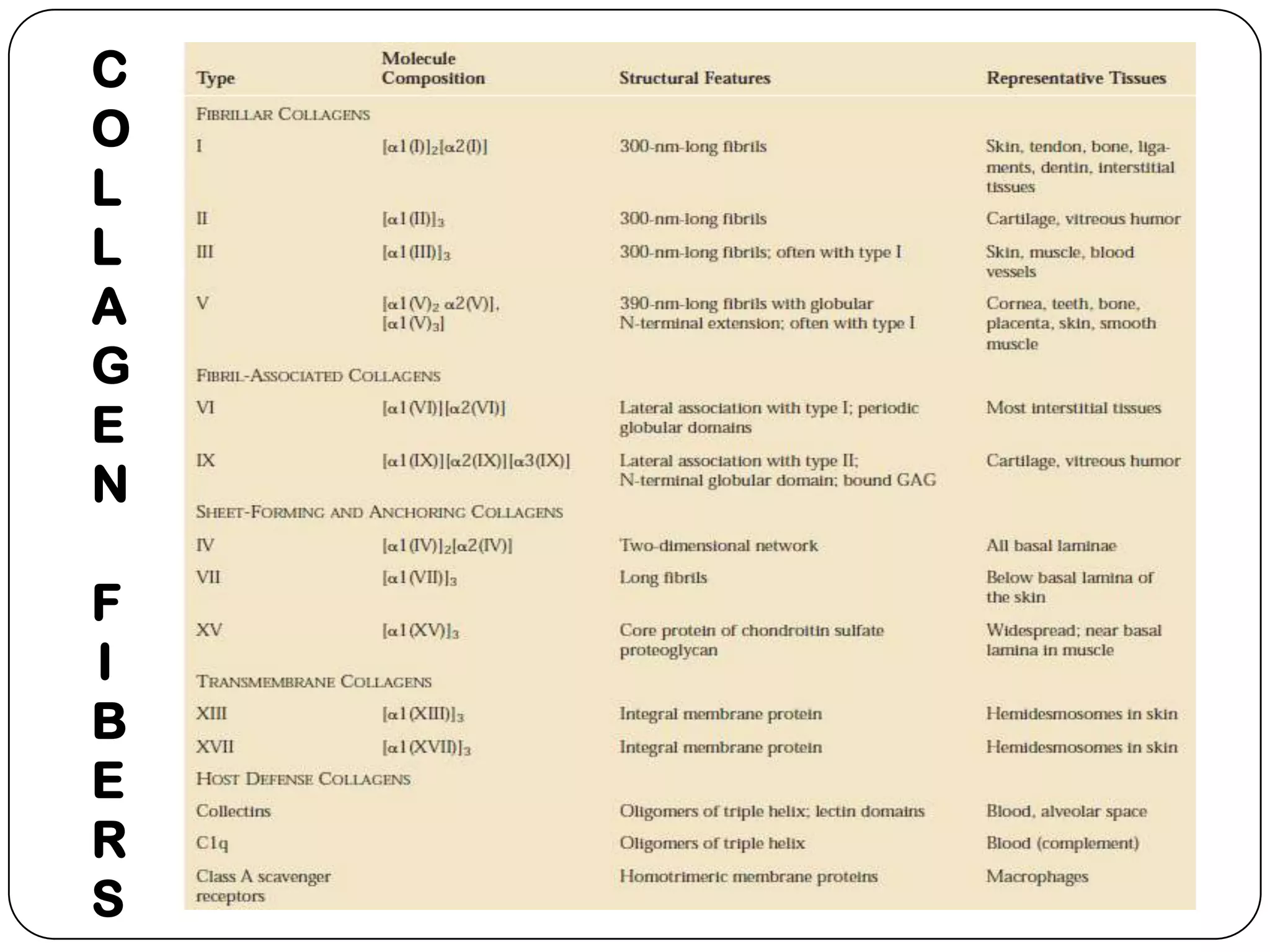
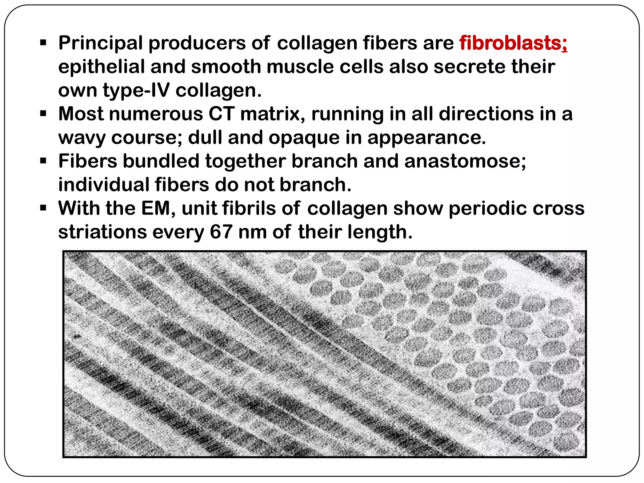
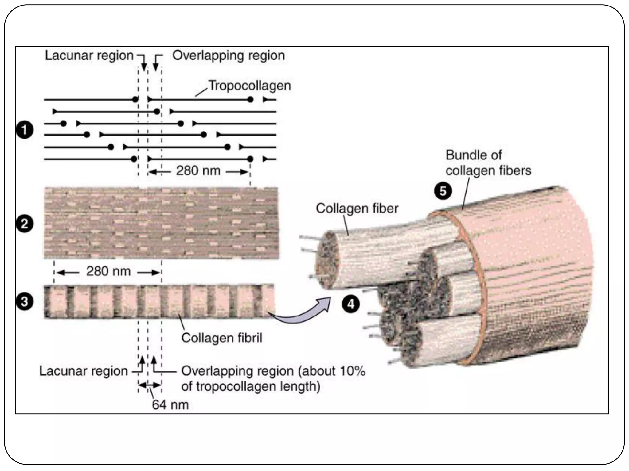
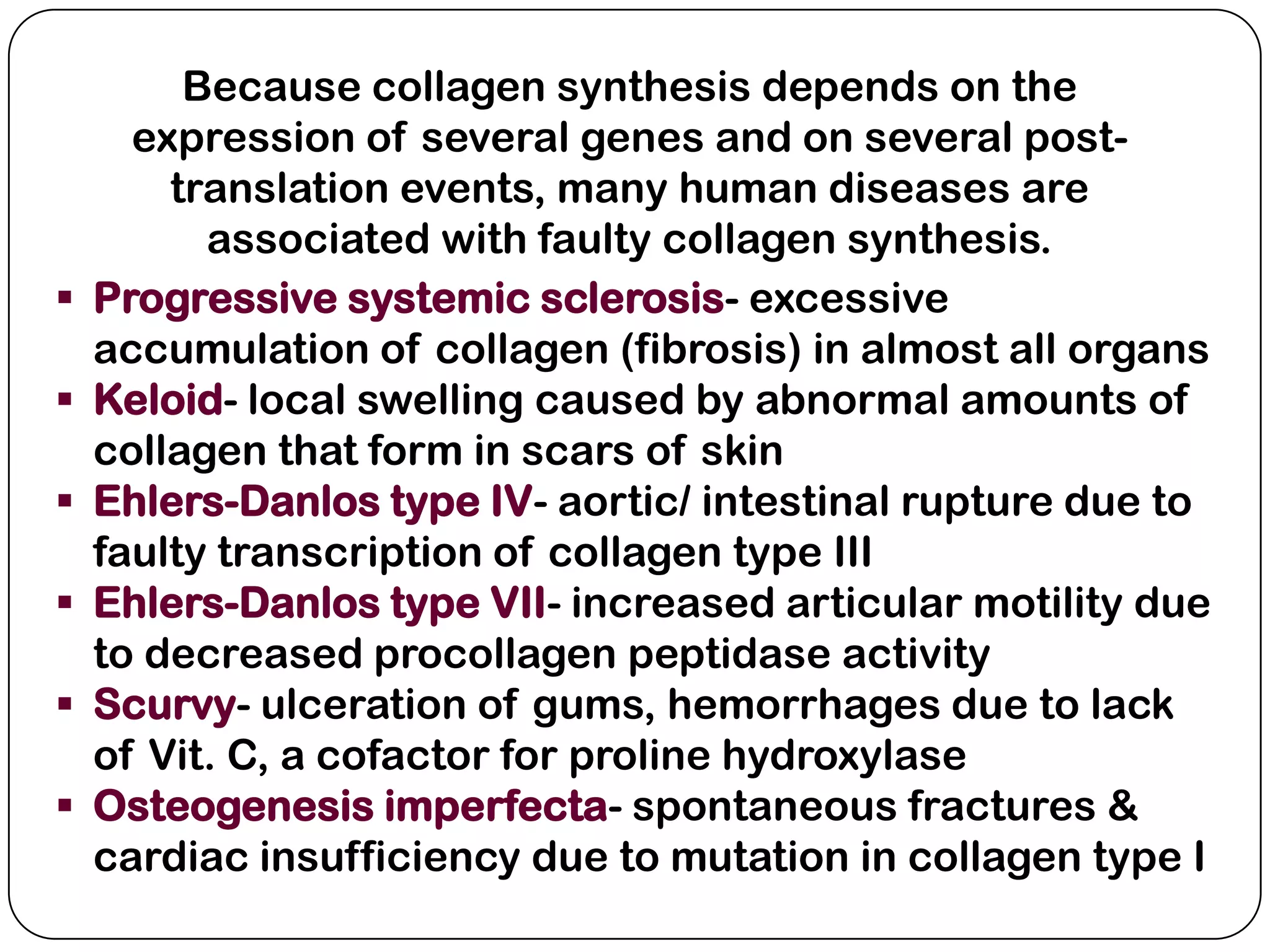
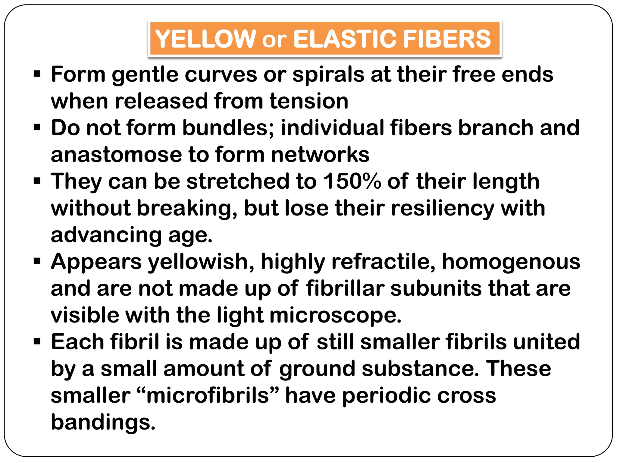
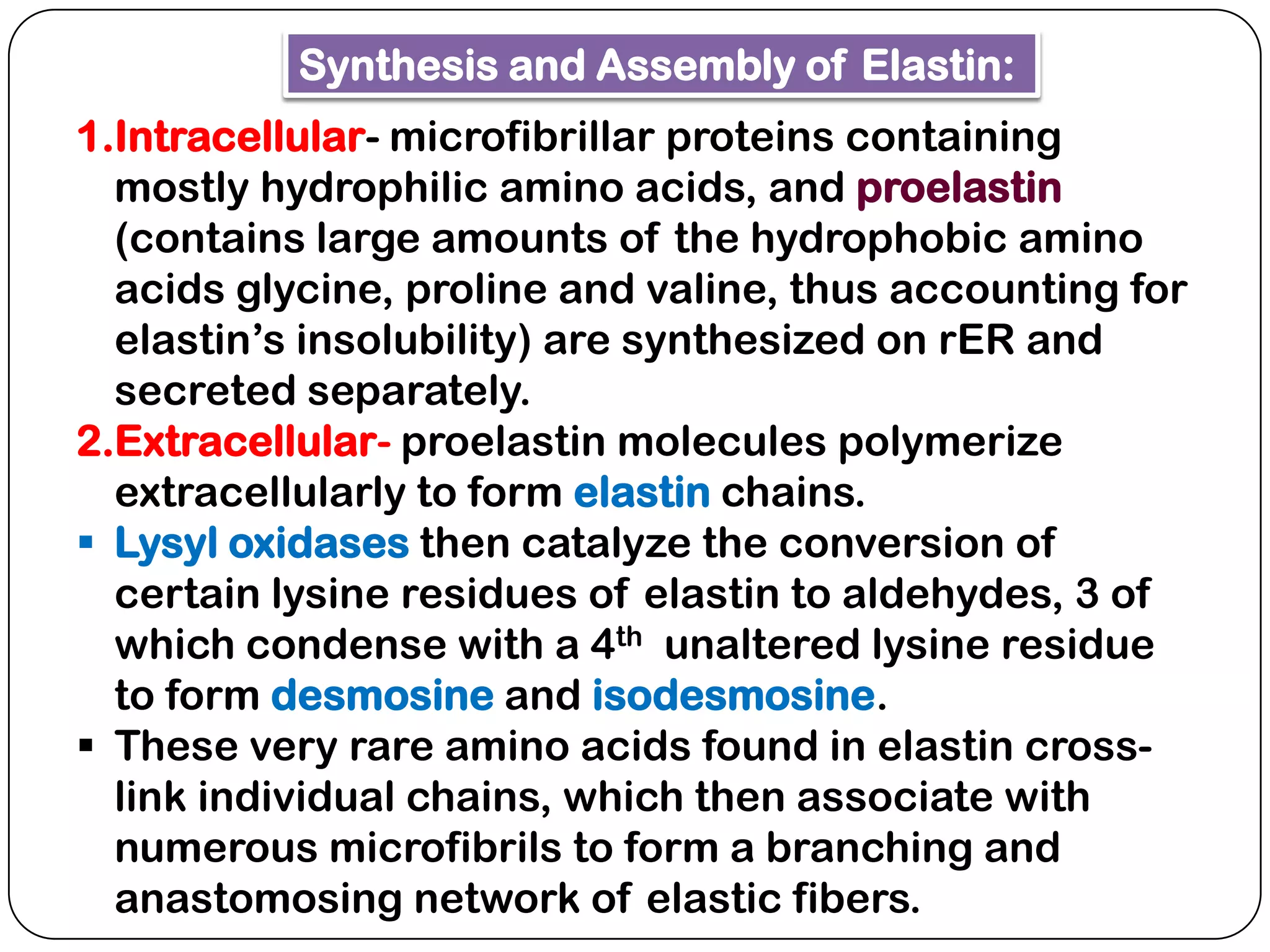
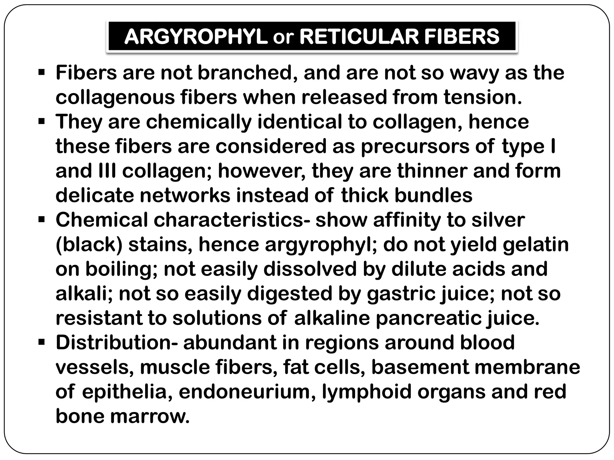
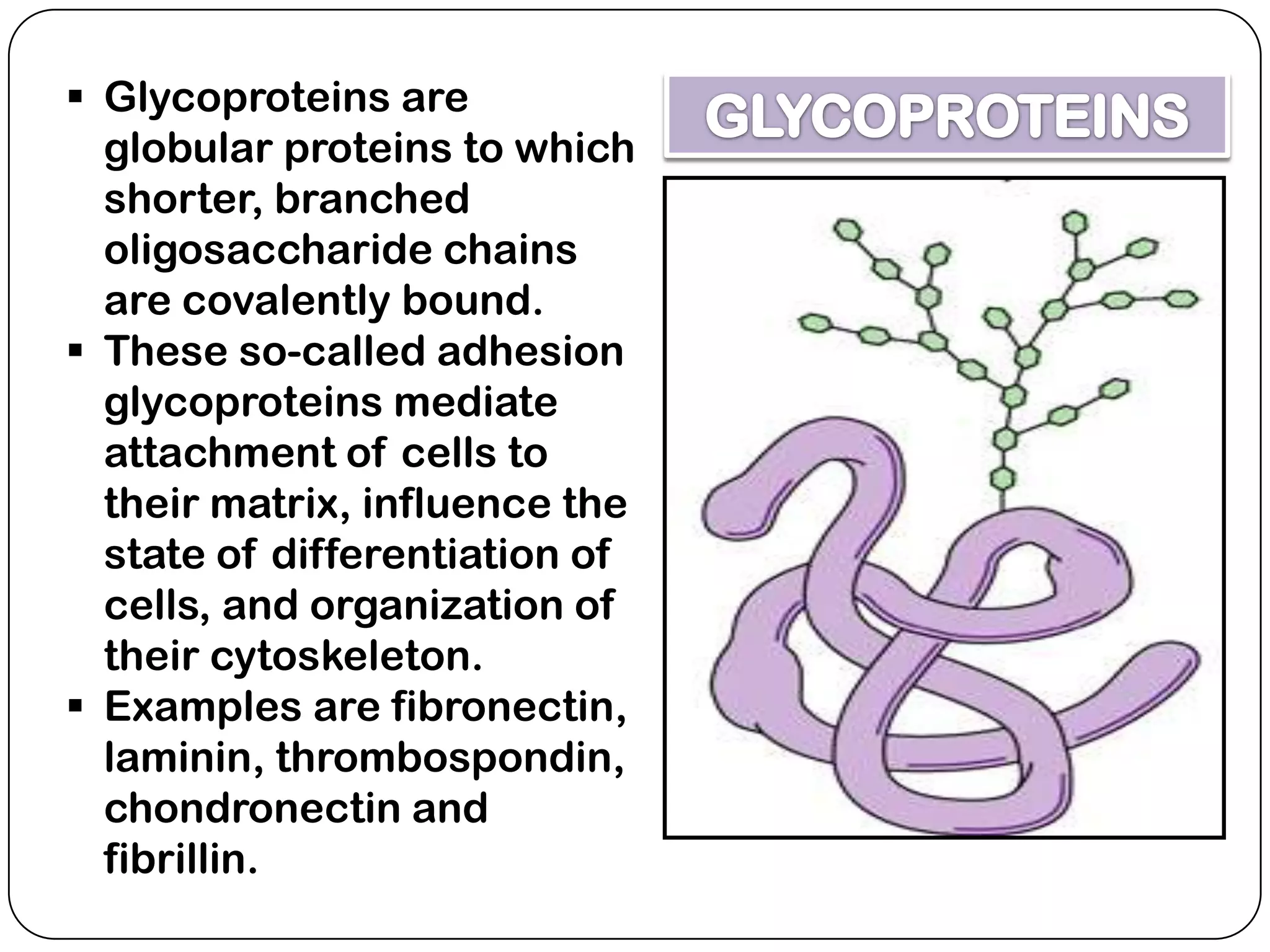
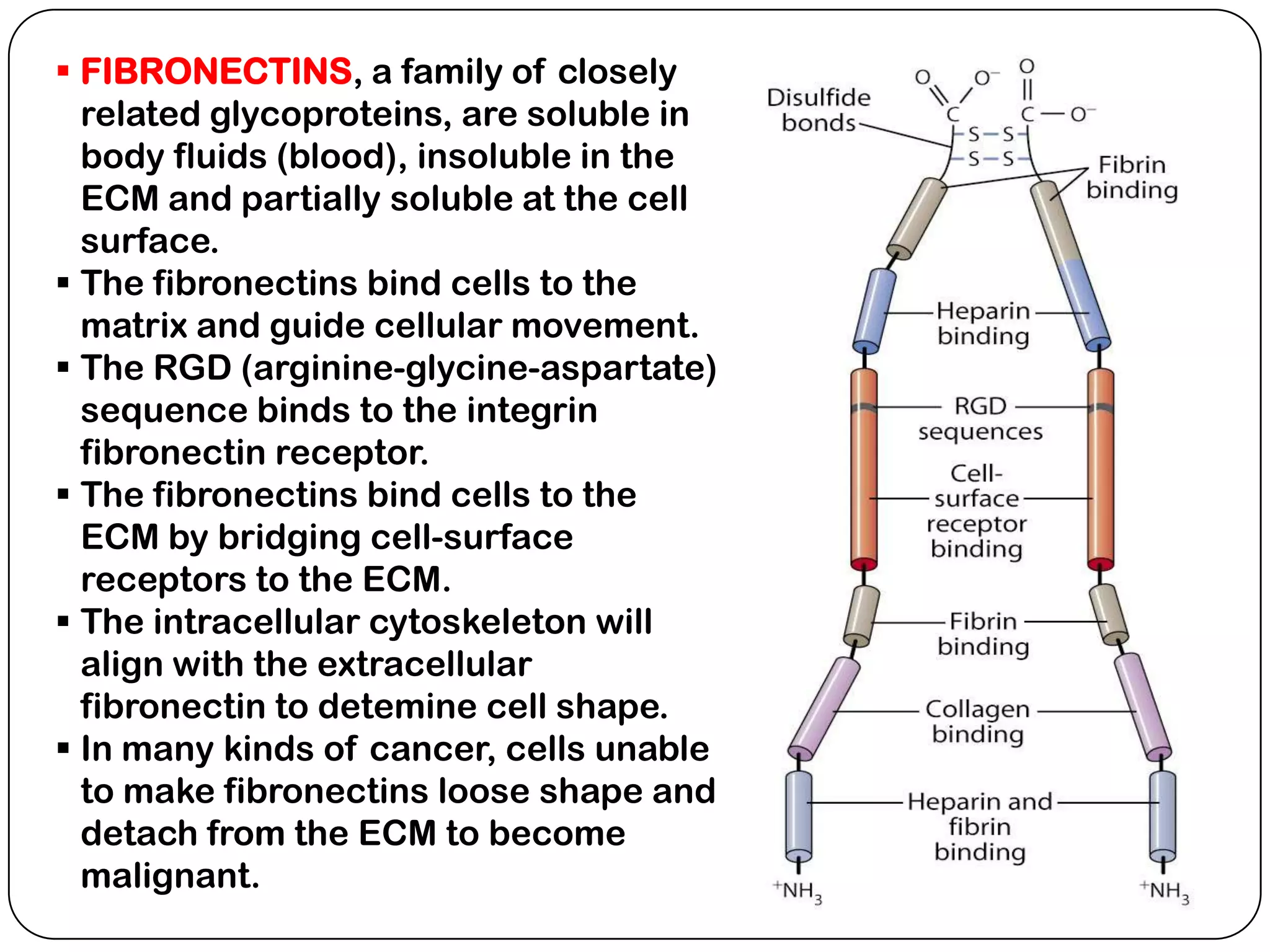
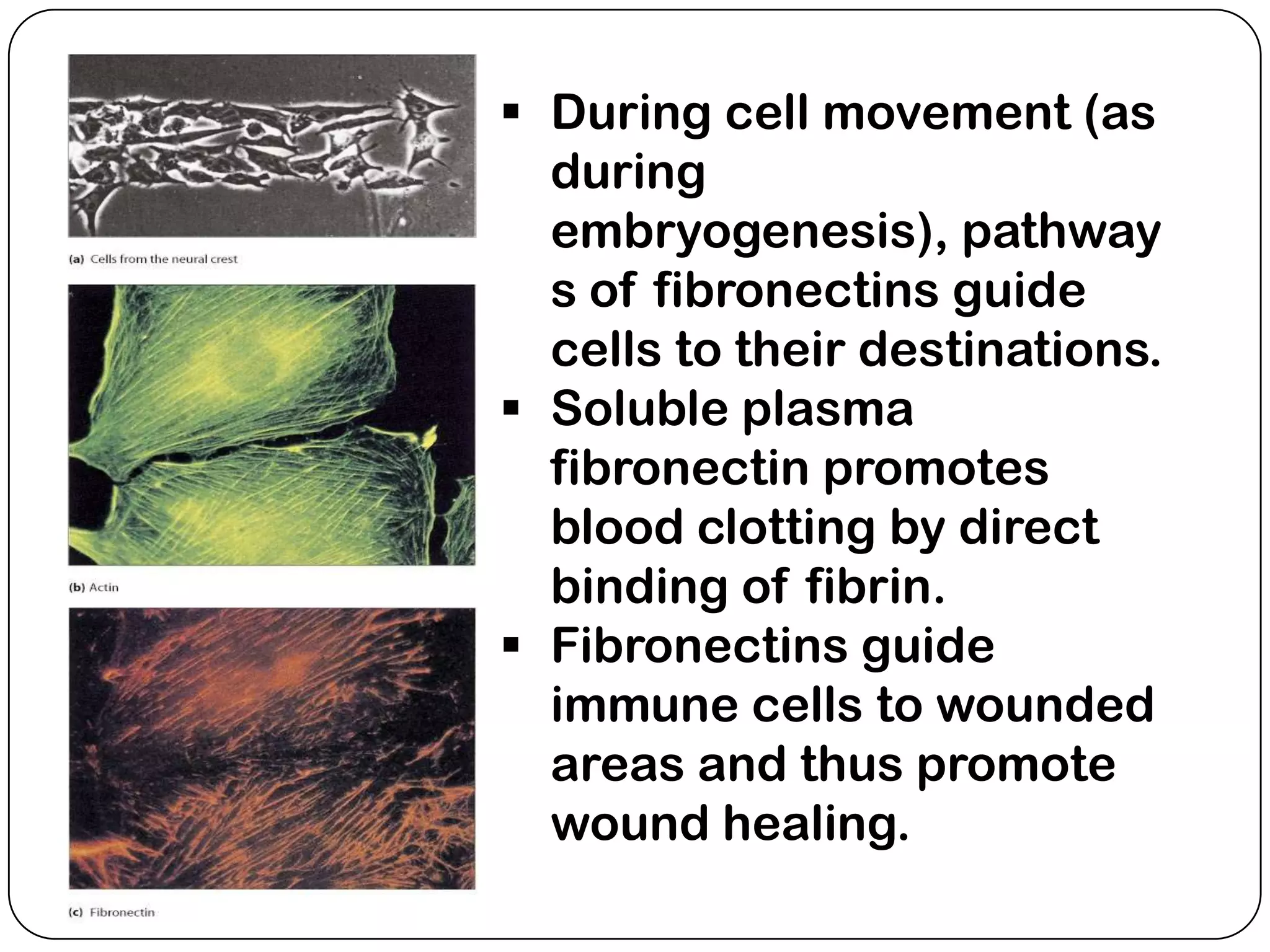
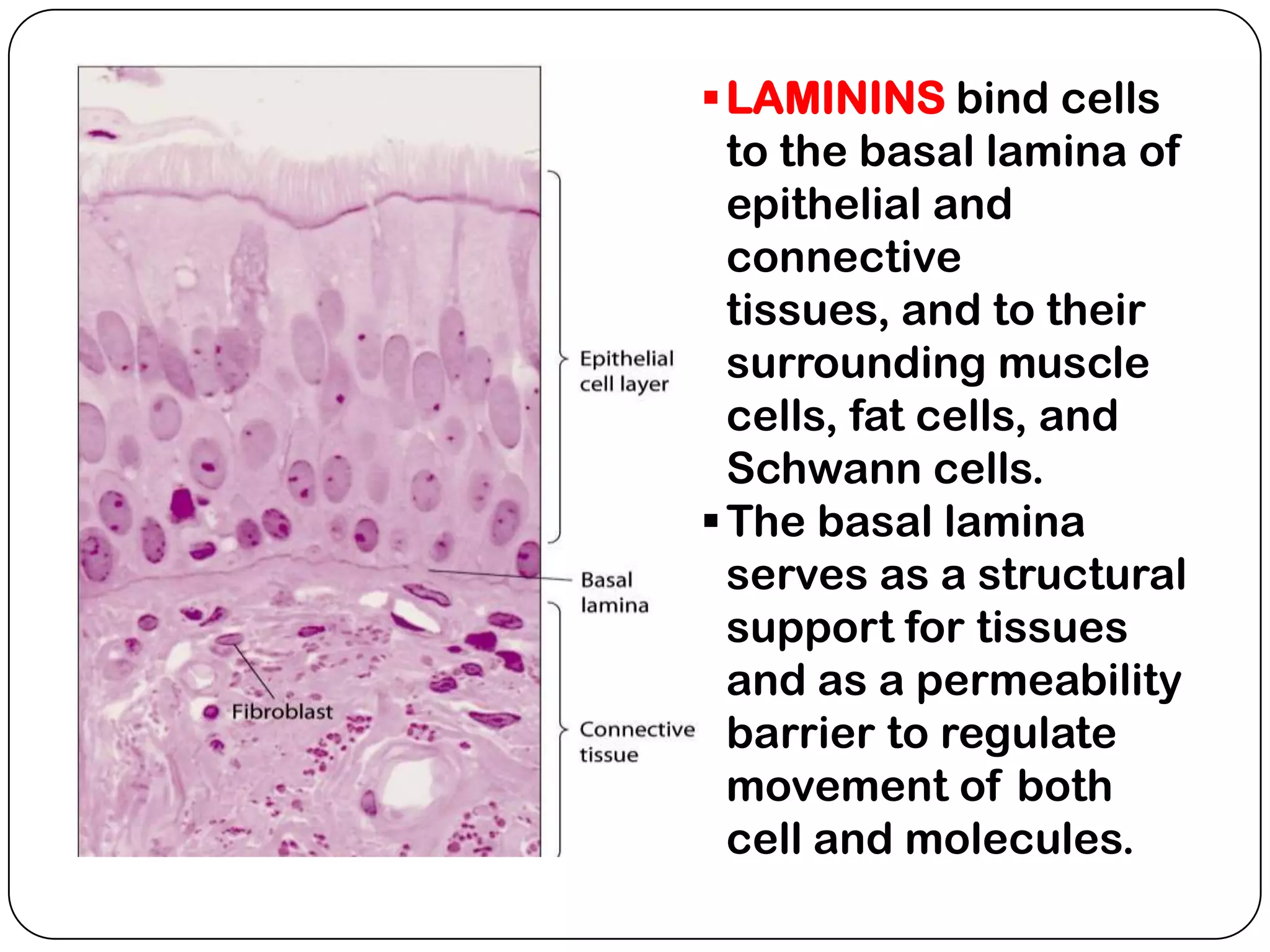
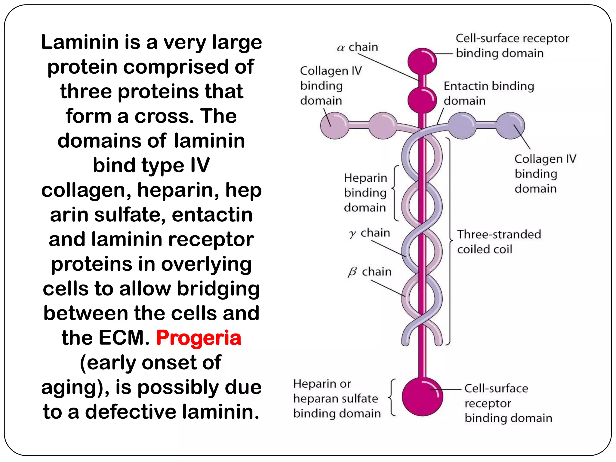
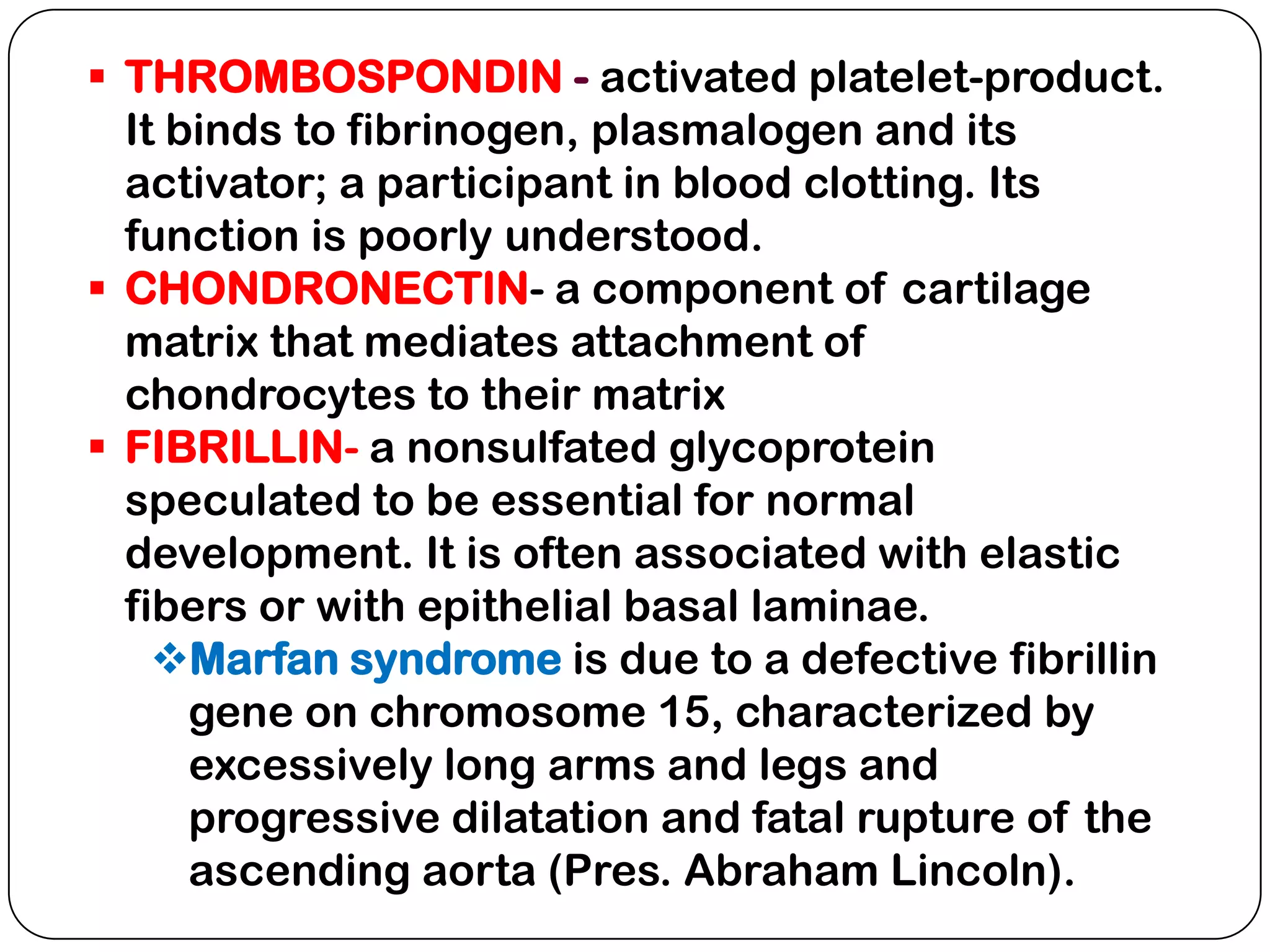
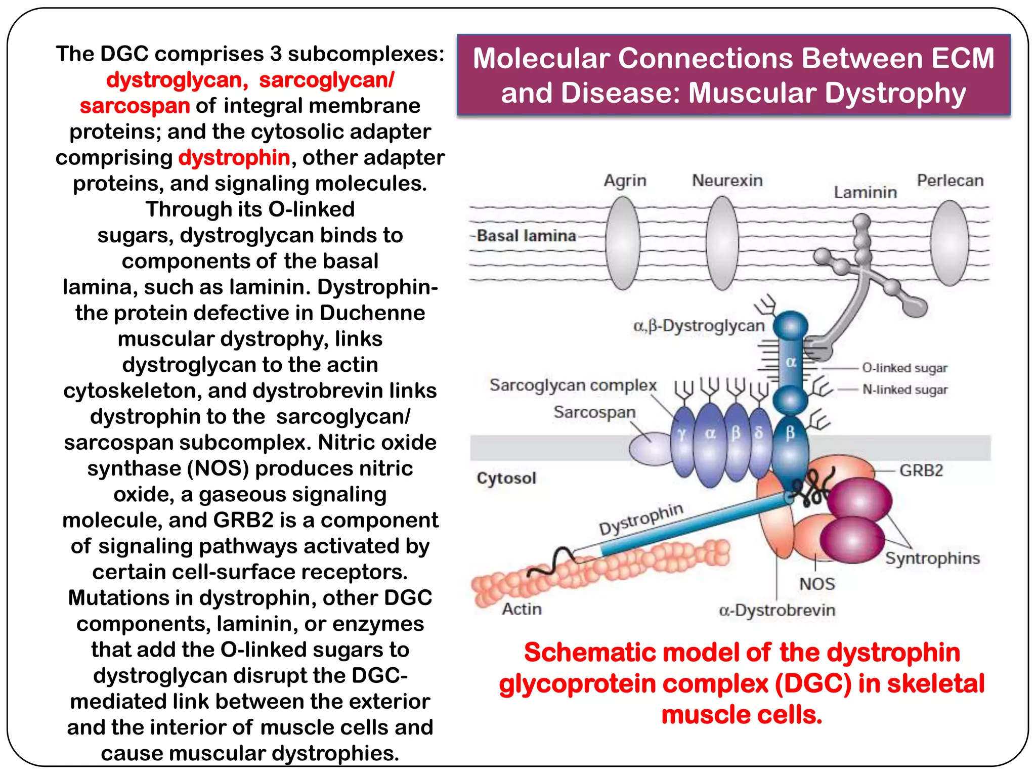
EXTRACELLULAR
   MATRIX


 Department of Natural Sciences
    University of St. La Salle
         Bacolod City
 Many animal cells are intrinsically linked to other
  cells and to the extracellular matrix (ECM).
 Cell surface molecules bind to other cells, or to
  other components of the ECM. They also play a role
  in mutual recognition of similar cell types.
 Bone and cartilage are mostly ECM plus a very few
  cells. Connective tissue that surrounds glands and
  blood vessels, is a gelatinous matrix containing
  many fibroblast cells.
The ECM contains 3 classes of molecules:
 structural proteins (collagens and elastins)
 protein-polysaccharide complexes to embed the
  structural
  proteins
  (proteoglycans)
 adhesive
  glycoproteins
  to attach cells
  to matrix
  (fibronectins
  and laminins).
PROTEOGLYCANS
1. PROTEOGLYCANS are composed of a core protein to which
   glycosaminolycans (GAGs) are attached. GAGs consist of repeating
   disaccharide subunits.
     One of the two sugars in the disaccharide is often an amino sugar
      (N-acetyl-glucosamine or N-acetyl-galactosamine; usually with an
      attached sulfate group) and the other is a sugar or sugar acid
      (galactose or glucuronate).
     Chondroitin sulfate, keratan sulfate, heparan sulfate and
      hyaluronate are the most common GAGs.
Each of the four classes of GAGs is
 formed by polymerization of monomer
    units into repeats of a particular
     disaccharide and subsequent
   modifications, including addition of
   sulfate groups and inversion of the
    carboxyl group on carbon 5 of D-
glucuronic acid to yield L-iduronic acid.


                                                  Heparin is generated by
                                                 hypersulfation of heparan
                                               sulfate, whereas hyaluronan is
                                               unsulfated. The squiggly lines
                                             represent covalent bonds that are
                                            oriented either above (D-glucuronic
                                                        acid) or below
                                                 (L-iduronic acid) the ring.
 Most GAGs in the ECM are bound to proteins
  to form proteoglycans or mucoproteins.
 Numerous GAGs (1-200 per
  molecule, average length of 800
  monosaccharide units) are attached to a core
  protein and different kinds of proteoglycans
  can be made by varying the combination of
  core proteins and GAGs.
 Proteoglycans (MW of~ 1 million) can be
  individual or attached to long hyaluronate
  molecules to form complexes (as in cartilage).
 They can be embedded in the plasma
  membrane or covalently linked to membrane
  phospholipids or bound to receptor proteins.
 Proteoglycans and collagen may bind to receptor
  proteins (often integrins) which are reinforced by
  adhesive glycoproteins, such as fibronectins and
  laminins, to anchor cells to the ECM.
 GAGs in CT are highly sulfated which attracts
  water of hydration. They trap water (up to 50x
  their weight) to act as extracellular sponges
  resistant to physical forces in cartilage and joints.
 If fluid is injected into CT, it remains
  localized, walled off by a viscous ground
  substance. This property acts as barrier to the
  spread of bacteria that gains access to the
  tissues.
 Some bacteria secrete hyaluronidase (Staphylo/
  Strepto/ Pneumococci), and collagenase
  (Clostridium perfringens) that breakdown matrix
  components.
EDEMA is a condition characterized by
 accumulation of excess tissue fluid.
 Edema accompanies pathological conditions that
  cause:
    Increased hydrostatic pressure
     in capillaries by obstructing
     venous blood flow
     (e.g. congestive heart failure)
    Decreased colloid osmotic pressure in the blood
     caused by lack of blood proteins (e.g. starvation)
    Increased hydrostatic pressure in the tissue
     caused by blockage of lymphatic
     drainage by parasites or tumor cells
    Increased colloid osmotic pressure in
     the tissue caused by excessive
     accumulation of GAGs in the matrix.
 Hypothyroidism resulting from this
  condition is referred to as myxedema.
C
O
L
L
A
G
E
N

F
I
B
E
R
S
 Principal producers of collagen fibers are fibroblasts;
  epithelial and smooth muscle cells also secrete their
  own type-IV collagen.
 Most numerous CT matrix, running in all directions in a
  wavy course; dull and opaque in appearance.
 Fibers bundled together branch and anastomose;
  individual fibers do not branch.
 With the EM, unit fibrils of collagen show periodic cross
  striations every 67 nm of their length.
(a) In tendons, type I
  fibrils are all oriented in the      Interactions of fibrous
direction of the stress applied      collagens with nonfibrous
 to the tendon. Proteoglycans
    and type VI collagen bind
                                    fibril-associated collagens.
          noncovalently to
   fibrils, coating the surface.
    The microfibrils of type VI
     collagen, which contain
    globular and triple-helical
     segments, bind to type I
 fibrils and link them together
     into thicker fibers. (b) In
    cartilage, type IX collagen
    molecules are covalently
    bound at regular intervals
       along type II fibrils. A
    chondroitin sulfate chain,
covalently linked to the 2 type
      IX chains at the flexible
  kink, projects outward from
the fibril, as does the globular
         N-terminal region.
1.INTRACELLULAR – free polysomes reading collagen mRNA
  attach to the rER, and protocollagen or precollagen-chains
  are deposited in the cisternae. Each chain has about 250
  amino acids; every 3rd amino acid is glycine.
 The signal peptide is clipped off. Proline and lysine
  residues within the chains are then hydroxylated in the ER
  to form hydroxyproline and hydroxylysine (unusual amino
  acids present in large amounts in collagen).
 Core sugars (galactose and glucose) attach to the
  hydroxylysine residues in the ER.
 Each chain is synthesized with an extra length of peptides
  known as registration peptides, which ensure that the
  appropriate chains assemble in their correct position in the
  resulting triple helical molecule called procollagen.
 Further glycosylation may occur in the Golgi
  complex, where procollagen is packaged for secretion.
  Golgi vesicles release procollagen into the extracellular
  space by exocytosis.
2.EXTRACELLULAR- in the extracellular
  space, the enzyme procollagen peptidase
  cleaves the registration peptides from
  procollagen, converting it to tropocollagen.
 Catalyzed by lysyl oxidase, these become
  aligned in staggered fashion to form collagen
  fibers, possibly under the control of adjacent
  fiber-producing cells.
 The turnover of collagen is slowest in
  tendons, fastest in loose CT. Macrophages and
  neutrophils break down old collagen, and
  replaced by fibroblasts.
 As humans age, extracellular collagen becomes
  increasingly cross-linked, & turn-over slows
  down in CT.
Because collagen synthesis depends on the
      expression of several genes and on several post-
        translation events, many human diseases are
          associated with faulty collagen synthesis.
   Progressive systemic sclerosis- excessive
    accumulation of collagen (fibrosis) in almost all organs
   Keloid- local swelling caused by abnormal amounts of
    collagen that form in scars of skin
   Ehlers-Danlos type IV- aortic/ intestinal rupture due to
    faulty transcription of collagen type III
   Ehlers-Danlos type VII- increased articular motility due
    to decreased procollagen peptidase activity
   Scurvy- ulceration of gums, hemorrhages due to lack
    of Vit. C, a cofactor for proline hydroxylase
   Osteogenesis imperfecta- spontaneous fractures &
    cardiac insufficiency due to mutation in collagen type I
YELLOW or ELASTIC FIBERS
 Form gentle curves or spirals at their free ends
  when released from tension
 Do not form bundles; individual fibers branch and
  anastomose to form networks
 They can be stretched to 150% of their length
  without breaking, but lose their resiliency with
  advancing age.
 Appears yellowish, highly refractile, homogenous
  and are not made up of fibrillar subunits that are
  visible with the light microscope.
 Each fibril is made up of still smaller fibrils united
  by a small amount of ground substance. These
  smaller “microfibrils” have periodic cross
  bandings.
Synthesis and Assembly of Elastin:
1.Intracellular- microfibrillar proteins containing
  mostly hydrophilic amino acids, and proelastin
  (contains large amounts of the hydrophobic amino
  acids glycine, proline and valine, thus accounting for
  elastin’s insolubility) are synthesized on rER and
  secreted separately.
2.Extracellular- proelastin molecules polymerize
  extracellularly to form elastin chains.
 Lysyl oxidases then catalyze the conversion of
  certain lysine residues of elastin to aldehydes, 3 of
  which condense with a 4th unaltered lysine residue
  to form desmosine and isodesmosine.
 These very rare amino acids found in elastin cross-
  link individual chains, which then associate with
  numerous microfibrils to form a branching and
  anastomosing network of elastic fibers.
ARGYROPHYL or RETICULAR FIBERS
 Fibers are not branched, and are not so wavy as the
  collagenous fibers when released from tension.
 They are chemically identical to collagen, hence
  these fibers are considered as precursors of type I
  and III collagen; however, they are thinner and form
  delicate networks instead of thick bundles
 Chemical characteristics- show affinity to silver
  (black) stains, hence argyrophyl; do not yield gelatin
  on boiling; not easily dissolved by dilute acids and
  alkali; not so easily digested by gastric juice; not so
  resistant to solutions of alkaline pancreatic juice.
 Distribution- abundant in regions around blood
  vessels, muscle fibers, fat cells, basement membrane
  of epithelia, endoneurium, lymphoid organs and red
  bone marrow.
 Glycoproteins are
  globular proteins to which
  shorter, branched
  oligosaccharide chains
  are covalently bound.
 These so-called adhesion
  glycoproteins mediate
  attachment of cells to
  their matrix, influence the
  state of differentiation of
  cells, and organization of
  their cytoskeleton.
 Examples are fibronectin,
  laminin, thrombospondin,
  chondronectin and
  fibrillin.
 FIBRONECTINS, a family of closely
  related glycoproteins, are soluble in
  body fluids (blood), insoluble in the
  ECM and partially soluble at the cell
  surface.
 The fibronectins bind cells to the
  matrix and guide cellular movement.
 The RGD (arginine-glycine-aspartate)
  sequence binds to the integrin
  fibronectin receptor.
 The fibronectins bind cells to the
  ECM by bridging cell-surface
  receptors to the ECM.
 The intracellular cytoskeleton will
  align with the extracellular
  fibronectin to detemine cell shape.
 In many kinds of cancer, cells unable
  to make fibronectins loose shape and
  detach from the ECM to become
  malignant.
 During cell movement (as
  during
  embryogenesis), pathway
  s of fibronectins guide
  cells to their destinations.
 Soluble plasma
  fibronectin promotes
  blood clotting by direct
  binding of fibrin.
 Fibronectins guide
  immune cells to wounded
  areas and thus promote
  wound healing.
 LAMININS bind cells
  to the basal lamina of
  epithelial and
  connective
  tissues, and to their
  surrounding muscle
  cells, fat cells, and
  Schwann cells.
 The basal lamina
  serves as a structural
  support for tissues
  and as a permeability
  barrier to regulate
  movement of both
  cell and molecules.
Laminin is a very large
 protein comprised of
  three proteins that
   form a cross. The
  domains of laminin
      bind type IV
collagen, heparin, hep
 arin sulfate, entactin
 and laminin receptor
 proteins in overlying
cells to allow bridging
between the cells and
  the ECM. Progeria
     (early onset of
aging), is possibly due
to a defective laminin.
 THROMBOSPONDIN - activated platelet-product.
  It binds to fibrinogen, plasmalogen and its
  activator; a participant in blood clotting. Its
  function is poorly understood.
 CHONDRONECTIN- a component of cartilage
  matrix that mediates attachment of
  chondrocytes to their matrix
 FIBRILLIN- a nonsulfated glycoprotein
  speculated to be essential for normal
  development. It is often associated with elastic
  fibers or with epithelial basal laminae.
    Marfan syndrome is due to a defective fibrillin
      gene on chromosome 15, characterized by
      excessively long arms and legs and
      progressive dilatation and fatal rupture of the
      ascending aorta (Pres. Abraham Lincoln).
The DGC comprises 3 subcomplexes:        Molecular Connections Between ECM
       dystroglycan, sarcoglycan/
    sarcospan of integral membrane        and Disease: Muscular Dystrophy
  proteins; and the cytosolic adapter
comprising dystrophin, other adapter
  proteins, and signaling molecules.
           Through its O-linked
      sugars, dystroglycan binds to
         components of the basal
 lamina, such as laminin. Dystrophin-
   the protein defective in Duchenne
        muscular dystrophy, links
         dystroglycan to the actin
 cytoskeleton, and dystrobrevin links
     dystrophin to the sarcoglycan/
 sarcospan subcomplex. Nitric oxide
     synthase (NOS) produces nitric
        oxide, a gaseous signaling
 molecule, and GRB2 is a component
  of signaling pathways activated by
     certain cell-surface receptors.
  Mutations in dystrophin, other DGC
   components, laminin, or enzymes
     that add the O-linked sugars to        Schematic model of the dystrophin
      dystroglycan disrupt the DGC-       glycoprotein complex (DGC) in skeletal
  mediated link between the exterior
  and the interior of muscle cells and
                                                       muscle cells.
       cause muscular dystrophies.
Extracellular matrix

Extracellular matrix

  • 1.
    EXTRACELLULAR MATRIX Department of Natural Sciences University of St. La Salle Bacolod City
  • 2.
     Many animalcells are intrinsically linked to other cells and to the extracellular matrix (ECM).  Cell surface molecules bind to other cells, or to other components of the ECM. They also play a role in mutual recognition of similar cell types.  Bone and cartilage are mostly ECM plus a very few cells. Connective tissue that surrounds glands and blood vessels, is a gelatinous matrix containing many fibroblast cells.
  • 3.
    The ECM contains3 classes of molecules:  structural proteins (collagens and elastins)  protein-polysaccharide complexes to embed the structural proteins (proteoglycans)  adhesive glycoproteins to attach cells to matrix (fibronectins and laminins).
  • 4.
    PROTEOGLYCANS 1. PROTEOGLYCANS arecomposed of a core protein to which glycosaminolycans (GAGs) are attached. GAGs consist of repeating disaccharide subunits.  One of the two sugars in the disaccharide is often an amino sugar (N-acetyl-glucosamine or N-acetyl-galactosamine; usually with an attached sulfate group) and the other is a sugar or sugar acid (galactose or glucuronate).  Chondroitin sulfate, keratan sulfate, heparan sulfate and hyaluronate are the most common GAGs.
  • 5.
    Each of thefour classes of GAGs is formed by polymerization of monomer units into repeats of a particular disaccharide and subsequent modifications, including addition of sulfate groups and inversion of the carboxyl group on carbon 5 of D- glucuronic acid to yield L-iduronic acid. Heparin is generated by hypersulfation of heparan sulfate, whereas hyaluronan is unsulfated. The squiggly lines represent covalent bonds that are oriented either above (D-glucuronic acid) or below (L-iduronic acid) the ring.
  • 6.
     Most GAGsin the ECM are bound to proteins to form proteoglycans or mucoproteins.  Numerous GAGs (1-200 per molecule, average length of 800 monosaccharide units) are attached to a core protein and different kinds of proteoglycans can be made by varying the combination of core proteins and GAGs.  Proteoglycans (MW of~ 1 million) can be individual or attached to long hyaluronate molecules to form complexes (as in cartilage).  They can be embedded in the plasma membrane or covalently linked to membrane phospholipids or bound to receptor proteins.
  • 7.
     Proteoglycans andcollagen may bind to receptor proteins (often integrins) which are reinforced by adhesive glycoproteins, such as fibronectins and laminins, to anchor cells to the ECM.  GAGs in CT are highly sulfated which attracts water of hydration. They trap water (up to 50x their weight) to act as extracellular sponges resistant to physical forces in cartilage and joints.  If fluid is injected into CT, it remains localized, walled off by a viscous ground substance. This property acts as barrier to the spread of bacteria that gains access to the tissues.  Some bacteria secrete hyaluronidase (Staphylo/ Strepto/ Pneumococci), and collagenase (Clostridium perfringens) that breakdown matrix components.
  • 8.
    EDEMA is acondition characterized by accumulation of excess tissue fluid.
  • 9.
     Edema accompaniespathological conditions that cause:  Increased hydrostatic pressure in capillaries by obstructing venous blood flow (e.g. congestive heart failure)  Decreased colloid osmotic pressure in the blood caused by lack of blood proteins (e.g. starvation)  Increased hydrostatic pressure in the tissue caused by blockage of lymphatic drainage by parasites or tumor cells  Increased colloid osmotic pressure in the tissue caused by excessive accumulation of GAGs in the matrix.  Hypothyroidism resulting from this condition is referred to as myxedema.
  • 10.
  • 11.
     Principal producersof collagen fibers are fibroblasts; epithelial and smooth muscle cells also secrete their own type-IV collagen.  Most numerous CT matrix, running in all directions in a wavy course; dull and opaque in appearance.  Fibers bundled together branch and anastomose; individual fibers do not branch.  With the EM, unit fibrils of collagen show periodic cross striations every 67 nm of their length.
  • 13.
    (a) In tendons,type I fibrils are all oriented in the Interactions of fibrous direction of the stress applied collagens with nonfibrous to the tendon. Proteoglycans and type VI collagen bind fibril-associated collagens. noncovalently to fibrils, coating the surface. The microfibrils of type VI collagen, which contain globular and triple-helical segments, bind to type I fibrils and link them together into thicker fibers. (b) In cartilage, type IX collagen molecules are covalently bound at regular intervals along type II fibrils. A chondroitin sulfate chain, covalently linked to the 2 type IX chains at the flexible kink, projects outward from the fibril, as does the globular N-terminal region.
  • 15.
    1.INTRACELLULAR – freepolysomes reading collagen mRNA attach to the rER, and protocollagen or precollagen-chains are deposited in the cisternae. Each chain has about 250 amino acids; every 3rd amino acid is glycine.  The signal peptide is clipped off. Proline and lysine residues within the chains are then hydroxylated in the ER to form hydroxyproline and hydroxylysine (unusual amino acids present in large amounts in collagen).  Core sugars (galactose and glucose) attach to the hydroxylysine residues in the ER.  Each chain is synthesized with an extra length of peptides known as registration peptides, which ensure that the appropriate chains assemble in their correct position in the resulting triple helical molecule called procollagen.  Further glycosylation may occur in the Golgi complex, where procollagen is packaged for secretion. Golgi vesicles release procollagen into the extracellular space by exocytosis.
  • 16.
    2.EXTRACELLULAR- in theextracellular space, the enzyme procollagen peptidase cleaves the registration peptides from procollagen, converting it to tropocollagen.  Catalyzed by lysyl oxidase, these become aligned in staggered fashion to form collagen fibers, possibly under the control of adjacent fiber-producing cells.  The turnover of collagen is slowest in tendons, fastest in loose CT. Macrophages and neutrophils break down old collagen, and replaced by fibroblasts.  As humans age, extracellular collagen becomes increasingly cross-linked, & turn-over slows down in CT.
  • 17.
    Because collagen synthesisdepends on the expression of several genes and on several post- translation events, many human diseases are associated with faulty collagen synthesis.  Progressive systemic sclerosis- excessive accumulation of collagen (fibrosis) in almost all organs  Keloid- local swelling caused by abnormal amounts of collagen that form in scars of skin  Ehlers-Danlos type IV- aortic/ intestinal rupture due to faulty transcription of collagen type III  Ehlers-Danlos type VII- increased articular motility due to decreased procollagen peptidase activity  Scurvy- ulceration of gums, hemorrhages due to lack of Vit. C, a cofactor for proline hydroxylase  Osteogenesis imperfecta- spontaneous fractures & cardiac insufficiency due to mutation in collagen type I
  • 18.
    YELLOW or ELASTICFIBERS  Form gentle curves or spirals at their free ends when released from tension  Do not form bundles; individual fibers branch and anastomose to form networks  They can be stretched to 150% of their length without breaking, but lose their resiliency with advancing age.  Appears yellowish, highly refractile, homogenous and are not made up of fibrillar subunits that are visible with the light microscope.  Each fibril is made up of still smaller fibrils united by a small amount of ground substance. These smaller “microfibrils” have periodic cross bandings.
  • 19.
    Synthesis and Assemblyof Elastin: 1.Intracellular- microfibrillar proteins containing mostly hydrophilic amino acids, and proelastin (contains large amounts of the hydrophobic amino acids glycine, proline and valine, thus accounting for elastin’s insolubility) are synthesized on rER and secreted separately. 2.Extracellular- proelastin molecules polymerize extracellularly to form elastin chains.  Lysyl oxidases then catalyze the conversion of certain lysine residues of elastin to aldehydes, 3 of which condense with a 4th unaltered lysine residue to form desmosine and isodesmosine.  These very rare amino acids found in elastin cross- link individual chains, which then associate with numerous microfibrils to form a branching and anastomosing network of elastic fibers.
  • 20.
    ARGYROPHYL or RETICULARFIBERS  Fibers are not branched, and are not so wavy as the collagenous fibers when released from tension.  They are chemically identical to collagen, hence these fibers are considered as precursors of type I and III collagen; however, they are thinner and form delicate networks instead of thick bundles  Chemical characteristics- show affinity to silver (black) stains, hence argyrophyl; do not yield gelatin on boiling; not easily dissolved by dilute acids and alkali; not so easily digested by gastric juice; not so resistant to solutions of alkaline pancreatic juice.  Distribution- abundant in regions around blood vessels, muscle fibers, fat cells, basement membrane of epithelia, endoneurium, lymphoid organs and red bone marrow.
  • 21.
     Glycoproteins are globular proteins to which shorter, branched oligosaccharide chains are covalently bound.  These so-called adhesion glycoproteins mediate attachment of cells to their matrix, influence the state of differentiation of cells, and organization of their cytoskeleton.  Examples are fibronectin, laminin, thrombospondin, chondronectin and fibrillin.
  • 22.
     FIBRONECTINS, afamily of closely related glycoproteins, are soluble in body fluids (blood), insoluble in the ECM and partially soluble at the cell surface.  The fibronectins bind cells to the matrix and guide cellular movement.  The RGD (arginine-glycine-aspartate) sequence binds to the integrin fibronectin receptor.  The fibronectins bind cells to the ECM by bridging cell-surface receptors to the ECM.  The intracellular cytoskeleton will align with the extracellular fibronectin to detemine cell shape.  In many kinds of cancer, cells unable to make fibronectins loose shape and detach from the ECM to become malignant.
  • 23.
     During cellmovement (as during embryogenesis), pathway s of fibronectins guide cells to their destinations.  Soluble plasma fibronectin promotes blood clotting by direct binding of fibrin.  Fibronectins guide immune cells to wounded areas and thus promote wound healing.
  • 24.
     LAMININS bindcells to the basal lamina of epithelial and connective tissues, and to their surrounding muscle cells, fat cells, and Schwann cells.  The basal lamina serves as a structural support for tissues and as a permeability barrier to regulate movement of both cell and molecules.
  • 25.
    Laminin is avery large protein comprised of three proteins that form a cross. The domains of laminin bind type IV collagen, heparin, hep arin sulfate, entactin and laminin receptor proteins in overlying cells to allow bridging between the cells and the ECM. Progeria (early onset of aging), is possibly due to a defective laminin.
  • 26.
     THROMBOSPONDIN -activated platelet-product. It binds to fibrinogen, plasmalogen and its activator; a participant in blood clotting. Its function is poorly understood.  CHONDRONECTIN- a component of cartilage matrix that mediates attachment of chondrocytes to their matrix  FIBRILLIN- a nonsulfated glycoprotein speculated to be essential for normal development. It is often associated with elastic fibers or with epithelial basal laminae. Marfan syndrome is due to a defective fibrillin gene on chromosome 15, characterized by excessively long arms and legs and progressive dilatation and fatal rupture of the ascending aorta (Pres. Abraham Lincoln).
  • 27.
    The DGC comprises3 subcomplexes: Molecular Connections Between ECM dystroglycan, sarcoglycan/ sarcospan of integral membrane and Disease: Muscular Dystrophy proteins; and the cytosolic adapter comprising dystrophin, other adapter proteins, and signaling molecules. Through its O-linked sugars, dystroglycan binds to components of the basal lamina, such as laminin. Dystrophin- the protein defective in Duchenne muscular dystrophy, links dystroglycan to the actin cytoskeleton, and dystrobrevin links dystrophin to the sarcoglycan/ sarcospan subcomplex. Nitric oxide synthase (NOS) produces nitric oxide, a gaseous signaling molecule, and GRB2 is a component of signaling pathways activated by certain cell-surface receptors. Mutations in dystrophin, other DGC components, laminin, or enzymes that add the O-linked sugars to Schematic model of the dystrophin dystroglycan disrupt the DGC- glycoprotein complex (DGC) in skeletal mediated link between the exterior and the interior of muscle cells and muscle cells. cause muscular dystrophies.

Editor's Notes

  • #9 Movement of fluid through connective tissue. There is a decrease in hydrostatic pressure and an increase in osmotic pressure from the arterial to the venous ends of blood capillaries (upper part of drawing). Fluid leaves the capillary through its arterial end and repenetrates the blood at the venous end. Some fluid is drained by the lymphatic capillaries
  • #12 EM of human collagen fibrils in cross and longitudinal sections. Each fibril consists of regular alternating dark and light bands that are further divided by cross-striations. Ground substance completely surrounds the fibrils.