Name – Pradeep Kumar
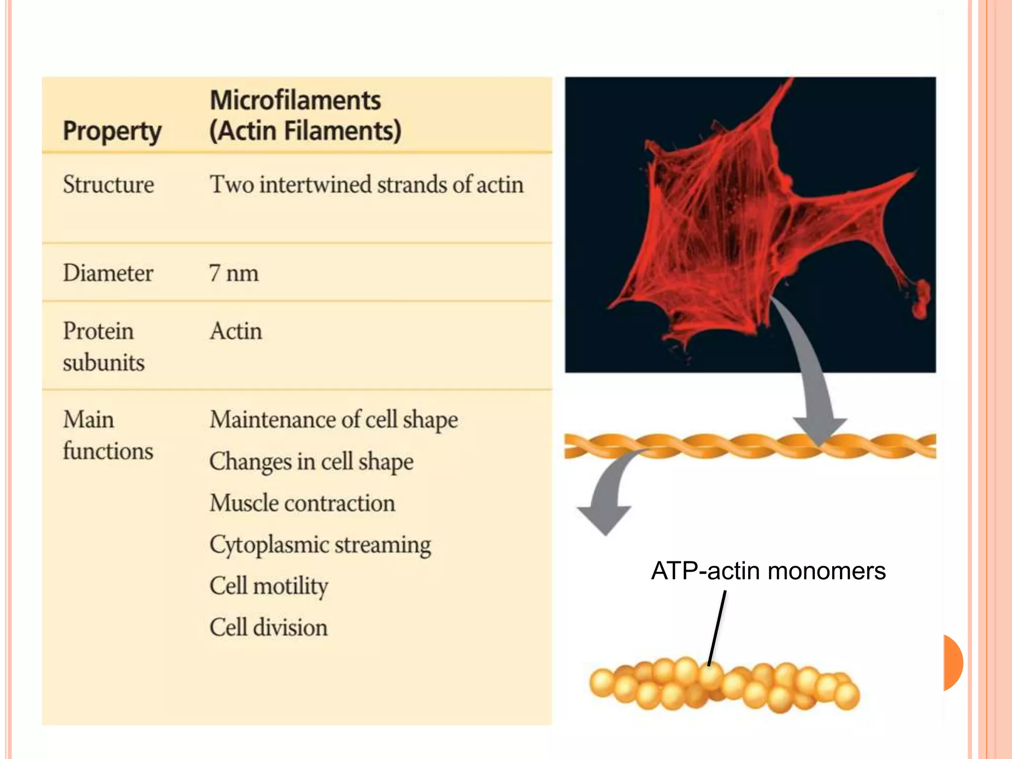
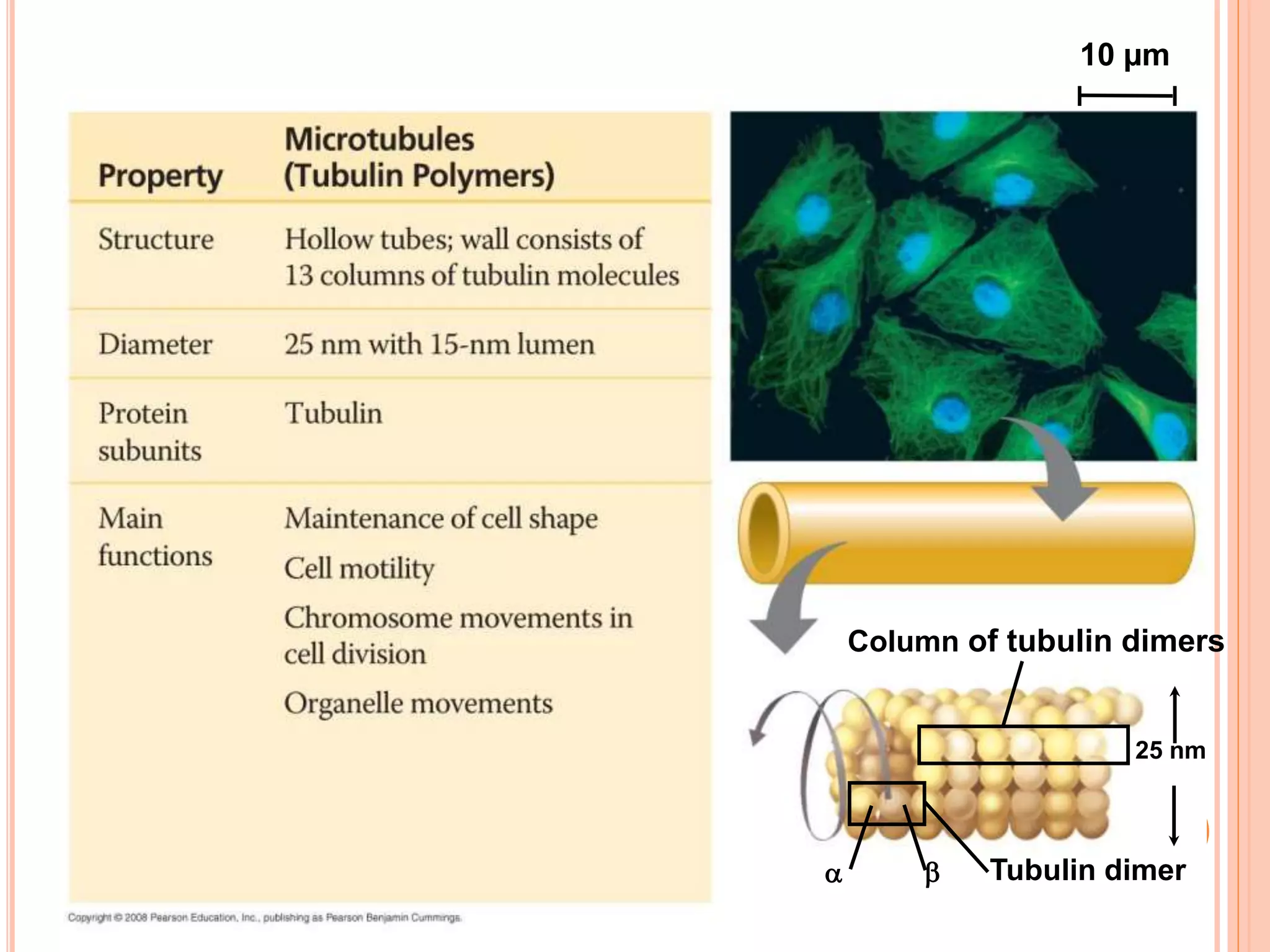
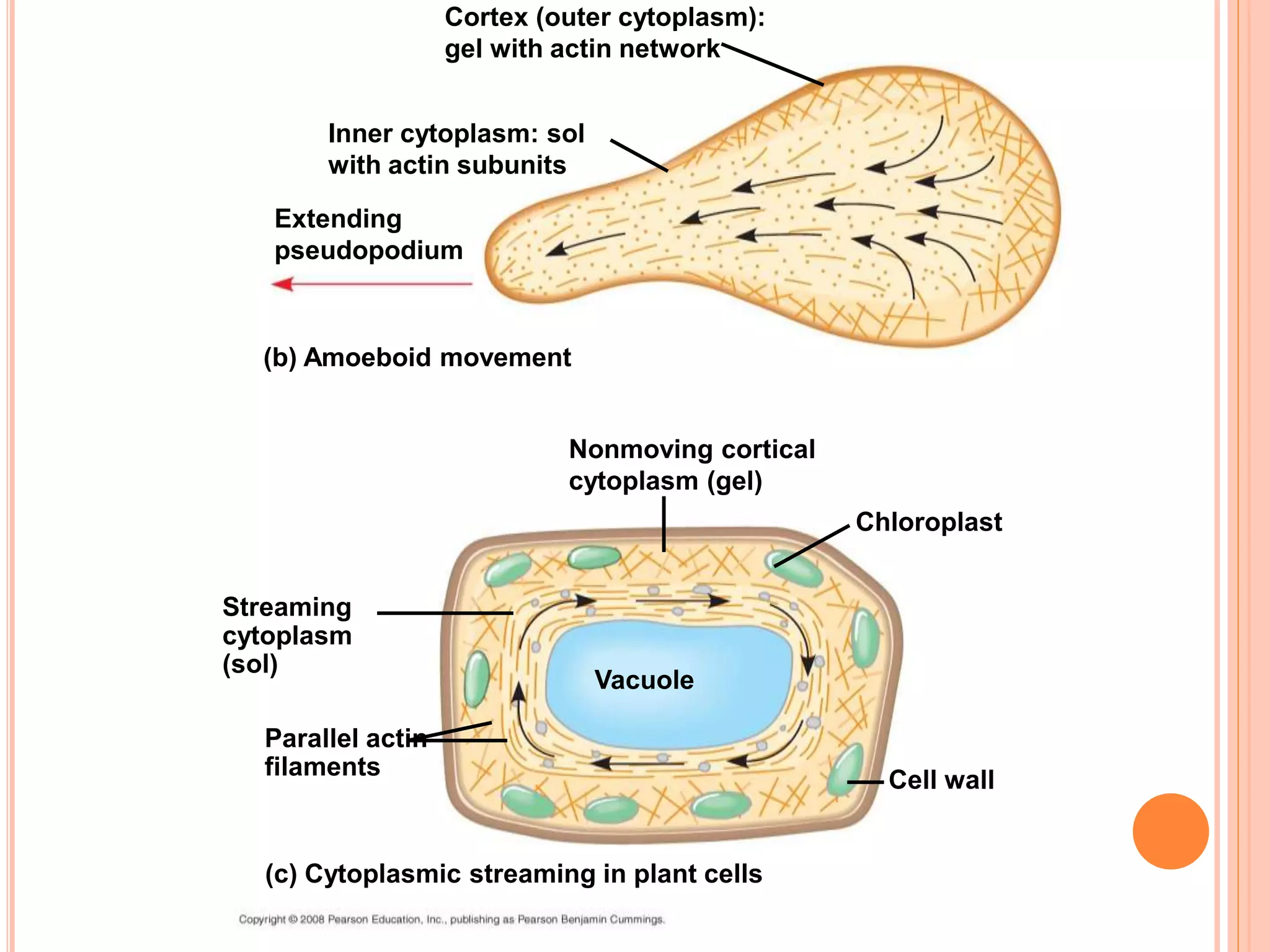
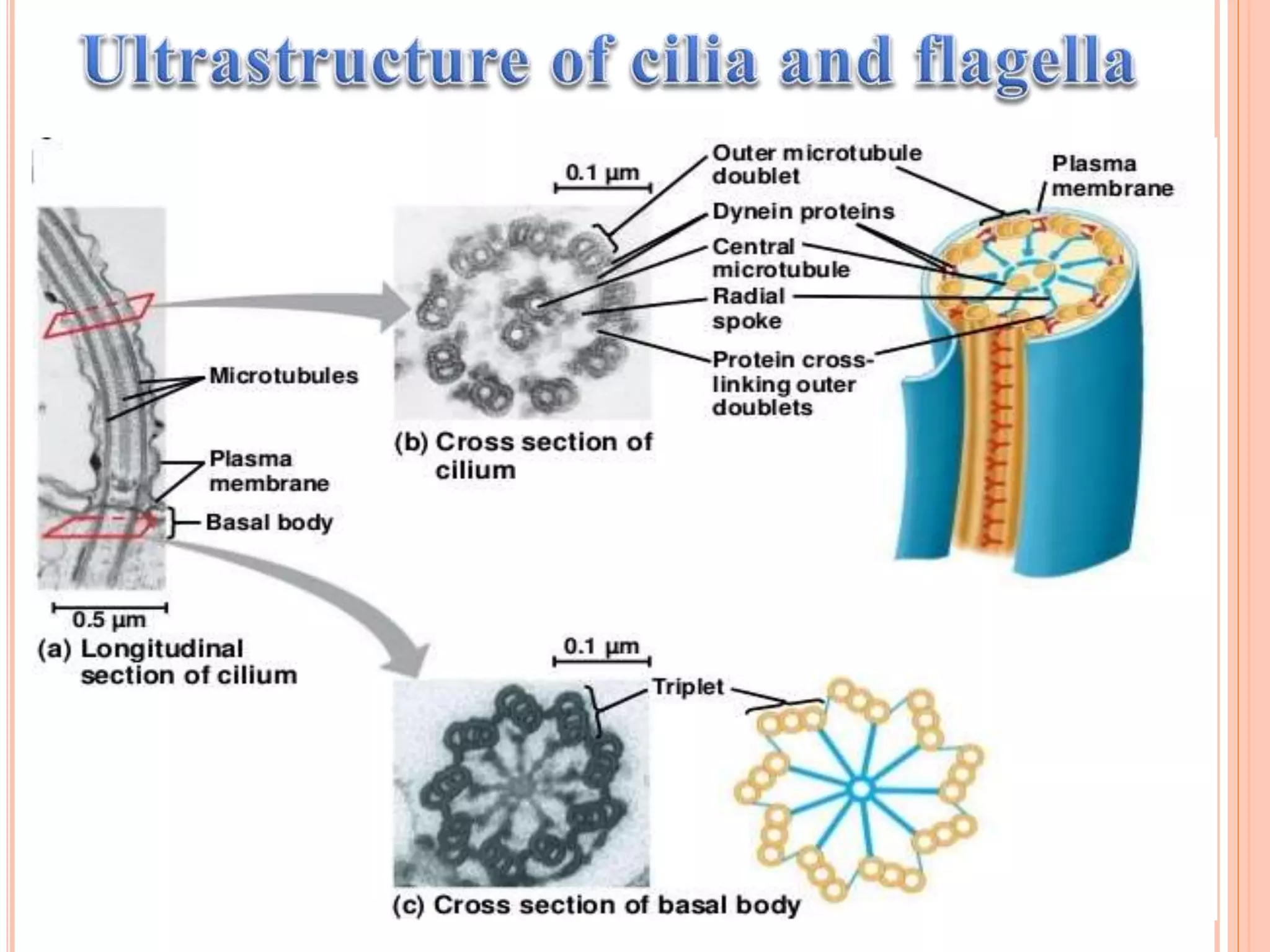
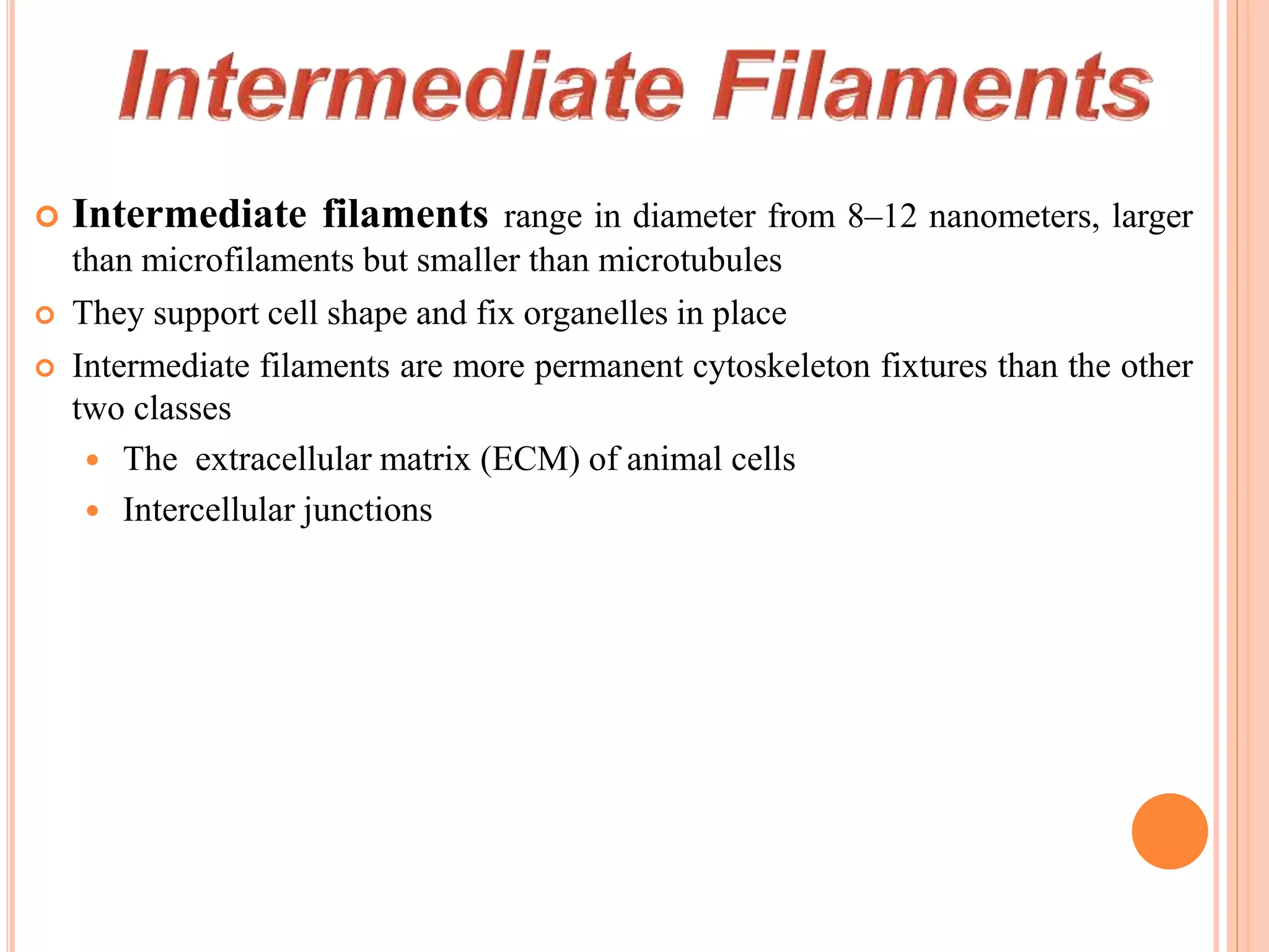
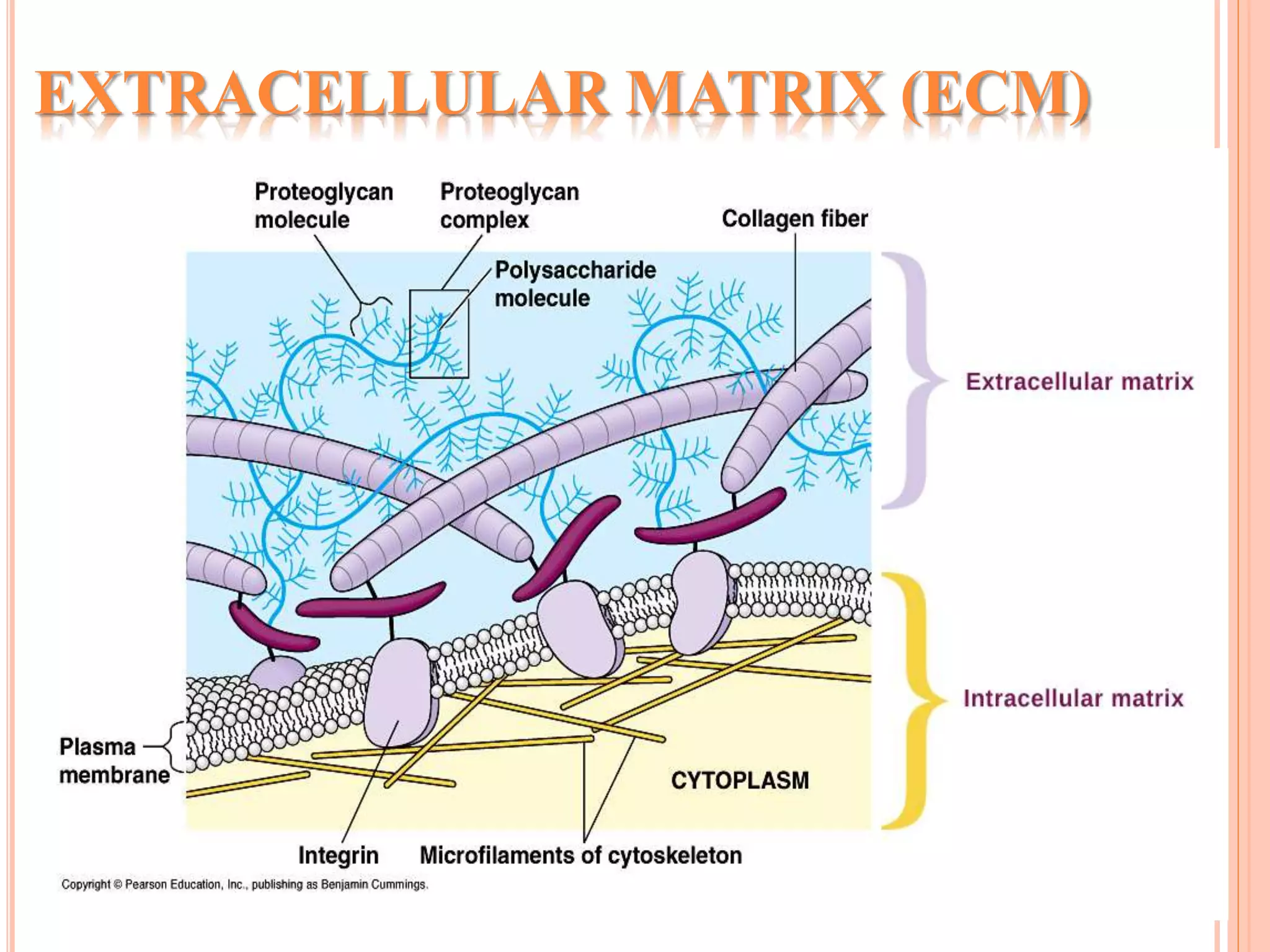
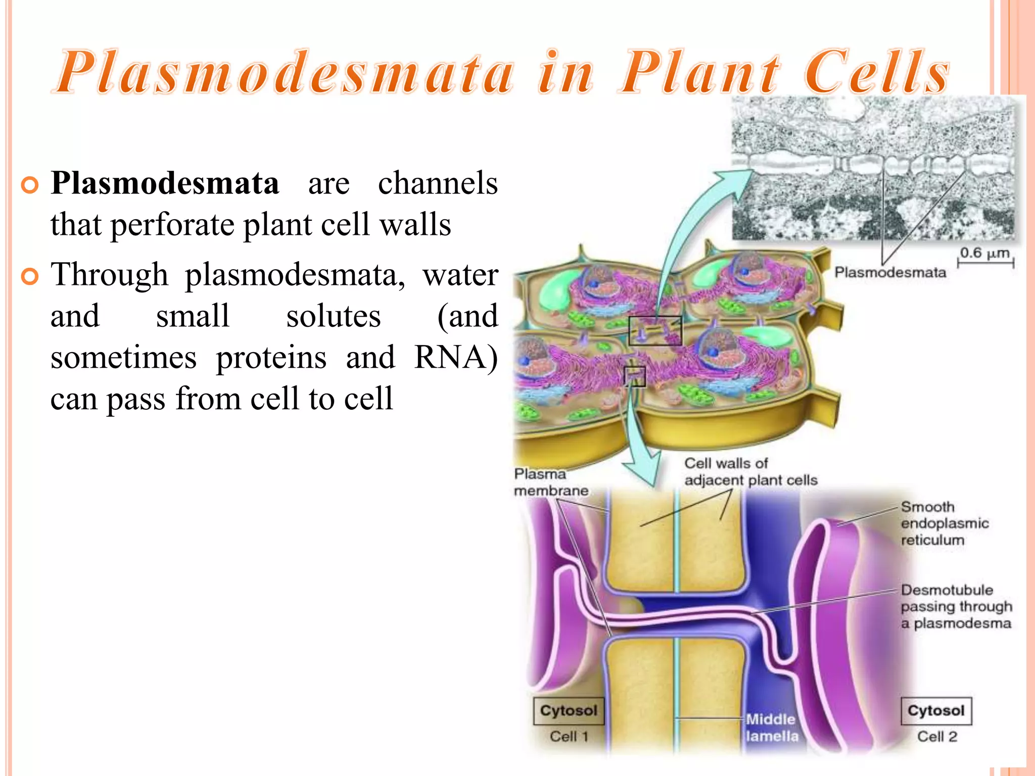
 The cytoskeleton is a network of fibers extending throughout the
cytoplasm
 It organizes the cell’s structures and activities, anchoring many
organelles
 It is composed of three types of molecular structures:
 Microfilaments
 Microtubules
 Intermediate filaments
Microfilaments:
 Actin
7nm
Microtubules:
 Tubulins (a, b)
25 nm
Intermediate filaments:
 Lamin
 cell specific prot. (e.g.
Vimentin)
8-12 nm
Microfilament
 The cytoskeleton helps to support the cell and maintain
its shape
 It interacts with motor proteins to produce motility
 Inside the cell, vesicles can travel along “monorails”
provided by the cytoskeleton
 Recent evidence suggests that the cytoskeleton may
help regulate biochemical activities
Properties Microfilament Microtubules Intermediate
filaments
Subunits incorporated
into polymer
ATP - actin
monomers
GTP - αβ - tubulin
heterodimer
Various globular
proteins
Preferential site of
incorporation
+ End barbed + End β - tubulin Internal
Polarity Yes Yes No
Enzymatic activity ATPase GTPase None
Motor proteins Myosin's Kinesins, Dyneins None
Major group of
associated proteins
Actin – binding
proteins
Maps Plakins
Structure
Flexible, helical
filament
Stiff, hollow tube Tough, ropelike fibers
Dimensions 8 nm diam. 25 nm outer diam. 10 nm diam.
Distribution All eukaryotes All eukaryotes Animals
Primary functions Motility, contractility
Support, intracellular
transport, cell
organization
Structural support
ATP-actin monomers
10 µm
Column of tubulin dimers
Tubulin dimer 
25 nm
Fibrous subunit (keratins
coiled together)
8–12 nm
5 µm
Keratin proteins
Tissue level
Muscle
movement
Determines shape of
the cell
Motility of the cells
Cell adhesion
 Mitosis, meiosis
Cellular level
Subcellular level
Anchors organelles
Organization of organelles
Provides tensile strength
 Movement of
chromosomes
Organizing cell polarity
Intracellular movement of
vesicles
 Endocytosis – clathrin-
mediated endocytosis and
phagocytosis
Dynamic
Adaptable
Stable
Strong
o Cytoskeletal filaments are all constructed
from smaller protein subunits
o All form as helical assemblies
of subunits
o Noncovalent interactions: rapid assembly
and disassembly
Intermediate filaments- resistant to stretching forces
 Microfilaments are solid rods about 7 nm in diameter,
built as a twisted double chain of actin subunits
 The structural role of microfilaments is to bear tension,
resisting pulling forces within the cell
 They form a 3-D network called the cortex just inside
the plasma membrane to help support the cell’s shape
 Bundles of microfilaments make up the core of
microvilli of intestinal cells.
Microvillus
Plasma membrane
Microfilaments (actin
filaments)
Intermediate filaments
0.25 µm
F-actin
G-actin
Two parallel protofilaments
that twist around each other in
a right-handed helix
 Microfilaments that function in cellular motility contain the
protein myosin in addition to actin
 In muscle cells, thousands of actin filaments are arranged parallel
to one another
 Thicker filaments composed of myosin interdigitate with the
thinner actin fibers
 Myosin molecules walk along the actin filament, pulling stacks of
actin fibers together and shortening
the cell.
o Localized contraction brought about by actin and myosin also
drives amoeboid movement
o Pseudopodia (cellular extensions) extend and contract through
the reversible assembly and contraction of actin subunits into
microfilaments
 Cytoplasmic streaming is a circular flow of cytoplasm within
cells
 This streaming speeds distribution of materials within the cell
 In plant cells, actin-myosin interactions and sol-gel
transformations drive cytoplasmic streaming
Cortex (outer cytoplasm):
gel with actin network
Inner cytoplasm: sol
with actin subunits
Extending
pseudopodium
(b) Amoeboid movement
Nonmoving cortical
cytoplasm (gel)
Chloroplast
Cell wall
Streaming
cytoplasm
(sol)
Parallel actin
filaments
(c) Cytoplasmic streaming in plant cells
Vacuole
 Microtubules are hollow
rods about 25 nm in
diameter and about 200 nm
to 25 microns long
 Functions of microtubules:
 Shaping the cell
 Guiding movement of
organelles
 Separating chromosomes
during cell division
Interphase cell
Dividing cell
Neuron
centrosome
Basal body
Cilla
spindle
centrosome
axon
-Centrosome
-Cilia / flagellum
-Mitotic system
- Vesicular transport
o In many cells, microtubules grow out from a centrosome near the
nucleus
o The centrosome is a “microtubule-organizing center”
o In animal cells, the centrosome has a pair of centrioles, each with
nine triplets of microtubules arranged in a ring
5 µm
Direction of swimming
(a) Motion of flagella
Direction of organism’s
movement
Power stroke Recovery stroke
(b) Motion of cilia
15 µm
 Cilia and flagella share a
common ultrastructure:
 A core of microtubules sheathed
by the plasma membrane
 A basal body that anchors the
cilium or flagellum
 A motor protein called dynein,
which drives the bending
movements of a cilium or
flagellum
Role of the dynein arms in beating cilia
Telescopic effect Beating
 Intermediate filaments range in diameter from 8–12 nanometers, larger
than microfilaments but smaller than microtubules
 They support cell shape and fix organelles in place
 Intermediate filaments are more permanent cytoskeleton fixtures than the other
two classes
 The extracellular matrix (ECM) of animal cells
 Intercellular junctions
 Animal cells lack cell walls but are covered by an elaborate
extracellular matrix (ECM)
 The ECM is made up of glycoprotein's such as collagen,
proteoglycans, and fibronectin
 ECM proteins bind to receptor proteins in the plasma membrane
called integrins
 Functions of the ECM:
 Support
 Adhesion
 Movement
 Regulation
EXTRACELLULAR MATRIX (ECM)
 Neighboring cells in tissues, organs, or organ systems often
adhere, interact, and communicate through direct physical
contact
 Intercellular junctions facilitate this contact
 There are several types of intercellular junctions
 Plasmodesmata
 Tight junctions
 Desmosomes
 Gap junctions
 Plasmodesmata are channels
that perforate plant cell walls
 Through plasmodesmata, water
and small solutes (and
sometimes proteins and RNA)
can pass from cell to cell
 At tight junctions, membranes of neighboring cells are pressed
together, preventing leakage of extracellular fluid
 Desmosomes (anchoring junctions) fasten cells together into
strong sheets
 Gap junctions (communicating junctions) provide cytoplasmic
channels between adjacent cells
Thank you

Orginagition and role of cytoskeleton

  • 1.
  • 2.
     The cytoskeletonis a network of fibers extending throughout the cytoplasm  It organizes the cell’s structures and activities, anchoring many organelles  It is composed of three types of molecular structures:  Microfilaments  Microtubules  Intermediate filaments
  • 3.
    Microfilaments:  Actin 7nm Microtubules:  Tubulins(a, b) 25 nm Intermediate filaments:  Lamin  cell specific prot. (e.g. Vimentin) 8-12 nm Microfilament
  • 4.
     The cytoskeletonhelps to support the cell and maintain its shape  It interacts with motor proteins to produce motility  Inside the cell, vesicles can travel along “monorails” provided by the cytoskeleton  Recent evidence suggests that the cytoskeleton may help regulate biochemical activities
  • 5.
    Properties Microfilament MicrotubulesIntermediate filaments Subunits incorporated into polymer ATP - actin monomers GTP - αβ - tubulin heterodimer Various globular proteins Preferential site of incorporation + End barbed + End β - tubulin Internal Polarity Yes Yes No Enzymatic activity ATPase GTPase None Motor proteins Myosin's Kinesins, Dyneins None Major group of associated proteins Actin – binding proteins Maps Plakins Structure Flexible, helical filament Stiff, hollow tube Tough, ropelike fibers Dimensions 8 nm diam. 25 nm outer diam. 10 nm diam. Distribution All eukaryotes All eukaryotes Animals Primary functions Motility, contractility Support, intracellular transport, cell organization Structural support
  • 6.
  • 7.
    10 µm Column oftubulin dimers Tubulin dimer  25 nm
  • 8.
    Fibrous subunit (keratins coiledtogether) 8–12 nm 5 µm Keratin proteins
  • 9.
    Tissue level Muscle movement Determines shapeof the cell Motility of the cells Cell adhesion  Mitosis, meiosis Cellular level Subcellular level Anchors organelles Organization of organelles Provides tensile strength  Movement of chromosomes Organizing cell polarity Intracellular movement of vesicles  Endocytosis – clathrin- mediated endocytosis and phagocytosis Dynamic Adaptable Stable Strong
  • 10.
    o Cytoskeletal filamentsare all constructed from smaller protein subunits o All form as helical assemblies of subunits o Noncovalent interactions: rapid assembly and disassembly
  • 12.
  • 13.
     Microfilaments aresolid rods about 7 nm in diameter, built as a twisted double chain of actin subunits  The structural role of microfilaments is to bear tension, resisting pulling forces within the cell  They form a 3-D network called the cortex just inside the plasma membrane to help support the cell’s shape  Bundles of microfilaments make up the core of microvilli of intestinal cells.
  • 14.
  • 15.
    F-actin G-actin Two parallel protofilaments thattwist around each other in a right-handed helix
  • 16.
     Microfilaments thatfunction in cellular motility contain the protein myosin in addition to actin  In muscle cells, thousands of actin filaments are arranged parallel to one another  Thicker filaments composed of myosin interdigitate with the thinner actin fibers  Myosin molecules walk along the actin filament, pulling stacks of actin fibers together and shortening the cell.
  • 18.
    o Localized contractionbrought about by actin and myosin also drives amoeboid movement o Pseudopodia (cellular extensions) extend and contract through the reversible assembly and contraction of actin subunits into microfilaments  Cytoplasmic streaming is a circular flow of cytoplasm within cells  This streaming speeds distribution of materials within the cell  In plant cells, actin-myosin interactions and sol-gel transformations drive cytoplasmic streaming
  • 19.
    Cortex (outer cytoplasm): gelwith actin network Inner cytoplasm: sol with actin subunits Extending pseudopodium (b) Amoeboid movement Nonmoving cortical cytoplasm (gel) Chloroplast Cell wall Streaming cytoplasm (sol) Parallel actin filaments (c) Cytoplasmic streaming in plant cells Vacuole
  • 20.
     Microtubules arehollow rods about 25 nm in diameter and about 200 nm to 25 microns long  Functions of microtubules:  Shaping the cell  Guiding movement of organelles  Separating chromosomes during cell division
  • 21.
    Interphase cell Dividing cell Neuron centrosome Basalbody Cilla spindle centrosome axon -Centrosome -Cilia / flagellum -Mitotic system - Vesicular transport
  • 23.
    o In manycells, microtubules grow out from a centrosome near the nucleus o The centrosome is a “microtubule-organizing center” o In animal cells, the centrosome has a pair of centrioles, each with nine triplets of microtubules arranged in a ring
  • 24.
    5 µm Direction ofswimming (a) Motion of flagella Direction of organism’s movement Power stroke Recovery stroke (b) Motion of cilia 15 µm  Cilia and flagella share a common ultrastructure:  A core of microtubules sheathed by the plasma membrane  A basal body that anchors the cilium or flagellum  A motor protein called dynein, which drives the bending movements of a cilium or flagellum
  • 26.
    Role of thedynein arms in beating cilia Telescopic effect Beating
  • 27.
     Intermediate filamentsrange in diameter from 8–12 nanometers, larger than microfilaments but smaller than microtubules  They support cell shape and fix organelles in place  Intermediate filaments are more permanent cytoskeleton fixtures than the other two classes  The extracellular matrix (ECM) of animal cells  Intercellular junctions
  • 31.
     Animal cellslack cell walls but are covered by an elaborate extracellular matrix (ECM)  The ECM is made up of glycoprotein's such as collagen, proteoglycans, and fibronectin  ECM proteins bind to receptor proteins in the plasma membrane called integrins  Functions of the ECM:  Support  Adhesion  Movement  Regulation
  • 32.
  • 33.
     Neighboring cellsin tissues, organs, or organ systems often adhere, interact, and communicate through direct physical contact  Intercellular junctions facilitate this contact  There are several types of intercellular junctions  Plasmodesmata  Tight junctions  Desmosomes  Gap junctions
  • 34.
     Plasmodesmata arechannels that perforate plant cell walls  Through plasmodesmata, water and small solutes (and sometimes proteins and RNA) can pass from cell to cell
  • 35.
     At tightjunctions, membranes of neighboring cells are pressed together, preventing leakage of extracellular fluid  Desmosomes (anchoring junctions) fasten cells together into strong sheets  Gap junctions (communicating junctions) provide cytoplasmic channels between adjacent cells
  • 37.