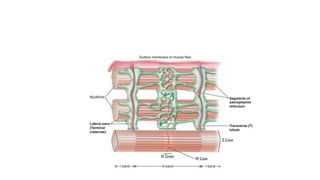
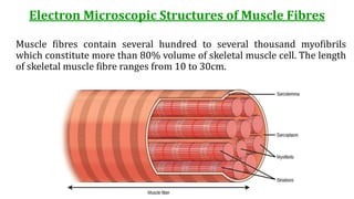
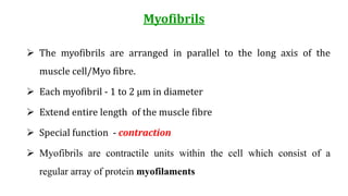
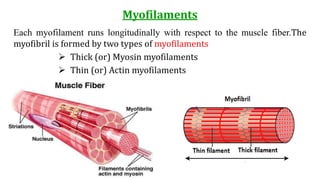
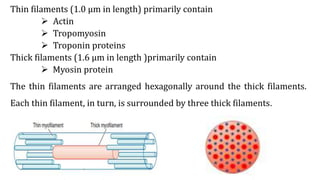
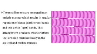
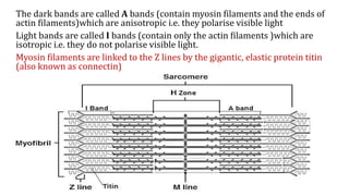
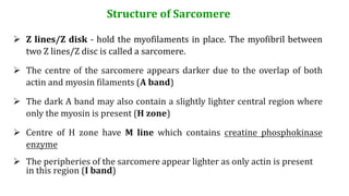
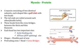
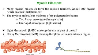
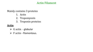
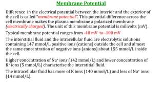
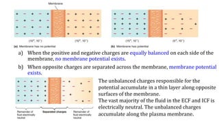
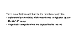
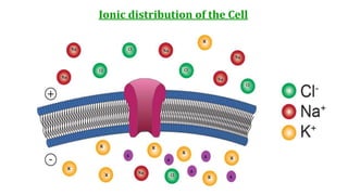
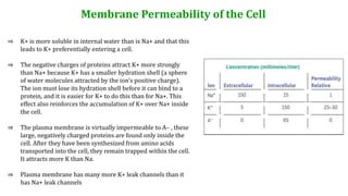
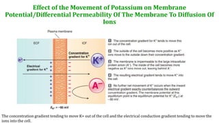
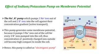
This document provides information about muscle physiology and the structure of muscle fibers at the microscopic level. It discusses the basic units of muscles including myofibrils, myofilaments, sarcomeres, and the proteins actin and myosin that make up the thin and thick filaments. It describes the striated banding pattern visible in muscle fibers due to the organized arrangement of these contractile proteins. It also covers concepts related to muscle membrane potential including ion gradients and the role of the sodium-potassium pump in establishing the resting potential.
i've used this note before this for my first year medicine in egypt. Fot those who taking this course(medicine) , I hope it'll give some ideas to you to study about this subject.
Goodluck :) !
All animal cells have a voltage across their cell membranes. Neurons and muscle cells can alter this potential and conduct impulses through their membranes, called nerve impulses. This is a comprehensive note on the "resting membrane potential" of a cell membrane, when no impulses are being conducted.
Anatomy of the masticatory machine. It consists of a fixed and a movable member. The movable member is activated by a series of voluntary muscles, and its efficiency is increased by another set of voluntary muscles that feed the machine.
1st year medical school physiology essay:
Describe the effects between the action potential arriving at the axon terminal and skeletal muscle contraction.
Chemical and molecular basis of muscle contractionChirag Dhankhar
here in this ppt I have told about the different types of muscles their biological cycle of muscle contraction, needs of contraction, neural network working for muscle contraction, atp and cp energy use in muscles , how energy is used and made by muscles in middle of the exercise, anatomy of muscles, working of muscles, different types of bands and proteins needed for muscle contraction
Muscle movement plays an important role in day to day life where the contraction and relaxation of muscle is significant. The current slide has been developed with the focus on different phases during muscle contraction and the physiological change involved on it.
Muscle organization and structure Characteristics of muscle - give s.pdfarshicollection001
Muscle organization and structure Characteristics of muscle - give specific example of each of
those terms Kinds of muscle - overall, know skeletal in and out. Know basics of cardiac and
smooth, esp in compare/contrast with skel or in a way that makes it work the best organization
and hierarchy of macro to micro structures Larger to smaller bundles, wrapped in own sheath
Muscle fibers - organization and function of these components sarcoplasm (surrounds/contains
mitos, nuclei, glycogen, myoglobin) Myofibril Sarcomere - thick and thin filaments, actin,
troponin, tropomyosin, myosin, Z disk. RELATED TO LAB: ID kinds of muscle and what the
light/dark bands are Triad - T tubules, SR and Terminal cisternae - WHY IS THIS
IMPORTANT? Electrophys basics Voltage - what is it, how does it relate to membrane?
Electrochem gradients - be able to predict direction ion would flow given vm and E(ion) Finish
elcetrophys basics, start muscle contraction Electrophys basics Electrochem gradients - be able
to predict direction ion would flow given vm and E(ion). Know WHAT Nernst is used for and
what the important values are for calculation concentrations in and out), but don\'t memorize
equation; won\'t be asked to use it. Movement of ions Ion channels Different kinds, stimuli to
open Pumps - active Why is Na/K super important? (and, later, why would Ca pump back into
SR be super important?) Again: Voltage - what is it, how does it relate to membrane? AP v at
rest v Equilib potential (and later, post synaptic potential) NMJ and Steps to muscle contraction -
Understand organization and how all the parts work together. We went really into depth here;
fair to expect some fluency with it. To test yourself, think about what would happen if you
messed with individual components, ion channels, signaling molecules. Axon terminal ACh
(and -esterase) Synapse Post-synaptic receptors and junctional folds Sarcomere - thick and thin
filaments, actin, troponin, tropomyosin, myosin, Z disk. RELATED TO LAB: ID kinds of
muscle and what the light/dark bands are Triad - T tubules, SR and Terminal cisternae - WHY
IS THIS IMPORTANT? Muscle contraction, con\'t - see Week 5 Write! Fluent in the major
steps - what changes concentration, shape/length, moves, binds
Solution
The questions are huge and broad, I have tried to answer some.
Kinds of Muscles
There are three types of muscle - Skeletal, smooth and cardiac. Skeletal muscle is attached to
bones and moves and supports the skeleton. Smooth muscle surrounds hollow cavities and tubes.
Cardiac muscle is the muscle of the heart.
Structure of skeletal muscles
The most striking feature seen when observing a muscle fiber through a light microscope is a
series of light and dark bands perpendicular to the long axis of the fiber. Both skeletal- and
cardiac- muscle fibers have this characteristic banding and are known as striated muscles.
Smooth muscles do not show banding pattern. The striated pattern in skeletal and cardiac fibers
results from .
Francesca Gottschalk - How can education support child empowerment.pptxEduSkills OECD
Francesca Gottschalk from the OECD’s Centre for Educational Research and Innovation presents at the Ask an Expert Webinar: How can education support child empowerment?
Embracing GenAI - A Strategic ImperativePeter Windle
Artificial Intelligence (AI) technologies such as Generative AI, Image Generators and Large Language Models have had a dramatic impact on teaching, learning and assessment over the past 18 months. The most immediate threat AI posed was to Academic Integrity with Higher Education Institutes (HEIs) focusing their efforts on combating the use of GenAI in assessment. Guidelines were developed for staff and students, policies put in place too. Innovative educators have forged paths in the use of Generative AI for teaching, learning and assessments leading to pockets of transformation springing up across HEIs, often with little or no top-down guidance, support or direction.
This Gasta posits a strategic approach to integrating AI into HEIs to prepare staff, students and the curriculum for an evolving world and workplace. We will highlight the advantages of working with these technologies beyond the realm of teaching, learning and assessment by considering prompt engineering skills, industry impact, curriculum changes, and the need for staff upskilling. In contrast, not engaging strategically with Generative AI poses risks, including falling behind peers, missed opportunities and failing to ensure our graduates remain employable. The rapid evolution of AI technologies necessitates a proactive and strategic approach if we are to remain relevant.
June 3, 2024 Anti-Semitism Letter Sent to MIT President Kornbluth and MIT Cor...Levi Shapiro
Letter from the Congress of the United States regarding Anti-Semitism sent June 3rd to MIT President Sally Kornbluth, MIT Corp Chair, Mark Gorenberg
Dear Dr. Kornbluth and Mr. Gorenberg,
The US House of Representatives is deeply concerned by ongoing and pervasive acts of antisemitic
harassment and intimidation at the Massachusetts Institute of Technology (MIT). Failing to act decisively to ensure a safe learning environment for all students would be a grave dereliction of your responsibilities as President of MIT and Chair of the MIT Corporation.
This Congress will not stand idly by and allow an environment hostile to Jewish students to persist. The House believes that your institution is in violation of Title VI of the Civil Rights Act, and the inability or
unwillingness to rectify this violation through action requires accountability.
Postsecondary education is a unique opportunity for students to learn and have their ideas and beliefs challenged. However, universities receiving hundreds of millions of federal funds annually have denied
students that opportunity and have been hijacked to become venues for the promotion of terrorism, antisemitic harassment and intimidation, unlawful encampments, and in some cases, assaults and riots.
The House of Representatives will not countenance the use of federal funds to indoctrinate students into hateful, antisemitic, anti-American supporters of terrorism. Investigations into campus antisemitism by the Committee on Education and the Workforce and the Committee on Ways and Means have been expanded into a Congress-wide probe across all relevant jurisdictions to address this national crisis. The undersigned Committees will conduct oversight into the use of federal funds at MIT and its learning environment under authorities granted to each Committee.
• The Committee on Education and the Workforce has been investigating your institution since December 7, 2023. The Committee has broad jurisdiction over postsecondary education, including its compliance with Title VI of the Civil Rights Act, campus safety concerns over disruptions to the learning environment, and the awarding of federal student aid under the Higher Education Act.
• The Committee on Oversight and Accountability is investigating the sources of funding and other support flowing to groups espousing pro-Hamas propaganda and engaged in antisemitic harassment and intimidation of students. The Committee on Oversight and Accountability is the principal oversight committee of the US House of Representatives and has broad authority to investigate “any matter” at “any time” under House Rule X.
• The Committee on Ways and Means has been investigating several universities since November 15, 2023, when the Committee held a hearing entitled From Ivory Towers to Dark Corners: Investigating the Nexus Between Antisemitism, Tax-Exempt Universities, and Terror Financing. The Committee followed the hearing with letters to those institutions on January 10, 202
2. Muscle Physiology-Syllabus
Basic muscle unit characteristic
Electrical phenomenon in muscle cell
Muscle action potential, excitation and propagation of impulse
Latent period, refractiveness, threshold level
All and none law
Contractile mechanism - excitation - contraction coupling
Neuro-muscular transmission
Types of muscle contraction
Phenomenon of Fatigue, Rigor mortis
3.
4. Electron Microscopic Structures of Muscle Fibres
Muscle fibres contain several hundred to several thousand myofibrils
which constitute more than 80% volume of skeletal muscle cell. The length
of skeletal muscle fibre ranges from 10 to 30cm.
5. Myofibrils
The myofibrils are arranged in parallel to the long axis of the
muscle cell/Myo fibre.
Each myofibril - 1 to 2 µm in diameter
Extend entire length of the muscle fibre
Special function - contraction
Myofibrils are contractile units within the cell which consist of a
regular array of protein myofilaments
6. Myofilaments
Each myofilament runs longitudinally with respect to the muscle fiber.The
myofibril is formed by two types of myofilaments
Thick (or) Myosin myofilaments
Thin (or) Actin myofilaments
7. Thin filaments (1.0 μm in length) primarily contain
Actin
Tropomyosin
Troponin proteins
Thick filaments (1.6 μm in length )primarily contain
Myosin protein
The thin filaments are arranged hexagonally around the thick filaments.
Each thin filament, in turn, is surrounded by three thick filaments.
8. The myofilaments are arranged in an
orderly manner which results in regular
repetition of dense (dark) cross-bands
and less dense (light) bands. This
arrangement produces cross-striations
that are seen microscopically in the
skeletal and cardiac muscles.
9. The dark bands are called A bands (contain myosin filaments and the ends of
actin filaments)which are anisotropic i.e. they polarise visible light
Light bands are called I bands (contain only the actin filaments )which are
isotropic i.e. they do not polarise visible light.
Myosin filaments are linked to the Z lines by the gigantic, elastic protein titin
(also known as connectin)
10. Structure of Sarcomere
Z lines/Z disk - hold the myofilaments in place. The myofibril between
two Z lines/Z disc is called a sarcomere.
The centre of the sarcomere appears darker due to the overlap of both
actin and myosin filaments (A band)
The dark A band may also contain a slightly lighter central region where
only the myosin is present (H zone)
Centre of H zone have M line which contains creatine phosphokinase
enzyme
The peripheries of the sarcomere appear lighter as only actin is present
in this region (I band)
11. Myosin - Protein
A myosin consisting of two identical
subunits, each shaped like a golf club with
two heads
The tail ends are coiled around each
other(double helix).
These heads form the cross bridges.
between the thick and thin
filaments
Each head has two important sites
Actin binding site
ATPase (ATP-splitting) site
Hinges – Flexible part of arm
Protruding arms + Head = Cross Bridges
12. Myosin Filament
Many myosin molecules form the myosin filament. About 500 myosin
heads on each thick filament
The myosin molecule is made up of six polypeptide chains:
o Two heavy meromysin (heavy chain)
o Four light meromysin. (light chain)
Light Meromysin (LMM) makeup the major part of the tail
Heavy Meromysin (HMM) makeup the globular head and neck region.
14. Membrane Potential
Difference in the electrical potential between the interior and the exterior of
the cell is called “membrane potential”. This potential difference across the
cell membrane makes the plasma membrane a polarized membrane
(electrically charged). The unit of this membrane potential is milivolts (mV).
Typical membrane potential ranges from -40 mV to -100 mV
The interstitial fluid and the intracellular fluid are electrolytic solutions
containing 147 mmol/L positive ions (cations) outside the cell and almost
the same concentration of negative ions (anions) about 155 mmol/L inside
the cell.
Higher concentration of Na+ ions (142 mmol/L) and lower concentration of
K+ ions (5 mmol/L) characterise the interstitial fluid.
The intracellular fluid has more of K ions (140 mmol/L) and less of Na+ ions
(14 mmol/L).
15. a) When the positive and negative charges are equally balanced on each side of the
membrane, no membrane potential exists.
b) When opposite charges are separated across the membrane, membrane potential
exists.
The unbalanced charges responsible for the
potential accumulate in a thin layer along opposite
surfaces of the membrane.
The vast majority of the fluid in the ECF and ICF is
electrically neutral. The unbalanced charges
accumulate along the plasma membrane.
16. Three major factors contribute to the membrane potential.
• Differential permeability of the membrane to diffusion of ions
• The Na+, K+ pump
• Negatively charged anions are trapped inside the cell
18. Membrane Permeability of the Cell
⇒ K+ is more soluble in internal water than is Na+ and that this
leads to K+ preferentially entering a cell.
⇒ The negative charges of proteins attract K+ more strongly
than Na+ because K+ has a smaller hydration shell (a sphere
of water molecules attracted by the ion’s positive charge).
The ion must lose its hydration shell before it can bind to a
protein, and it is easier for K+ to do this than for Na+. This
effect also reinforces the accumulation of K+ over Na+ inside
the cell.
⇒ The plasma membrane is virtually impermeable to A– , these
large, negatively charged proteins are found only inside the
cell. After they have been synthesized from amino acids
transported into the cell, they remain trapped within the cell.
It attracts more K than Na.
⇒ Plasma membrane has many more K+ leak channels than it
has Na+ leak channels
19. Effect of the Movement of Potassium on Membrane
Potential/Differential Permeability Of The Membrane To Diffusion Of
Ions
The concentration gradient tending to move K+ out of the cell and the electrical conduction gradient tending to move the
ions into the cell.
20. Effect of Sodium/Potassium Pump on Membrane Potential
The Na+, K+ pump which pumps 3 Na+ ions out of
the cell and 2 K+ ions into the cell against their
concentration gradient (active transport).
This pump generates some membrane potential,
because it pumps 3 Na+ ions out of the cell for
every 2 K+ ions pumped into the cell, thus
concentration of positively charged ions outside
cell become high compare to inside the cell.
Hence, this pump is called as “electrogenic pump”