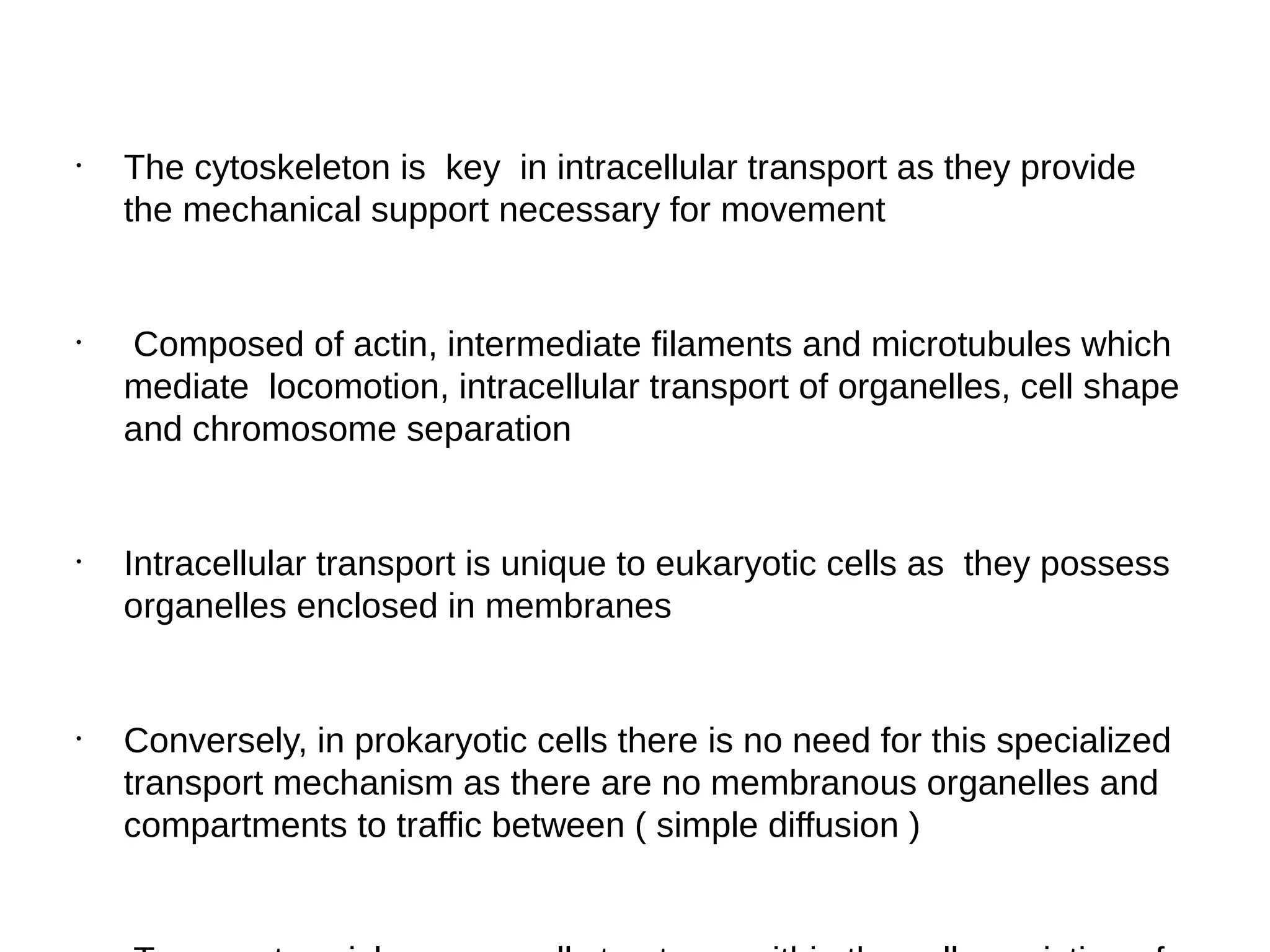
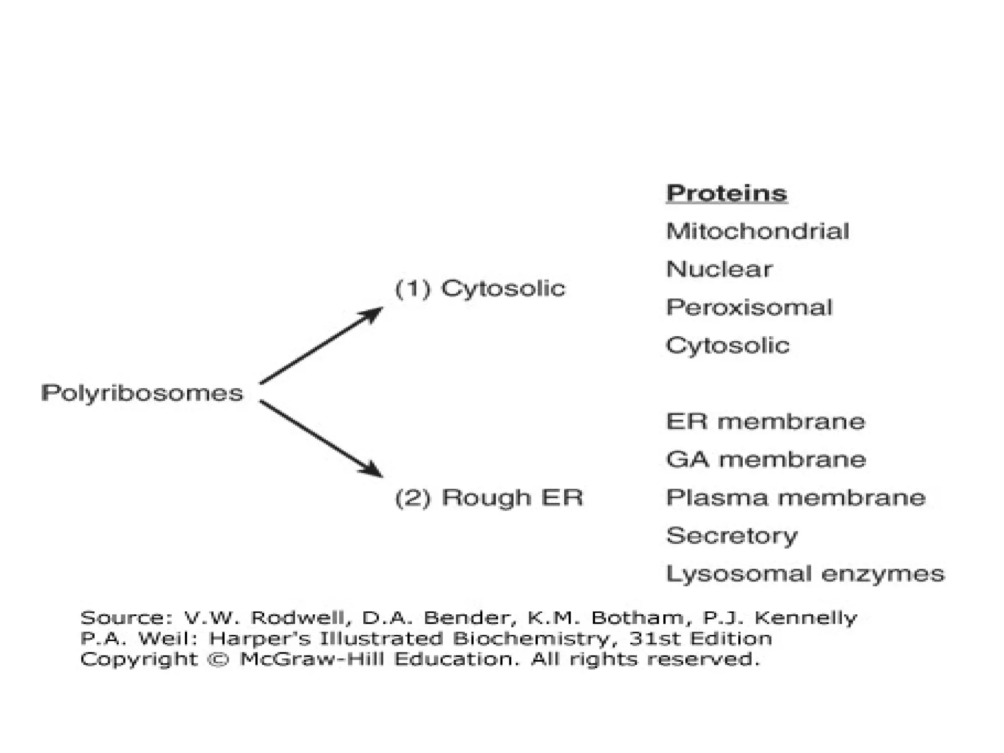
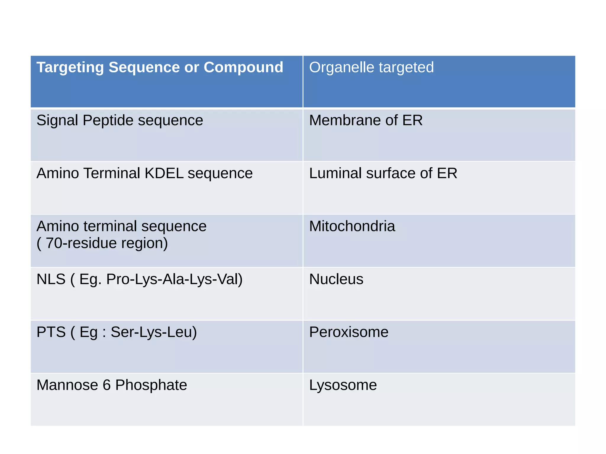
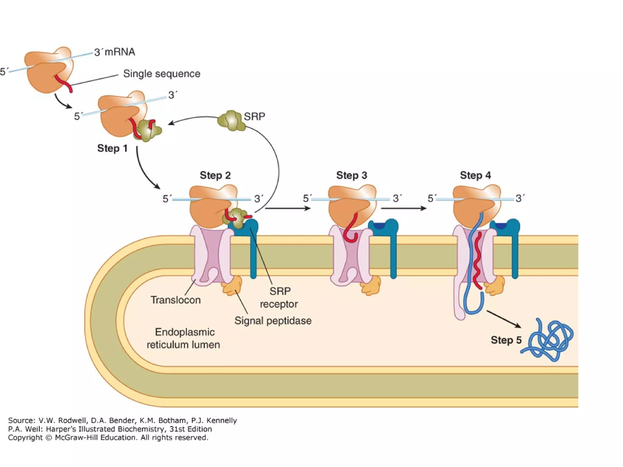
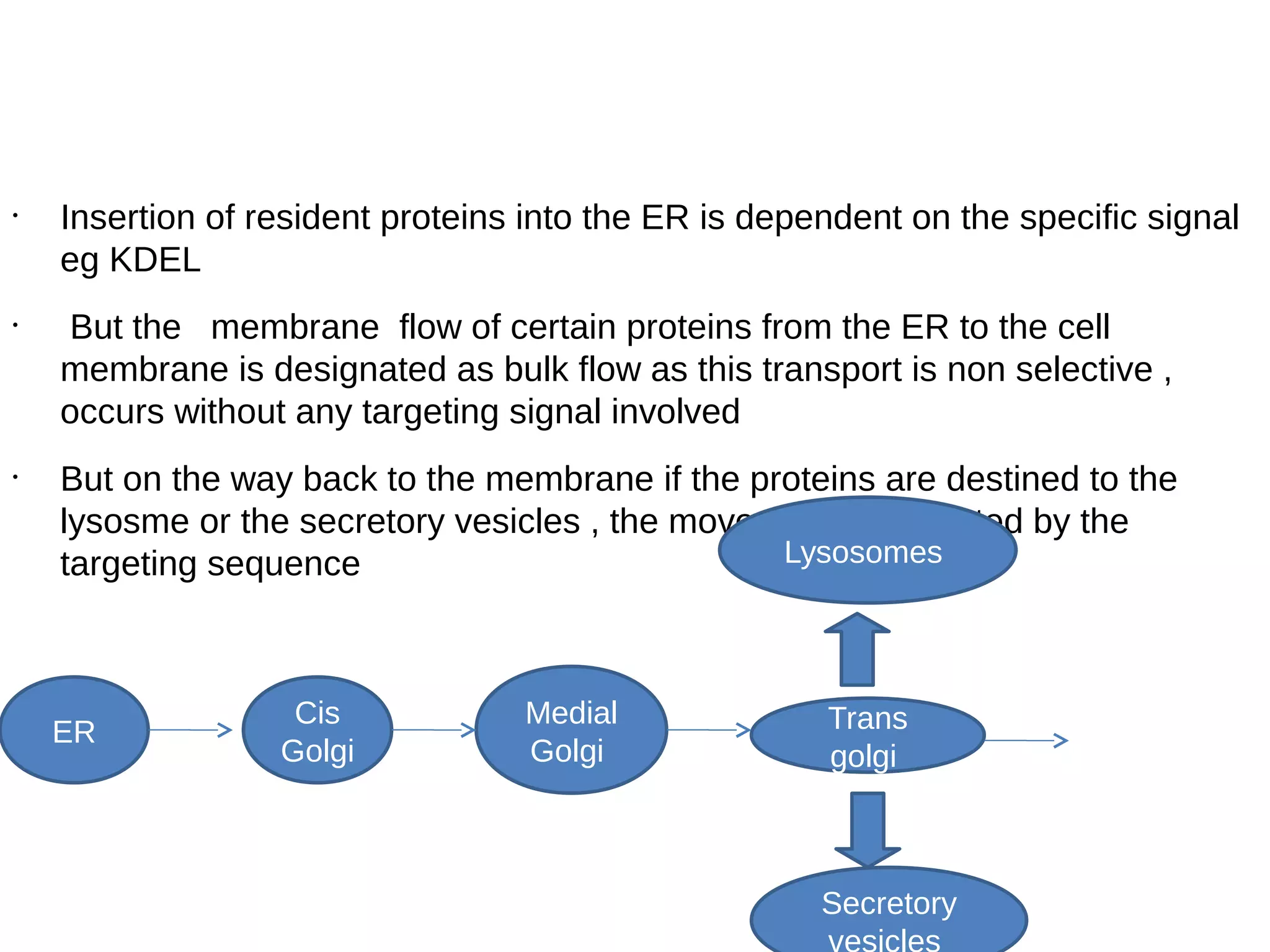
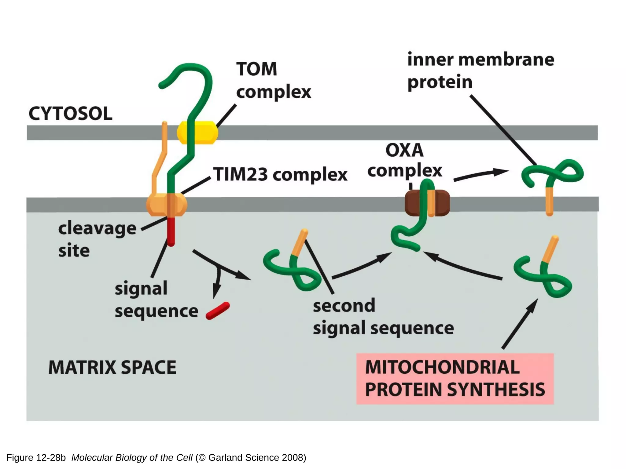
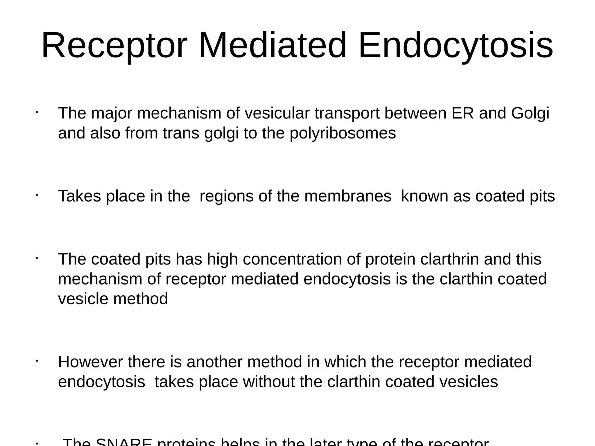
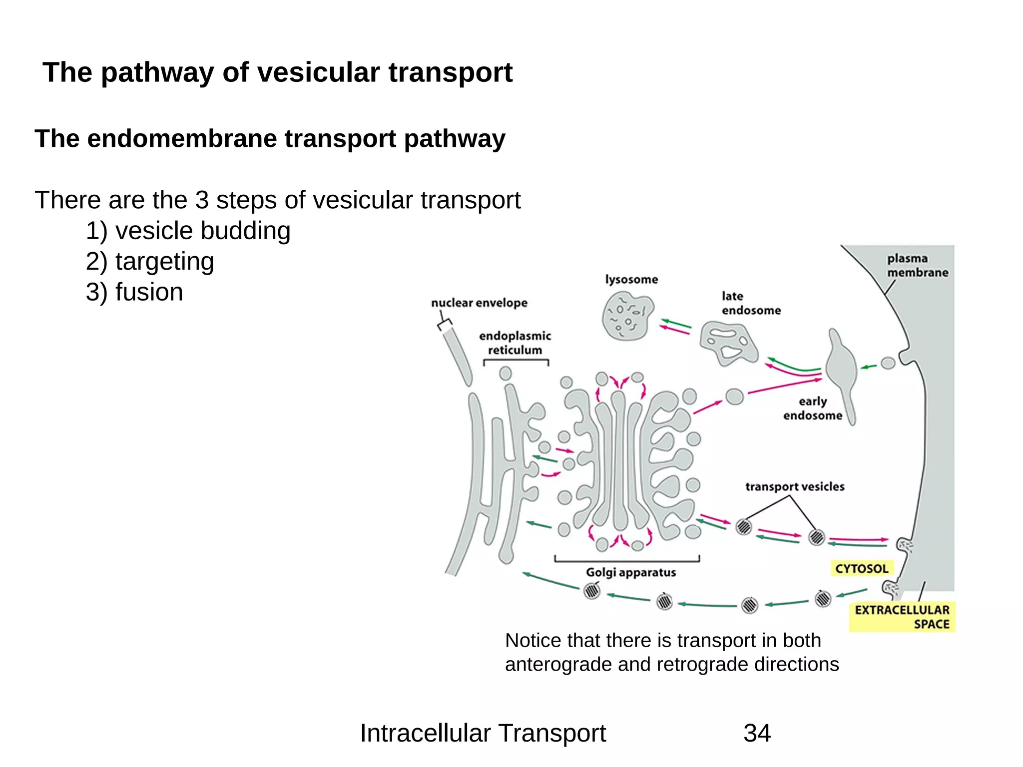
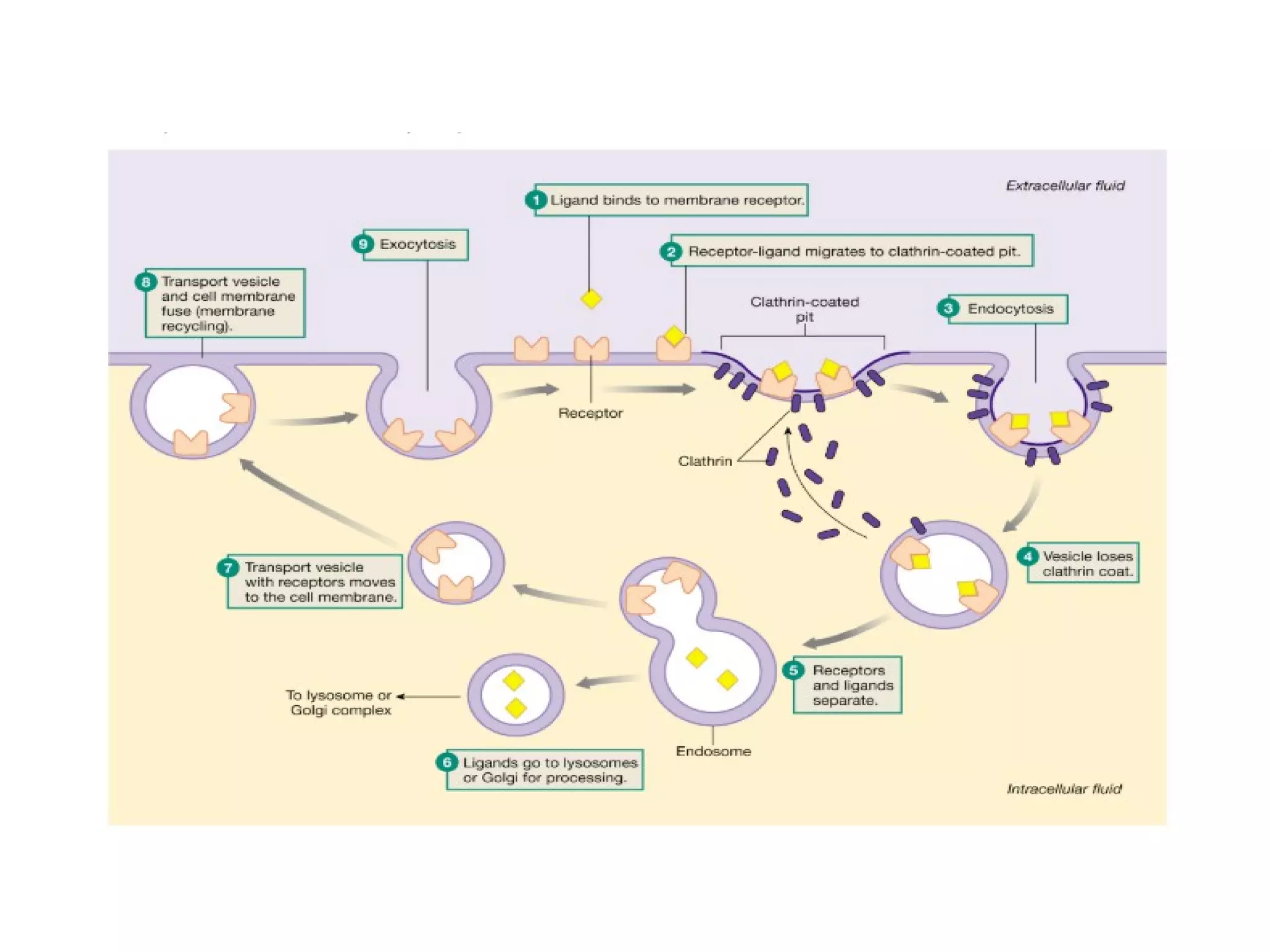
Intracellular Trafficking
Rakesh Pokhrel
MSc. Clinical
Biochemistry
Institute of Medicine
Overview
•
Introduction
•
Protein Sorting
•
Nuclear Pore Complex
•
Protein Translocation
Introduction
•
It is the movement of the solutes and the vesicles within the cell
•
Eukaryotic cells transport packets of components (membrane‐bound
vesicles and organelles, protein rafts, mRNA, chromosomes)
•
Mechanism of movement is the attachment to molecular
motors that haul them along microtubules and actin filaments
•
This is distinct from intercellular transport, which deals solely with
the movement of cargo between cells not the net movement within a
cell
•
Intracellular transport involves the movement of various
components within the cells
•
Paracellular transport refers to the transfer of substances across
an epithelium by passing through the intercellular
space between the cells
•
Transcellular transport, where the substances travel through the
cell, passing through both the apical membrane and basolateral
membrane
•
The cytoskeleton is key in intracellular transport as they provide
the mechanical support necessary for movement
•
Composed of actin, intermediate filaments and microtubules which
mediate locomotion, intracellular transport of organelles, cell shape
and chromosome separation
•
Intracellular transport is unique to eukaryotic cells as they possess
organelles enclosed in membranes
•
Conversely, in prokaryotic cells there is no need for this specialized
transport mechanism as there are no membranous organelles and
compartments to traffic between ( simple diffusion )
Protein Sorting
•
The process by which the proteins are transported to their
appropriate target in the cell or outside it
•
Proteins can be targeted to the inner space of an organelle, different
intracellular membranes, plasma membrane, or to exterior of the cell
via secretion
•
Many Proteins carry the signals that target them to their destinations
•
The signals are the fundamental component of the sorting system
•
Sorting decision made early in the process of biosynthesis
Targeting Sequence or Compound Organelle targeted
Signal Peptide sequence Membrane of ER
Amino Terminal KDEL sequence Luminal surface of ER
Amino terminal sequence
( 70-residue region)
Mitochondria
NLS ( Eg. Pro-Lys-Ala-Lys-Val) Nucleus
PTS ( Eg : Ser-Lys-Leu) Peroxisome
Mannose 6 Phosphate Lysosome
Mechanisms of Intracellular
Transport•
Nuclear Pores ( Nucleus )
•
Protein Translocators (ER, mitochondria,
chloroplasts, peroxisomes)
•
Vesicular Transport
( Lysosome , cell surface ,golgi )
Nuclear Pores
•
Nuclear pore complexes are large protein complexes spanning
the nuclear envelope
•
The proteins that make up the nuclear pore complex are known
as nucleoporins
•
About half of the nucleoporins typically contain solenoid protein
domains
•
This transport includes RNA and ribosomal proteins moving from
nucleus to the cytoplasm
•
The diameter of NPC is 9mm but can increase upto 28 mm
•
Molecules smaller than 28nm can pass through the NPC by
diffusion
•
Larger ones require special translocation mechanisms
•
The proteins to be imported carry a nuclear localization signal eg
Pro-Lys-Ala-Lys
•
Depending on the NLS it contains the cargo protein interacts with
the importins and the complex docks at the NPC
•
Another family of protein RAN comes into play and helps in the
translocation
•
Ran are the small monomeric nuclear GTPase and exist in either
GTP bound or GDP bound state
•
The GTP bound state of Ran is favored in the nucleus and the GDP
bound state is favored in the cytoplasm
•
The cargo molecules once released into the nucleus , the importins
again recirculate in the cytoplasm for another cargo molecule
•
The proteins similar to importins called exportins are involved in the
transport of macromolecules from the nucleus
•
Similar to NLS the cargo proteins to be exported carry NES
•
Ran protein is involved in the export of the cargo molecules from the
nucleus as well
Protein Translocators
•
Proteins moving from the cytosol into the ER, mitochondria,
chloroplasts, or peroxisomes
•
Protein movement is mediated by specialized proteins
termed protein translocators
•
Unlike passage through nuclear pores, translocation
requires unfolding or co-translational transport
Protein Translocation Into the
ER•
Translocation of the proteins into the ER is mainly guided by the
signal hypothesis
•
It was proposed by the Bolbel and Sabatini
•
Proteins synthesized on the membrane bound polyribosomes
contained a peptide extension called signal peptide
•
Responsible for mediating their attachment with the ER membrane
•
In contrast the protein being synthesized on the free polyribosomes
lack this signal peptide
Ribosomes are directed to the ER by the
SRP and ER signal
•
Insertion of resident proteins into the ER is dependent on the specific signal
eg KDEL
•
But the membrane flow of certain proteins from the ER to the cell
membrane is designated as bulk flow as this transport is non selective ,
occurs without any targeting signal involved
•
But on the way back to the membrane if the proteins are destined to the
lysosme or the secretory vesicles , the movement is mediated by the
targeting sequence
ER
Cis
Golgi
Medial
Golgi
Trans
golgi
Lysosomes
Secretory
vesicles
Mitochondrial Protein Import
*
Most mitochondrial proteins are synthesized on cytoplasmic ribosomes
and are post-translationally imported into this organelle.
*
Because of the double membrane surrounding this organelle, there are
four targets for mitochondrial proteins:
1. Outer membrane 3. Inner membrane
2. Intermembrane space 4. Matrix space
*
Mitochondrial proteins usually contain an N-terminal targeting sequence
that is capable of forming an amphipathic -helix;
*
Positively-charged residues are clustered on one side of the helix and
uncharged residues are present on the other.
*
The mitochondrial outer membrane contains specific receptor proteins
that bind to the mitochondrial targeting signal.
Figure 12-22
*
Signal sequence for
mitochondrial protein
import.
*
Note the amphipathic nature
Figure 12-23
Protein translocators in
mitochondrial membranes
(Matrix/Inner Membrane)
(Inner Membrane)
*
Translocation into the mitochondrial matrix requires both ATP hydrolysis
and an electrochemical gradient across the inner mitochondrial
membrane.
*
Translocation occurs at sites where the inner and outer membrane are
in close apposition. These regions are known as contact sites.
*
Proteins are imported into the mitochondria in an unfolded state.
*
Maintenance in an unfolded state is mediated by hsp70 proteins that act
as molecular chaperones.
*
Protein transport into the inner membrane or intermembrane space
Figure 12-25 Molecular Biology of the Cell (© Garland Science 2008)
Protein import into mitochondria
Figure 12-28b Molecular Biology of the Cell (© Garland Science 2008)
Figure 6-86 Molecular Biology of the Cell (© Garland Science 2008)
The hsp70 family of molecular chaperones
Vesicular Transport
•
Vesicular transport is the predominant mechanism for exchange of
proteins and lipids between membrane-bound organelles in
eukaryotic cells
•
This form of transport involves the movement of various elements
with the aid of the bubble like vesicles created from the cell
membrane
•
It is fundamentally divided into endocytosis and exocytosis
•
Endocytosis is divided into 3 distinct mechanisms : Phagocytosis ,
Pinocytosis and receptor mediated endocytosis
Receptor Mediated Endocytosis
•
The major mechanism of vesicular transport between ER and Golgi
and also from trans golgi to the polyribosomes
•
Takes place in the regions of the membranes known as coated pits
•
The coated pits has high concentration of protein clarthrin and this
mechanism of receptor mediated endocytosis is the clarthin coated
vesicle method
•
However there is another method in which the receptor mediated
endocytosis takes place without the clarthin coated vesicles
•
At least four types of coated vesicles has been distinguished
•
Clarthin coated vesicles : Trans golgi to prelysosomes and from
plasma membrane to endosomes
•
COPI: Bi-directional transport from ER to golgi and in the reverse
•
COPII : From ER to golgi
Intracellular Transport 34
The pathway of vesicular transport
The endomembrane transport pathway
There are the 3 steps of vesicular transport
1) vesicle budding
2) targeting
3) fusion
Notice that there is transport in both
anterograde and retrograde directions
Clathrin Independent Pathway
•
The pathway proposed by Rothman and colleagues
•
The pathway plays the major role in the anterograde transport of
proteins into the lysosome or the cell membrane from the ER
•
Each transport vesicle bears one specific target marker consisting of
one or more v-SNARE proteins
•
Each target membrane bears t-SNARE proteins with which the v-
SNARE proteins interact specifically
•
This pathway is complex and has been proposed to occur in
8 Steps of Clathrin Independent
Transport Pathway
ü
Coat Assembly
ü
Recruitment of coat proteins
ü
Bud pinching off
ü
Coat disassembly
ü
Vesicle targeting
ü
Fusion
Rab Protein Family
•
Are small monomeric GTPase
•
Attached to cytosolic faces of membrane via geranyl geranyl chains
•
They attach in the GTP bound state
•
They mediate the fusion of v-SNARE and t-SNARE by displacing a
protein sec1
•
Rab and sec1 proteins regulates the speed of vesicle fusion by
opposing action
SNARE protein family
•
SNARE proteins — "SNAP" (Soluble NSF Attachment
Protein) REceptor" — are a large protein complex consisting of at
least 24 members in yeasts and more than 60 members in
mammalian cells
•
The primary role of SNARE proteins is to mediate vesicle fusion,
that is, the fusion of vesicles with their target membrane bound
compartments (such as a lysosome)
•
Are the machinery required for the membrane fusion
•
Vesicle SNARE and Target SNARE are two divisions
Botulinum Toxin
•
Most lethal toxin known
•
Most serious cause of food poisoning
•
One component of the toxin is a protease specific only to the
synaptobrevin
•
Thus by inhibiting the v-SNARE the release of acetylcholine into the
NMJ is halted
Brefeldin –A
§
An anti viral produced by fungus Penicillium brefeldianum
§
Prevents GTP from binding to ARF in the step 1 of the anterograde
pathway that Is the step of Coat assembly
§
So in the presence of this fungal metabolite the golgi apparatus
appears to disintegrate and fragments are lost
§
Disorders Related to
Intracellular Transport
Familial Hypercholesteremia
•
Familial hypercholesterolemia, FH (type II hyperlipoproteinemia) is
an autosomal dominant disorder
•
Results from mutations affecting the structure and function of the
cell-surface receptor that binds plasma LDLs (low density
lipoproteins) removing them from the circulation
•
The defects in LDL-receptor (LDLR) interaction result in lifelong
elevation of LDL-cholesterol in the blood
1.Receptor null mutation ( lack of receptor synthesis in the ER
2.Defective intracellular transport to golgi apparatus
3.Defective extracellular ligand binding
4.Defective endocytosis
5. Failure to release LDL molecules inside the endosome
Another finding underlying autosomal dominant
Mucolipodosis
•
Mucolipidosis II (I-cell disease) and related milder disorders,
characterized by leakage of multiple lysosomal hydrolases from
cells
•
In these diseases, the activity of the Golgi enzyme N-
acetylglucosamine-1-phosphotransferase is missing, reduced or
altered
•
Inheritence is atosomal recessive
•
Not generating common phosphomannosyl recognition marker of
acid hydrolases
Clinical Features
§
Puffy eyelids with slight exophthalmia
§
Excessive prominence of the epicanthal folds
§
Depressed nasal bridge
§
Full cheeks exhibiting multiple fine telangiectasia
§
Gingival hyperplasia and alveolar enlargement with buried teeth
§
Thick tongue
§
Low birth weight
Hermansky-Pudlak syndrome
•
Exhibit defects in the generation of transport intermediates is
Hermansky-Pudlak syndrome (HPS), a cluster of diseases
characterized by defective biogenesis of lysosome-related
organelles
•
In HPS type 1 and types 3-8, the defects have been pinpointed to
subunits of three distinct protein assemblies: biogenesis of
lysosome-related organelles complex (BLOC) 1, BLOC2 and
BLOC3
•
Results in oculocutaneous albinism (decreased pigmentation),
bleeding problems due to a platelet abnormality (platelet storage
pool defect), and respiratory issues due to the pulmonary fibrosis
•
The clinical manifestations of the disease are due to the effect on
the lysosome related organelles (LROs) , ie : melanosomes, platelet
dense granuels ,MHC class II compartments
Summary
References
•
Harper’s Illustrated Biochemistry 26th edition.
•
Rink J, Ghigo E, Kalaidzidis Y, et al. Rab conversion as a
mechanism of progression from early to late
endosomes. Cell. 2005;122(5):735–49.
•
Prydz K, Dick G, Tveit H. How many ways through the Golgi
maze Traffic.
•
Mostov KE, Verges M, Altschuler Y. Membrane traffic in
polarized epithelial cells. Curr Opin Cell Biol. 2000;12(4):483–
90.
THANK
YOU!

Intracellular trafficing

  • 1.
    Intracellular Trafficking Rakesh Pokhrel MSc.Clinical Biochemistry Institute of Medicine
  • 2.
  • 3.
    Introduction • It is themovement of the solutes and the vesicles within the cell • Eukaryotic cells transport packets of components (membrane‐bound vesicles and organelles, protein rafts, mRNA, chromosomes) • Mechanism of movement is the attachment to molecular motors that haul them along microtubules and actin filaments • This is distinct from intercellular transport, which deals solely with the movement of cargo between cells not the net movement within a cell
  • 4.
    • Intracellular transport involvesthe movement of various components within the cells • Paracellular transport refers to the transfer of substances across an epithelium by passing through the intercellular space between the cells • Transcellular transport, where the substances travel through the cell, passing through both the apical membrane and basolateral membrane
  • 6.
    • The cytoskeleton iskey in intracellular transport as they provide the mechanical support necessary for movement • Composed of actin, intermediate filaments and microtubules which mediate locomotion, intracellular transport of organelles, cell shape and chromosome separation • Intracellular transport is unique to eukaryotic cells as they possess organelles enclosed in membranes • Conversely, in prokaryotic cells there is no need for this specialized transport mechanism as there are no membranous organelles and compartments to traffic between ( simple diffusion )
  • 7.
    Protein Sorting • The processby which the proteins are transported to their appropriate target in the cell or outside it • Proteins can be targeted to the inner space of an organelle, different intracellular membranes, plasma membrane, or to exterior of the cell via secretion • Many Proteins carry the signals that target them to their destinations • The signals are the fundamental component of the sorting system • Sorting decision made early in the process of biosynthesis
  • 9.
    Targeting Sequence orCompound Organelle targeted Signal Peptide sequence Membrane of ER Amino Terminal KDEL sequence Luminal surface of ER Amino terminal sequence ( 70-residue region) Mitochondria NLS ( Eg. Pro-Lys-Ala-Lys-Val) Nucleus PTS ( Eg : Ser-Lys-Leu) Peroxisome Mannose 6 Phosphate Lysosome
  • 11.
    Mechanisms of Intracellular Transport• NuclearPores ( Nucleus ) • Protein Translocators (ER, mitochondria, chloroplasts, peroxisomes) • Vesicular Transport ( Lysosome , cell surface ,golgi )
  • 12.
    Nuclear Pores • Nuclear porecomplexes are large protein complexes spanning the nuclear envelope • The proteins that make up the nuclear pore complex are known as nucleoporins • About half of the nucleoporins typically contain solenoid protein domains • This transport includes RNA and ribosomal proteins moving from nucleus to the cytoplasm
  • 13.
    • The diameter ofNPC is 9mm but can increase upto 28 mm • Molecules smaller than 28nm can pass through the NPC by diffusion • Larger ones require special translocation mechanisms • The proteins to be imported carry a nuclear localization signal eg Pro-Lys-Ala-Lys • Depending on the NLS it contains the cargo protein interacts with the importins and the complex docks at the NPC
  • 14.
    • Another family ofprotein RAN comes into play and helps in the translocation • Ran are the small monomeric nuclear GTPase and exist in either GTP bound or GDP bound state • The GTP bound state of Ran is favored in the nucleus and the GDP bound state is favored in the cytoplasm • The cargo molecules once released into the nucleus , the importins again recirculate in the cytoplasm for another cargo molecule
  • 15.
    • The proteins similarto importins called exportins are involved in the transport of macromolecules from the nucleus • Similar to NLS the cargo proteins to be exported carry NES • Ran protein is involved in the export of the cargo molecules from the nucleus as well
  • 18.
    Protein Translocators • Proteins movingfrom the cytosol into the ER, mitochondria, chloroplasts, or peroxisomes • Protein movement is mediated by specialized proteins termed protein translocators • Unlike passage through nuclear pores, translocation requires unfolding or co-translational transport
  • 19.
    Protein Translocation Intothe ER• Translocation of the proteins into the ER is mainly guided by the signal hypothesis • It was proposed by the Bolbel and Sabatini • Proteins synthesized on the membrane bound polyribosomes contained a peptide extension called signal peptide • Responsible for mediating their attachment with the ER membrane • In contrast the protein being synthesized on the free polyribosomes lack this signal peptide
  • 21.
    Ribosomes are directedto the ER by the SRP and ER signal
  • 22.
    • Insertion of residentproteins into the ER is dependent on the specific signal eg KDEL • But the membrane flow of certain proteins from the ER to the cell membrane is designated as bulk flow as this transport is non selective , occurs without any targeting signal involved • But on the way back to the membrane if the proteins are destined to the lysosme or the secretory vesicles , the movement is mediated by the targeting sequence ER Cis Golgi Medial Golgi Trans golgi Lysosomes Secretory vesicles
  • 23.
    Mitochondrial Protein Import * Mostmitochondrial proteins are synthesized on cytoplasmic ribosomes and are post-translationally imported into this organelle. * Because of the double membrane surrounding this organelle, there are four targets for mitochondrial proteins: 1. Outer membrane 3. Inner membrane 2. Intermembrane space 4. Matrix space * Mitochondrial proteins usually contain an N-terminal targeting sequence that is capable of forming an amphipathic -helix; * Positively-charged residues are clustered on one side of the helix and uncharged residues are present on the other. * The mitochondrial outer membrane contains specific receptor proteins that bind to the mitochondrial targeting signal.
  • 24.
    Figure 12-22 * Signal sequencefor mitochondrial protein import. * Note the amphipathic nature
  • 25.
    Figure 12-23 Protein translocatorsin mitochondrial membranes (Matrix/Inner Membrane) (Inner Membrane)
  • 26.
    * Translocation into themitochondrial matrix requires both ATP hydrolysis and an electrochemical gradient across the inner mitochondrial membrane. * Translocation occurs at sites where the inner and outer membrane are in close apposition. These regions are known as contact sites. * Proteins are imported into the mitochondria in an unfolded state. * Maintenance in an unfolded state is mediated by hsp70 proteins that act as molecular chaperones. * Protein transport into the inner membrane or intermembrane space
  • 27.
    Figure 12-25 MolecularBiology of the Cell (© Garland Science 2008) Protein import into mitochondria
  • 28.
    Figure 12-28b MolecularBiology of the Cell (© Garland Science 2008)
  • 29.
    Figure 6-86 MolecularBiology of the Cell (© Garland Science 2008) The hsp70 family of molecular chaperones
  • 30.
    Vesicular Transport • Vesicular transportis the predominant mechanism for exchange of proteins and lipids between membrane-bound organelles in eukaryotic cells • This form of transport involves the movement of various elements with the aid of the bubble like vesicles created from the cell membrane • It is fundamentally divided into endocytosis and exocytosis • Endocytosis is divided into 3 distinct mechanisms : Phagocytosis , Pinocytosis and receptor mediated endocytosis
  • 32.
    Receptor Mediated Endocytosis • Themajor mechanism of vesicular transport between ER and Golgi and also from trans golgi to the polyribosomes • Takes place in the regions of the membranes known as coated pits • The coated pits has high concentration of protein clarthrin and this mechanism of receptor mediated endocytosis is the clarthin coated vesicle method • However there is another method in which the receptor mediated endocytosis takes place without the clarthin coated vesicles
  • 33.
    • At least fourtypes of coated vesicles has been distinguished • Clarthin coated vesicles : Trans golgi to prelysosomes and from plasma membrane to endosomes • COPI: Bi-directional transport from ER to golgi and in the reverse • COPII : From ER to golgi
  • 34.
    Intracellular Transport 34 Thepathway of vesicular transport The endomembrane transport pathway There are the 3 steps of vesicular transport 1) vesicle budding 2) targeting 3) fusion Notice that there is transport in both anterograde and retrograde directions
  • 36.
    Clathrin Independent Pathway • Thepathway proposed by Rothman and colleagues • The pathway plays the major role in the anterograde transport of proteins into the lysosome or the cell membrane from the ER • Each transport vesicle bears one specific target marker consisting of one or more v-SNARE proteins • Each target membrane bears t-SNARE proteins with which the v- SNARE proteins interact specifically • This pathway is complex and has been proposed to occur in
  • 37.
    8 Steps ofClathrin Independent Transport Pathway ü Coat Assembly ü Recruitment of coat proteins ü Bud pinching off ü Coat disassembly ü Vesicle targeting ü Fusion
  • 40.
    Rab Protein Family • Aresmall monomeric GTPase • Attached to cytosolic faces of membrane via geranyl geranyl chains • They attach in the GTP bound state • They mediate the fusion of v-SNARE and t-SNARE by displacing a protein sec1 • Rab and sec1 proteins regulates the speed of vesicle fusion by opposing action
  • 41.
    SNARE protein family • SNAREproteins — "SNAP" (Soluble NSF Attachment Protein) REceptor" — are a large protein complex consisting of at least 24 members in yeasts and more than 60 members in mammalian cells • The primary role of SNARE proteins is to mediate vesicle fusion, that is, the fusion of vesicles with their target membrane bound compartments (such as a lysosome) • Are the machinery required for the membrane fusion • Vesicle SNARE and Target SNARE are two divisions
  • 42.
    Botulinum Toxin • Most lethaltoxin known • Most serious cause of food poisoning • One component of the toxin is a protease specific only to the synaptobrevin • Thus by inhibiting the v-SNARE the release of acetylcholine into the NMJ is halted
  • 43.
    Brefeldin –A § An antiviral produced by fungus Penicillium brefeldianum § Prevents GTP from binding to ARF in the step 1 of the anterograde pathway that Is the step of Coat assembly § So in the presence of this fungal metabolite the golgi apparatus appears to disintegrate and fragments are lost §
  • 44.
    Disorders Related to IntracellularTransport Familial Hypercholesteremia • Familial hypercholesterolemia, FH (type II hyperlipoproteinemia) is an autosomal dominant disorder • Results from mutations affecting the structure and function of the cell-surface receptor that binds plasma LDLs (low density lipoproteins) removing them from the circulation • The defects in LDL-receptor (LDLR) interaction result in lifelong elevation of LDL-cholesterol in the blood
  • 45.
    1.Receptor null mutation( lack of receptor synthesis in the ER 2.Defective intracellular transport to golgi apparatus 3.Defective extracellular ligand binding 4.Defective endocytosis 5. Failure to release LDL molecules inside the endosome Another finding underlying autosomal dominant
  • 48.
    Mucolipodosis • Mucolipidosis II (I-celldisease) and related milder disorders, characterized by leakage of multiple lysosomal hydrolases from cells • In these diseases, the activity of the Golgi enzyme N- acetylglucosamine-1-phosphotransferase is missing, reduced or altered • Inheritence is atosomal recessive • Not generating common phosphomannosyl recognition marker of acid hydrolases
  • 49.
    Clinical Features § Puffy eyelidswith slight exophthalmia § Excessive prominence of the epicanthal folds § Depressed nasal bridge § Full cheeks exhibiting multiple fine telangiectasia § Gingival hyperplasia and alveolar enlargement with buried teeth § Thick tongue § Low birth weight
  • 50.
    Hermansky-Pudlak syndrome • Exhibit defectsin the generation of transport intermediates is Hermansky-Pudlak syndrome (HPS), a cluster of diseases characterized by defective biogenesis of lysosome-related organelles • In HPS type 1 and types 3-8, the defects have been pinpointed to subunits of three distinct protein assemblies: biogenesis of lysosome-related organelles complex (BLOC) 1, BLOC2 and BLOC3 • Results in oculocutaneous albinism (decreased pigmentation), bleeding problems due to a platelet abnormality (platelet storage pool defect), and respiratory issues due to the pulmonary fibrosis • The clinical manifestations of the disease are due to the effect on the lysosome related organelles (LROs) , ie : melanosomes, platelet dense granuels ,MHC class II compartments
  • 51.
  • 52.
    References • Harper’s Illustrated Biochemistry26th edition. • Rink J, Ghigo E, Kalaidzidis Y, et al. Rab conversion as a mechanism of progression from early to late endosomes. Cell. 2005;122(5):735–49. • Prydz K, Dick G, Tveit H. How many ways through the Golgi maze Traffic. • Mostov KE, Verges M, Altschuler Y. Membrane traffic in polarized epithelial cells. Curr Opin Cell Biol. 2000;12(4):483– 90.
  • 53.