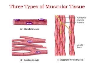
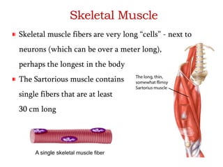
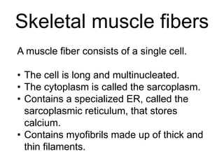
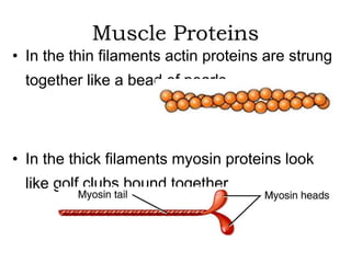
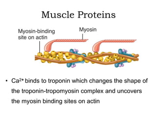
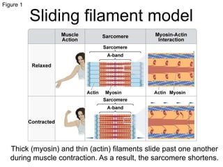
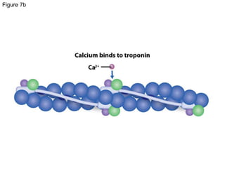
MO Figure 
Muscle Function at 
the Cellular Level 
Michael Patrick O'Neill/Science Source.
Functions of Muscular Tissue 
• Like nervous tissue, muscles are excitable 
or "irritable” 
they have the ability to respond to a stimulus 
• Unlike nerves, however, muscles are also: 
Contractible (they can shorten 
in length) 
Extensible (they can extend or 
stretch) 
Elastic (they can return to their 
original shape)
Functions of Muscular Tissue 
• Muscle makes up a large percentage of the 
body’s weight 
• Their main functions are to: 
Create motion – muscles work with nerves, 
bones, and joints to produce body movements 
Stabilize body positions and maintain posture 
Store substances within the body using 
sphincters
Three Types of Muscular 
Location Function Appearance Control 
Skeletal 
skeleton 
movement, 
heat, posture 
striated, multi-nucleated 
(eccentric), 
fibers parallel 
voluntary 
Cardiac 
heart 
pump blood 
continuously 
striated, one central 
nucleus 
involuntary 
Visceral 
(smooth muscle) 
G.I. tract, 
uterus, eye, 
blood vessels 
Peristalsis, 
blood pressure, 
pupil size, 
erects hairs 
no striations, one 
central nucleus 
involuntary 
Tissue
Three Types of Muscular Tissue 
(a) Skeletal muscle 
(b) Cardiac muscle (c) Visceral smooth muscle
Location Function Appearance Control 
Skeletal 
skeleton 
movement, 
heat, posture 
striated, multi-nucleated 
(eccentric), 
fibers parallel 
voluntary 
Cardiac 
heart 
pump blood 
continuously 
striated, one central 
nucleus 
involuntary 
Visceral 
(smooth muscle) 
G.I. tract, 
uterus, eye, 
blood vessels 
Peristalsis, 
blood pressure, 
pupil size, 
erects hairs 
no striations, one 
central nucleus 
involuntary 
Skeletal Muscle
Skeletal Muscle
Skeletal Muscle 
Skeletal muscle fibers are very long “cells” - next to 
neurons (which can be over a meter long), 
perhaps the longest in the body 
The Sartorious muscle contains 
single fibers that are at least 
30 cm long 
A single skeletal muscle fiber
Motor neuron 
Sarcolemma 
Skeletal Muscle 
The terminal processes of a motor 
neuron in close proximity to the 
sarcolemma of a skeletal muscle fiber
Skeletal muscle fibers 
A muscle fiber consists of a single cell. 
• The cell is long and multinucleated. 
• The cytoplasm is called the sarcoplasm. 
• Contains a specialized ER, called the 
sarcoplasmic reticulum, that stores 
calcium. 
• Contains myofibrils made up of thick and 
thin filaments.
The Skeletal Muscle Fiber 
Increasing the level of magnification, the myofibrils are 
seen to be composed 
of filaments 
Thick filaments 
Thing filaments
The Skeletal Muscle Fiber 
• The basic functional unit of skeletal muscle 
fibers is the sarcomere: An arrangement of 
thick and thin filaments sandwiched between 
two Z discs 
A scanning electron micrograph of a sarcomere
The Skeletal Muscle Fiber 
• Muscle contraction occurs in the sarcomeres 
The “Z line” is really a Z disc when considered in 3 
dimensions. A sarcomere extends from Z disc to Z disc.
Muscle Proteins 
• Myofibrils are built from three groups of 
proteins 
Contractile proteins generate force during 
contraction 
Regulatory proteins help switch the contraction 
process on and off 
Structural proteins keep the thick and thin 
filaments in proper alignment and link the
Muscle Proteins 
• The thin filaments are comprised mostly of 
the structural protein actin, and the thick 
filaments are comprised mostly of the 
structural protein myosin
Muscle Proteins 
• In the thin filaments actin proteins are strung 
together like a bead of pearls 
• In the thick filaments myosin proteins look 
like golf clubs bound together
Muscle Proteins 
In this first graphic, the myosin binding sites on the actin 
proteins are readily visible. 
The regulatory proteins troponin and tropomyosin have 
been added to the bottom graphic: The myosin binding 
sites have been 
covered
Muscle Proteins 
In this graphic the troponin-tropomyosin complex has 
slid down into the “gutters” of the actin molecule 
unblocking the myosin binding site 
Myosin binding site exposed 
The troponin-tropomyosin complex can slide back and 
forth depending on the presence of Ca2+
Muscle Proteins 
• Ca2+ binds to troponin which changes the shape of 
the troponin-tropomyosin complex and uncovers 
the myosin binding sites on actin
Muscle Proteins 
• Besides contractile and regulatory proteins, muscle 
contains about a dozen structural proteins which 
contribute to the alignment, stability, elasticity, and 
extensibility of myofibrils 
• Titan is the third most plentiful protein in muscle, 
after actin and myosin - it extends from the Z disc and 
accounts for much of the elasticity of myofibrils 
• Dystrophin is discussed later as it relates to the disease 
of muscular dystrophy
The Sliding-Filament 
Mechanism 
• With exposure of the myosin binding sites on 
actin (the thin filaments)—in the presence of 
Ca2+ and ATP—the thick and thin filaments 
“slide” on one another and the sarcomere is 
shortened
The Sliding-Filament 
Mechanism 
• The “sliding” of actin on myosin (thick 
filaments on thin filaments) can be broken
Figure 1 
Sliding filament model 
Thick (myosin) and thin (actin) filaments slide past one another 
during muscle contraction. As a result, the sarcomere shortens.
Figure 2 
Sarcomeres 
C. F. Armstrong/Science Source. 
Z-discs form dark lines in electron micrographs and give 
skeletal muscle a striated appearance.
Figure 3
Figure 4 
Muscle anatomy
Figure 4a
Figure 4b
Muscle energy storage 
Glycogen 
• Glucose polymer. Glucose is used in 
cellular respiration to make ATP. 
Creatine phosphate 
• Transfers a phosphate to ADP to form 
ATP.
Figure 5 
Muscle innervation 
Muscle fibers can be innervated by a single neuron, which 
increases contraction strength at the expense of fine control, or by 
multiple neurons, which allows finer control but with little strength.
Figure 5a
Figure 5b
Figure 6 
Excitation-contraction coupling
Figure 7 
Regulation of contraction 
In resting muscle, 
tropomyosin covers 
myosin-binding sites 
on actin. The 
troponin complex 
holds tropomyosin in 
place. Ca2+ binding 
to troponin causes 
tropomyosin to shift, 
which exposes the 
myosin-binding sites. 
Myosin binds actin, 
and the muscle 
contracts.
Figure 7a
Figure 7b
Figure 7c
MO Figure 
Skeletal System 
and Locomotion 
Art Wolfe/Science Source
Skeletal systems 
Endoskeleton: inside the animal 
Exoskeleton: outside the animal 
Hydrostatic skeleton: composed of 
pressurized water in internal compartments.
Figure 1 
Hydrostatic skeletons 
© 2006 Nature Publishing Group Alvarado, A & Tsonis, P. Bridging the 
regeneration gap: genetic insights from diverse animal models. Nature 
Reviews Genetics 7, 873–884 (2006) doi:10.1038/nrg1923. Used with 
permission. 
This Hydra moves using a hydrostatic skeleton.
Figure 2 
Peristaltic movement 
Caused by 
alternating 
radial and 
longitudinal 
muscle 
contractions 
against a 
fluid-filled 
coelom.
Figure 2a
Figure 2b
Figure 3 
Exoskeletons 
© 2002 Nature Publishing Group GDC seeks members of new Appointing Body. British Dental Journal 193, 431– 
433 (2002), doi:10.1038/sj.bdj.4801590. Used with permission. 
Arthropod exoskeletons contain chitin.
Figure 4 
The human skeleton
Figure 5 
Antagonistic muscle pairs 
Coordinated 
movement 
enables flexion 
and extension 
of a limb.
Figure 5a
Figure 5b
Figure 6 
Balance and locomotion 
During walking and 
running, the feet are 
used for balancing, 
pushing off, and 
maintaining momentum.
Figure 7 
Terrestrial locomotion 
Stephen Dalton/Science Source. 
Energy stored in tendons enables this frog to jump.

Muscle funccellularlevel animal systems

  • 1.
    MO Figure MuscleFunction at the Cellular Level Michael Patrick O'Neill/Science Source.
  • 2.
    Functions of MuscularTissue • Like nervous tissue, muscles are excitable or "irritable” they have the ability to respond to a stimulus • Unlike nerves, however, muscles are also: Contractible (they can shorten in length) Extensible (they can extend or stretch) Elastic (they can return to their original shape)
  • 3.
    Functions of MuscularTissue • Muscle makes up a large percentage of the body’s weight • Their main functions are to: Create motion – muscles work with nerves, bones, and joints to produce body movements Stabilize body positions and maintain posture Store substances within the body using sphincters
  • 4.
    Three Types ofMuscular Location Function Appearance Control Skeletal skeleton movement, heat, posture striated, multi-nucleated (eccentric), fibers parallel voluntary Cardiac heart pump blood continuously striated, one central nucleus involuntary Visceral (smooth muscle) G.I. tract, uterus, eye, blood vessels Peristalsis, blood pressure, pupil size, erects hairs no striations, one central nucleus involuntary Tissue
  • 5.
    Three Types ofMuscular Tissue (a) Skeletal muscle (b) Cardiac muscle (c) Visceral smooth muscle
  • 6.
    Location Function AppearanceControl Skeletal skeleton movement, heat, posture striated, multi-nucleated (eccentric), fibers parallel voluntary Cardiac heart pump blood continuously striated, one central nucleus involuntary Visceral (smooth muscle) G.I. tract, uterus, eye, blood vessels Peristalsis, blood pressure, pupil size, erects hairs no striations, one central nucleus involuntary Skeletal Muscle
  • 7.
  • 8.
    Skeletal Muscle Skeletalmuscle fibers are very long “cells” - next to neurons (which can be over a meter long), perhaps the longest in the body The Sartorious muscle contains single fibers that are at least 30 cm long A single skeletal muscle fiber
  • 9.
    Motor neuron Sarcolemma Skeletal Muscle The terminal processes of a motor neuron in close proximity to the sarcolemma of a skeletal muscle fiber
  • 10.
    Skeletal muscle fibers A muscle fiber consists of a single cell. • The cell is long and multinucleated. • The cytoplasm is called the sarcoplasm. • Contains a specialized ER, called the sarcoplasmic reticulum, that stores calcium. • Contains myofibrils made up of thick and thin filaments.
  • 11.
    The Skeletal MuscleFiber Increasing the level of magnification, the myofibrils are seen to be composed of filaments Thick filaments Thing filaments
  • 12.
    The Skeletal MuscleFiber • The basic functional unit of skeletal muscle fibers is the sarcomere: An arrangement of thick and thin filaments sandwiched between two Z discs A scanning electron micrograph of a sarcomere
  • 13.
    The Skeletal MuscleFiber • Muscle contraction occurs in the sarcomeres The “Z line” is really a Z disc when considered in 3 dimensions. A sarcomere extends from Z disc to Z disc.
  • 14.
    Muscle Proteins •Myofibrils are built from three groups of proteins Contractile proteins generate force during contraction Regulatory proteins help switch the contraction process on and off Structural proteins keep the thick and thin filaments in proper alignment and link the
  • 15.
    Muscle Proteins •The thin filaments are comprised mostly of the structural protein actin, and the thick filaments are comprised mostly of the structural protein myosin
  • 16.
    Muscle Proteins •In the thin filaments actin proteins are strung together like a bead of pearls • In the thick filaments myosin proteins look like golf clubs bound together
  • 17.
    Muscle Proteins Inthis first graphic, the myosin binding sites on the actin proteins are readily visible. The regulatory proteins troponin and tropomyosin have been added to the bottom graphic: The myosin binding sites have been covered
  • 18.
    Muscle Proteins Inthis graphic the troponin-tropomyosin complex has slid down into the “gutters” of the actin molecule unblocking the myosin binding site Myosin binding site exposed The troponin-tropomyosin complex can slide back and forth depending on the presence of Ca2+
  • 19.
    Muscle Proteins •Ca2+ binds to troponin which changes the shape of the troponin-tropomyosin complex and uncovers the myosin binding sites on actin
  • 20.
    Muscle Proteins •Besides contractile and regulatory proteins, muscle contains about a dozen structural proteins which contribute to the alignment, stability, elasticity, and extensibility of myofibrils • Titan is the third most plentiful protein in muscle, after actin and myosin - it extends from the Z disc and accounts for much of the elasticity of myofibrils • Dystrophin is discussed later as it relates to the disease of muscular dystrophy
  • 21.
    The Sliding-Filament Mechanism • With exposure of the myosin binding sites on actin (the thin filaments)—in the presence of Ca2+ and ATP—the thick and thin filaments “slide” on one another and the sarcomere is shortened
  • 22.
    The Sliding-Filament Mechanism • The “sliding” of actin on myosin (thick filaments on thin filaments) can be broken
  • 23.
    Figure 1 Slidingfilament model Thick (myosin) and thin (actin) filaments slide past one another during muscle contraction. As a result, the sarcomere shortens.
  • 24.
    Figure 2 Sarcomeres C. F. Armstrong/Science Source. Z-discs form dark lines in electron micrographs and give skeletal muscle a striated appearance.
  • 25.
  • 26.
  • 27.
  • 28.
  • 29.
    Muscle energy storage Glycogen • Glucose polymer. Glucose is used in cellular respiration to make ATP. Creatine phosphate • Transfers a phosphate to ADP to form ATP.
  • 30.
    Figure 5 Muscleinnervation Muscle fibers can be innervated by a single neuron, which increases contraction strength at the expense of fine control, or by multiple neurons, which allows finer control but with little strength.
  • 31.
  • 32.
  • 33.
  • 34.
    Figure 7 Regulationof contraction In resting muscle, tropomyosin covers myosin-binding sites on actin. The troponin complex holds tropomyosin in place. Ca2+ binding to troponin causes tropomyosin to shift, which exposes the myosin-binding sites. Myosin binds actin, and the muscle contracts.
  • 35.
  • 36.
  • 37.
  • 38.
    MO Figure SkeletalSystem and Locomotion Art Wolfe/Science Source
  • 39.
    Skeletal systems Endoskeleton:inside the animal Exoskeleton: outside the animal Hydrostatic skeleton: composed of pressurized water in internal compartments.
  • 40.
    Figure 1 Hydrostaticskeletons © 2006 Nature Publishing Group Alvarado, A & Tsonis, P. Bridging the regeneration gap: genetic insights from diverse animal models. Nature Reviews Genetics 7, 873–884 (2006) doi:10.1038/nrg1923. Used with permission. This Hydra moves using a hydrostatic skeleton.
  • 41.
    Figure 2 Peristalticmovement Caused by alternating radial and longitudinal muscle contractions against a fluid-filled coelom.
  • 42.
  • 43.
  • 44.
    Figure 3 Exoskeletons © 2002 Nature Publishing Group GDC seeks members of new Appointing Body. British Dental Journal 193, 431– 433 (2002), doi:10.1038/sj.bdj.4801590. Used with permission. Arthropod exoskeletons contain chitin.
  • 45.
    Figure 4 Thehuman skeleton
  • 46.
    Figure 5 Antagonisticmuscle pairs Coordinated movement enables flexion and extension of a limb.
  • 47.
  • 48.
  • 49.
    Figure 6 Balanceand locomotion During walking and running, the feet are used for balancing, pushing off, and maintaining momentum.
  • 50.
    Figure 7 Terrestriallocomotion Stephen Dalton/Science Source. Energy stored in tendons enables this frog to jump.

Editor's Notes

  • #8 All muscle start as these spindly shaped cells from myoblasts – skeletal muscle cells fuse to form large fibers… Satellite cells are small mononuclear progenitor cells with virtually no cytoplasm found in mature muscle. They are found sandwiched between the basement membrane and sarcolemma (cell membrane) of individual muscle fibres, and can be difficult to distinguish from the sub-sarcolemmal nuclei of the fibres. Satellite cells are able to differentiate and fuse to augment existing muscle fibres and to form new fibres. These cells represent the oldest known adult stem cell niche, and are involved in the normal growth of muscle, as well as regeneration following injury or disease.
  • #9 It’s difficult to find the sartorius muscle in some people (cadavers)
  • #12 Sarcomeres are multi-protein complexes composed of three different filament systems. The thick filament system is composed of myosin protein which is connected from the M-line to the Z-disc by titin. It also contains myosin-binding protein C which binds at one end to the thick filament and the other to actin. The thin filaments are assembled by actin monomers bound to nebulin, which also involves tropomyosin (a dimer which coils itself around the F-actin core of the thin filament) and troponin. Nebulin and titin give stability and structure to the sarcomere.
  • #13  Z-line (from the German "Zwischenscheibe“). Each myofibril is made up of thin filament proteins and thick filament proteins, arranged (configured) in sarcomeres.
  • #14 Each myofibril is made up of thin filament proteins, and thick filament proteins, arranged (configured) in sarcomeres
  • #21 Titin is the largest known protein, consisting of 34,350 amino acids. Titin, also known as connectin is a protein that is important in the contraction of striated muscle tissues. Dystrophin, not Titin, is the protein absent in muscular dystrophy.
  • #26 This interactive can be found as a separate file in your downloaded folder. This slide is a placeholder only. The image is a screen shot taken from the digital book.