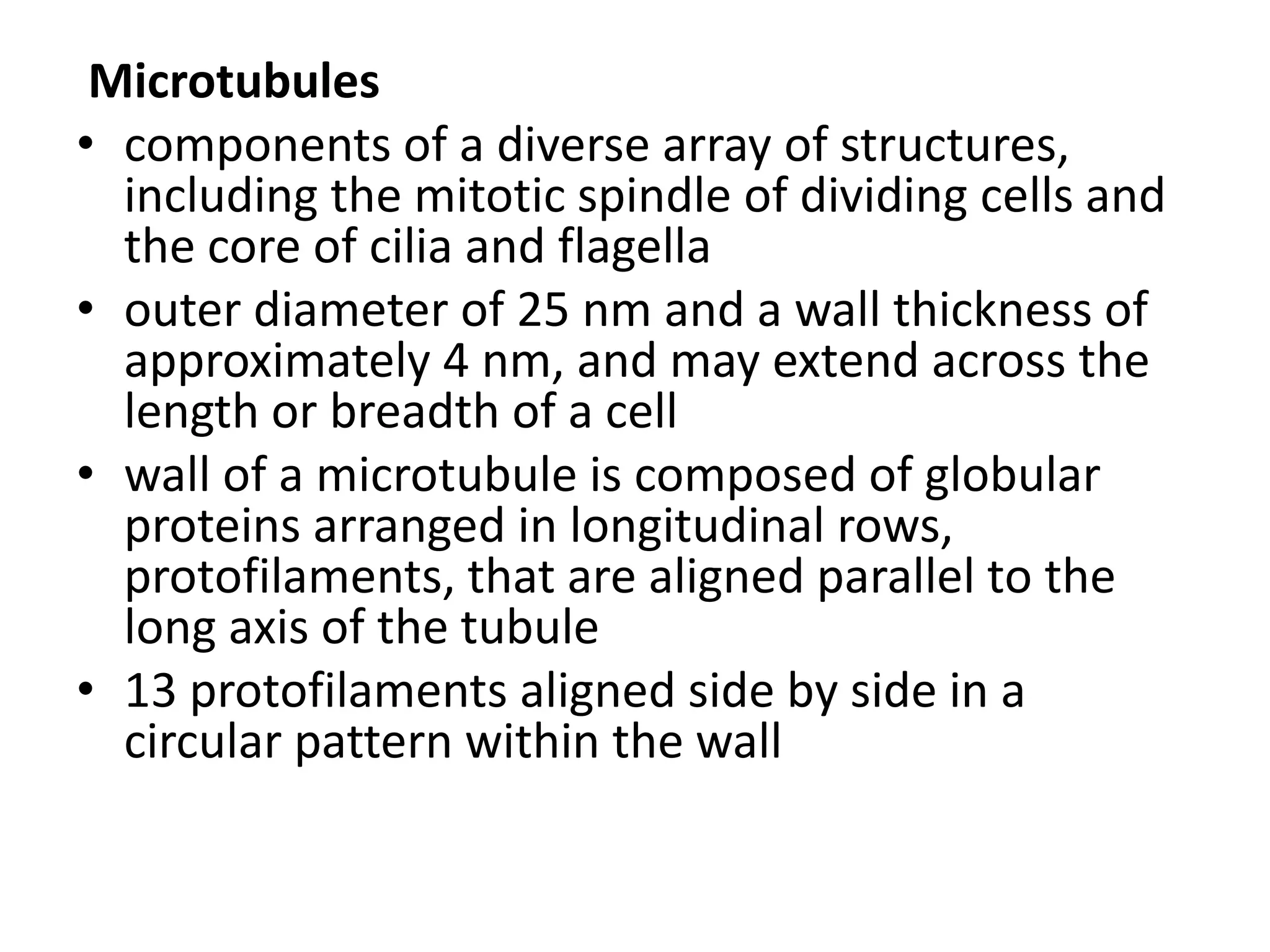
SlideShare a Scribd company logo
Cytoskeleton
• Form an elaborate interactive network of
  polymers of protein subunits held together by
  weak, noncovalent bonds
• Although they appear stationary in
  micrographs, they are highly dynamic
  structures capable of dramatic reorganization
• Recent study suggested that prokaryotes
  prokaryotes have proteins that carry
  cytoskeletal-like activities
Microtubules
• Long, hollow, and stiff unbranched tubes
  found in all eukaryotes that are distributed in
  the cytoplasm
• Functions in support, intracellular transport,
  and cell organization
• Composed of subunits of tubulin
Intermediate filaments
• Tough, ropelike fibers found in the nucleus
  and cytoplasm of animals
• Functions in structural support
• Composed of a variety of proteins
Microfilaments
• Solid, thin structures found in the cytoplasm
  of eukaryotes that are often organized into a
  branching network
• Functions in motility and contractility
• Composed of actin
Study of the cytoskeleton
Live-cell fluorescence imaging
• allows researchers to directly observe
  molecular processes in living cells—an
  approach known as live-cell imaging
FIGURE 9.4 Dynamic changes in length of microtubules within
      an epithelial cell. The cell was injected with a small volume of
tubulin that had been covalently linked to the fluorescent dye rhodamine.
         After allowing time for the cell to incorporate the labeled
  tubulin into microtubules, a small portion of the edge of the living cell
            was examined under the fluorescence microscope.
In Vitro and In Vivo Single-molecule Assays
• high-resolution video microscopy has led to
  the development of in vitro motility assays
• Single-molecule assays have allowed
  researchers to make measurements that were
  not possible with standard biochemical
  techniques that average the results obtained
  from large numbers of molecules
In some of the earlier assays, microtubules
were attached to a glass coverslip. Then,
microscopic beads containing attached motor
proteins were placed directly onto the
microtubules using aimed laser beams. The
laser beams are shone through the objective
lens of a microscope, producing a weak
attractive force near the point of focus.
Because it can grasp microscopic objects, this
apparatus is referred to as optical tweezers.
When ATP is present as an energy source, the
movements of a bead along a microtubule can
be followed by a video camera, revealing the
size of individual steps taken by the motor
protein. Focused laser beams can also be used
to “trap” a single bead and determine the
minute forces (measured as a few
piconewtons, pN) generated by a single motor
protein as it “tries” to move the bead against
the force exerted by the optical trap
In this experiment a purified GFP-labeled kinesin
  molecule is seen to move processively along a
  microtubule whose plus end is labeled with a
  red fluorescent dye named Cy5.
The clarity of these images is made possible by
  the use of a specialized type of laser-based
  fluorescence microscopy called TIRF (total
  internal reflection microscopy)
Atomic force microscopy

-measures the mechanical properties of the
cytoskeletal elements themselves. AFM is an
instrument that uses a nanosized tip to probe
the surface of a macromolecular specimen
- embed the tip of an AFM into a single intermediate
  filament and pull on the end or the middle of the
  filament to test its extensibility and tensile strength.
- a segment of filament can be mechanically stretched
  up to 3.5 times its normal length before it breaks into
  two pieces
Microtubules
• components of a diverse array of structures,
  including the mitotic spindle of dividing cells and
  the core of cilia and flagella
• outer diameter of 25 nm and a wall thickness of
  approximately 4 nm, and may extend across the
  length or breadth of a cell
• wall of a microtubule is composed of globular
  proteins arranged in longitudinal rows,
  protofilaments, that are aligned parallel to the
  long axis of the tubule
• 13 protofilaments aligned side by side in a
  circular pattern within the wall
• Each protofilament is assembled from dimeric building
  blocks consisting of one alpha-tubulin and one beta-
  tubulin subunit
• The tubulin dimers are organized in a linear array along
  the length of each protofilament
• All of the protofilaments of a microtubule have the
  same polarity. Consequently, the entire polymer has
  polarity. One end of a microtubule is known as the plus
  end and is terminated by a row of beta-tubulin
  subunits. The opposite end is the minus end and is
  terminated by a row of alpha- tubulin subunits(p325,
  d)
• contain additional proteins, called
  microtubule-associated proteins (MAPs)
• The binding of one of these MAPs to the
  surface of a microtubule connects
  microtubules to each other, thus maintaining
  their parallel alignment. MAPs generally
  increase the stability of microtubules and
  promote their assembly
Motor Proteins that Traverse the Microtubular
  skeleton
• convert chemical energy (stored in ATP) into
  mechanical energy, which is used to generate
  force or to move cellular cargo attached to the
  motor
• Collectively, motor proteins can be grouped
  into three broad superfamilies: kinesins,
  dyneins, and myosins. Kinesins and dyneins
  move along microtubules
Kinesins
  - discovered in 1985 by Ronald Vale and
  colleagues when they isolated a motor protein
  from the cytoplasm of squid giant axons
  - tetramer constructed from two identical
  heavy chains and two identical light chains
• The routes followed by cytoplasmic vesicles and
  organelles are largely defined by microtubules,
  and members of the kinesin superfamily are
  strongly implicated as force-generating agents
  that drive the movement of this membrane
  bounded cargo
Cytoplasmic Dynein
- discovered in 1963 as the protein responsible
for the movement of cilia and flagella
-a huge protein composed of two identical
heavy chains and a variety of intermediate
and light chains. Each dynein heavy chain
consists of a large globular head(force
generating agent) with an elongated
projection (stalk)
• As a force-generating agent in positioning the
  spindle and moving chromosomes during
  mitosis
• As a minus end–directed microtubular motor
  with a role in positioning the centrosome and
  Golgi complex and moving organelles, vesicles,
  and particles through the cytoplasm
Intermediate filaments
• strong, flexible ropelike fibers that provide
  mechanical strength to cells that are subjected
  to physical stress, including neurons, muscle
  cells, and the epithelial cells that line the
  body’s cavities with a diameter of 10–12 nm
• chemically heterogeneous group of structures
  that, in humans, are encoded by
  approximately 70 different genes
•   FIGURE 9.40 Cytoskeletal elements are connected to one another by protein
    cross-bridges. Electron micrograph of a replica of a small portion of the
    cytoskeleton of a fibroblast after selective removal of actin filaments. Individual
    components have been digitally colorized to assist visualization. Intermediate
    filaments (blue) are seen to be connected to microtubules (red) by long wispy
    cross-bridges consisting of the fibrous protein plectin (green).
Types of IF
Types I and II: Acidic Keratin and Basic Keratin, respectively. Produced
   by different types of epithelial cells (bladder, skin, etc)
Type III. Intermediate filaments are distributed in a number of cell
   types, including: Vimentin in fibroblasts, endothelial cells and
   leukocytes; desminin muscle; glial fibrillary acidic factor in
   astrocytes and other types of glia, and peripherin in peripheral
   nerve fibers
Type IV Neurofilament H (heavy), M (medium) and L (low). Modifiers
   refer to the molecular weight of the NF proteins. Another type IV
   is "internexin" and some nonstandard IV's are found in lens fibers of
   the eye (filensin and phakinin).
Type V are the lamins which have a nuclear signal sequence so they
   can form a filamentous support inside the inner nuclear
   membrane. Lamins are vital to the re-formation of the nuclear
   envelope after cell division

More Related Content

What's hot

Microtubules Structure and Function
Microtubules Structure and FunctionMicrotubules Structure and Function
Microtubules Structure and Function
Essence
 
CYTOSKELETON
CYTOSKELETONCYTOSKELETON
CYTOSKELETON
junaid ikram
 
chemical composition of plasma membrane
chemical composition of plasma membranechemical composition of plasma membrane
chemical composition of plasma membrane
Basharat Sangal
 
Microtubules
MicrotubulesMicrotubules
Microtubules
Sarita Nanda
 
Concurrent synthesis on the leading and lagging strands
Concurrent synthesis on the leading and lagging strandsConcurrent synthesis on the leading and lagging strands
Concurrent synthesis on the leading and lagging strands
Akanksha Rai
 
Endomembrane system
Endomembrane systemEndomembrane system
Endomembrane system
Lovnish Thakur
 
Cell cycle
Cell cycle Cell cycle
Cell cycle
Hamza Khan
 
Plasma membrane
Plasma membranePlasma membrane
Plasma membrane
Subramaniya Sharma
 
Membrane Trafficking
Membrane TraffickingMembrane Trafficking
Membrane Trafficking
Gokay-BIOTECH Ltd.
 
Cytoskeleton - microtubules ,microfilaments and intermediate filaments
Cytoskeleton - microtubules ,microfilaments and intermediate filamentsCytoskeleton - microtubules ,microfilaments and intermediate filaments
Cytoskeleton - microtubules ,microfilaments and intermediate filaments
BIOTECH SIMPLIFIED
 
Cytoskeleton
CytoskeletonCytoskeleton
Cytoskeleton
Subramaniya Sharma
 
Diversity of cell shape and size
Diversity of cell shape and sizeDiversity of cell shape and size
Diversity of cell shape and size
Akanksha Golchha
 
Cytoskeleton
Cytoskeleton Cytoskeleton
Cytoskeleton
Muhammadasif909
 
THE CYTOSKELETON
THE CYTOSKELETONTHE CYTOSKELETON
THE CYTOSKELETON
Mariya Raju
 
Cell adhesion molecules
Cell adhesion moleculesCell adhesion molecules
Cell adhesion molecules
Mital Chandegara
 
Cytoskeleton & Extracellular matrix
Cytoskeleton & Extracellular matrixCytoskeleton & Extracellular matrix
Cytoskeleton & Extracellular matrix
Pradeep Singh Narwat
 
Mitochondria structure and function
Mitochondria structure and functionMitochondria structure and function
Mitochondria structure and function
Rajnish Madhukar
 
Presentation1
Presentation1Presentation1
Presentation1
Tamanna Syeda
 
Cell membrane
Cell membraneCell membrane
Cell membrane
Akanksha Golchha
 
Miitochondria_Juken
Miitochondria_JukenMiitochondria_Juken
Miitochondria_Juken
School
 

What's hot (20)

Microtubules Structure and Function
Microtubules Structure and FunctionMicrotubules Structure and Function
Microtubules Structure and Function
 
CYTOSKELETON
CYTOSKELETONCYTOSKELETON
CYTOSKELETON
 
chemical composition of plasma membrane
chemical composition of plasma membranechemical composition of plasma membrane
chemical composition of plasma membrane
 
Microtubules
MicrotubulesMicrotubules
Microtubules
 
Concurrent synthesis on the leading and lagging strands
Concurrent synthesis on the leading and lagging strandsConcurrent synthesis on the leading and lagging strands
Concurrent synthesis on the leading and lagging strands
 
Endomembrane system
Endomembrane systemEndomembrane system
Endomembrane system
 
Cell cycle
Cell cycle Cell cycle
Cell cycle
 
Plasma membrane
Plasma membranePlasma membrane
Plasma membrane
 
Membrane Trafficking
Membrane TraffickingMembrane Trafficking
Membrane Trafficking
 
Cytoskeleton - microtubules ,microfilaments and intermediate filaments
Cytoskeleton - microtubules ,microfilaments and intermediate filamentsCytoskeleton - microtubules ,microfilaments and intermediate filaments
Cytoskeleton - microtubules ,microfilaments and intermediate filaments
 
Cytoskeleton
CytoskeletonCytoskeleton
Cytoskeleton
 
Diversity of cell shape and size
Diversity of cell shape and sizeDiversity of cell shape and size
Diversity of cell shape and size
 
Cytoskeleton
Cytoskeleton Cytoskeleton
Cytoskeleton
 
THE CYTOSKELETON
THE CYTOSKELETONTHE CYTOSKELETON
THE CYTOSKELETON
 
Cell adhesion molecules
Cell adhesion moleculesCell adhesion molecules
Cell adhesion molecules
 
Cytoskeleton & Extracellular matrix
Cytoskeleton & Extracellular matrixCytoskeleton & Extracellular matrix
Cytoskeleton & Extracellular matrix
 
Mitochondria structure and function
Mitochondria structure and functionMitochondria structure and function
Mitochondria structure and function
 
Presentation1
Presentation1Presentation1
Presentation1
 
Cell membrane
Cell membraneCell membrane
Cell membrane
 
Miitochondria_Juken
Miitochondria_JukenMiitochondria_Juken
Miitochondria_Juken
 

Viewers also liked

cytoskeleton
cytoskeletoncytoskeleton
cytoskeleton
ammara randhawa
 
Biology 12 - Cytoskeleton and Cell Membrane
Biology 12 - Cytoskeleton and Cell MembraneBiology 12 - Cytoskeleton and Cell Membrane
Biology 12 - Cytoskeleton and Cell Membrane
JEmmons
 
Cytoskeleton
CytoskeletonCytoskeleton
Cytoskeleton
Aftab Badshah
 
Cytoskeleton
CytoskeletonCytoskeleton
Cytoskeleton
Bharat Lodha
 
Cytoskeleton
CytoskeletonCytoskeleton
Cytoskeleton
Dilip Pandya
 
DNA structure, the bonds involved and it seperation
DNA structure, the bonds involved and it seperationDNA structure, the bonds involved and it seperation
DNA structure, the bonds involved and it seperation
Mohit Adhikary
 
Microfilaments and intermediate filaments
Microfilaments and intermediate filamentsMicrofilaments and intermediate filaments
Microfilaments and intermediate filamentsaljeirou
 
Cytoskeleton
CytoskeletonCytoskeleton
Cytoskeleton
Saleja Ngilay
 

Viewers also liked (12)

cytoskeleton
cytoskeletoncytoskeleton
cytoskeleton
 
Cytoskeleton
CytoskeletonCytoskeleton
Cytoskeleton
 
Biology 12 - Cytoskeleton and Cell Membrane
Biology 12 - Cytoskeleton and Cell MembraneBiology 12 - Cytoskeleton and Cell Membrane
Biology 12 - Cytoskeleton and Cell Membrane
 
Cytoskeleton
CytoskeletonCytoskeleton
Cytoskeleton
 
Cytoskeleton
CytoskeletonCytoskeleton
Cytoskeleton
 
Cytoskeleton
CytoskeletonCytoskeleton
Cytoskeleton
 
DNA structure, the bonds involved and it seperation
DNA structure, the bonds involved and it seperationDNA structure, the bonds involved and it seperation
DNA structure, the bonds involved and it seperation
 
Cytoskeleton
CytoskeletonCytoskeleton
Cytoskeleton
 
Microfilaments and intermediate filaments
Microfilaments and intermediate filamentsMicrofilaments and intermediate filaments
Microfilaments and intermediate filaments
 
Cilia and flagella
Cilia and flagellaCilia and flagella
Cilia and flagella
 
Cytoskeleton
CytoskeletonCytoskeleton
Cytoskeleton
 
Cytoskeleton
CytoskeletonCytoskeleton
Cytoskeleton
 

Similar to Cytoskeleton

Microtubules by jimmy
Microtubules by jimmyMicrotubules by jimmy
Microtubules by jimmy
Ahmad Ali
 
Cytoskeleton
CytoskeletonCytoskeleton
Cytoskeleton
Gauri Haval
 
Microtubules
MicrotubulesMicrotubules
Microtubules
Intesar Aba-Conding
 
Microtubules & filaments.pptx 2
Microtubules & filaments.pptx 2Microtubules & filaments.pptx 2
Microtubules & filaments.pptx 2
Arv080
 
microtubules and microfilaments
microtubules and microfilamentsmicrotubules and microfilaments
microtubules and microfilaments
karthika05
 
Cell movement
Cell movementCell movement
Cell movement
GOPAL KUMBHANI
 
Cytoskeleton[1]
Cytoskeleton[1]Cytoskeleton[1]
Cytoskeleton[1]
Dilip Pandya
 
cytosceleton
cytosceletoncytosceleton
cytosceleton
wayan sugiritama
 
Motor Proteins
Motor ProteinsMotor Proteins
Motor Proteins
Kautilya Nyalkalkar
 
CYTOSKELETON presentation by (Aneeq) (1).pptx
CYTOSKELETON presentation  by (Aneeq) (1).pptxCYTOSKELETON presentation  by (Aneeq) (1).pptx
CYTOSKELETON presentation by (Aneeq) (1).pptx
AneeqJaved
 
CYTOSKELETON Rohit kumar Hnb Garhwal university
CYTOSKELETON Rohit kumar Hnb Garhwal universityCYTOSKELETON Rohit kumar Hnb Garhwal university
CYTOSKELETON Rohit kumar Hnb Garhwal university
SkyeHarris3
 
Muscle Microanatomy
Muscle MicroanatomyMuscle Microanatomy
Muscle Microanatomy
Komal Parmar
 
Orginagition and role of cytoskeleton
Orginagition and role of cytoskeletonOrginagition and role of cytoskeleton
Orginagition and role of cytoskeleton
Pradeep Kumar
 
CMB_L28_2008s.ppt
CMB_L28_2008s.pptCMB_L28_2008s.ppt
CMB_L28_2008s.ppt
GazmendBojaj1
 
Cytoskeletal structures
Cytoskeletal structuresCytoskeletal structures
Cytoskeletal structures
HARINATHA REDDY ASWARTHA
 
lecture 3 b Cytoplasm and inclusion.pdf
lecture 3 b Cytoplasm and inclusion.pdflecture 3 b Cytoplasm and inclusion.pdf
lecture 3 b Cytoplasm and inclusion.pdf
elphaswalela
 
microfilaments and intermediate filaments.pptx
microfilaments and intermediate filaments.pptxmicrofilaments and intermediate filaments.pptx
microfilaments and intermediate filaments.pptx
tswathy0
 
microfilaments and intermediate filaments.pptx
microfilaments and intermediate filaments.pptxmicrofilaments and intermediate filaments.pptx
microfilaments and intermediate filaments.pptx
tswathy0
 
Cell's Cytoskeleton
Cell's CytoskeletonCell's Cytoskeleton
Cell's Cytoskeleton
DilveenOmer
 

Similar to Cytoskeleton (20)

Microtubules by jimmy
Microtubules by jimmyMicrotubules by jimmy
Microtubules by jimmy
 
Cytoskeleton
CytoskeletonCytoskeleton
Cytoskeleton
 
Microtubules
MicrotubulesMicrotubules
Microtubules
 
Microtubules & filaments.pptx 2
Microtubules & filaments.pptx 2Microtubules & filaments.pptx 2
Microtubules & filaments.pptx 2
 
microtubules and microfilaments
microtubules and microfilamentsmicrotubules and microfilaments
microtubules and microfilaments
 
Cell movement
Cell movementCell movement
Cell movement
 
Cytoskeleton[1]
Cytoskeleton[1]Cytoskeleton[1]
Cytoskeleton[1]
 
cytosceleton
cytosceletoncytosceleton
cytosceleton
 
Copy of cytoskeleton
Copy of cytoskeletonCopy of cytoskeleton
Copy of cytoskeleton
 
Motor Proteins
Motor ProteinsMotor Proteins
Motor Proteins
 
CYTOSKELETON presentation by (Aneeq) (1).pptx
CYTOSKELETON presentation  by (Aneeq) (1).pptxCYTOSKELETON presentation  by (Aneeq) (1).pptx
CYTOSKELETON presentation by (Aneeq) (1).pptx
 
CYTOSKELETON Rohit kumar Hnb Garhwal university
CYTOSKELETON Rohit kumar Hnb Garhwal universityCYTOSKELETON Rohit kumar Hnb Garhwal university
CYTOSKELETON Rohit kumar Hnb Garhwal university
 
Muscle Microanatomy
Muscle MicroanatomyMuscle Microanatomy
Muscle Microanatomy
 
Orginagition and role of cytoskeleton
Orginagition and role of cytoskeletonOrginagition and role of cytoskeleton
Orginagition and role of cytoskeleton
 
CMB_L28_2008s.ppt
CMB_L28_2008s.pptCMB_L28_2008s.ppt
CMB_L28_2008s.ppt
 
Cytoskeletal structures
Cytoskeletal structuresCytoskeletal structures
Cytoskeletal structures
 
lecture 3 b Cytoplasm and inclusion.pdf
lecture 3 b Cytoplasm and inclusion.pdflecture 3 b Cytoplasm and inclusion.pdf
lecture 3 b Cytoplasm and inclusion.pdf
 
microfilaments and intermediate filaments.pptx
microfilaments and intermediate filaments.pptxmicrofilaments and intermediate filaments.pptx
microfilaments and intermediate filaments.pptx
 
microfilaments and intermediate filaments.pptx
microfilaments and intermediate filaments.pptxmicrofilaments and intermediate filaments.pptx
microfilaments and intermediate filaments.pptx
 
Cell's Cytoskeleton
Cell's CytoskeletonCell's Cytoskeleton
Cell's Cytoskeleton
 

More from Geonyzl Alviola

Ginkgo biloba
Ginkgo bilobaGinkgo biloba
Ginkgo biloba
Geonyzl Alviola
 
family Equisetaceae
family Equisetaceaefamily Equisetaceae
family Equisetaceae
Geonyzl Alviola
 
Ginkgonrew
GinkgonrewGinkgonrew
Ginkgonrew
Geonyzl Alviola
 
Biogerographyand ecology of ferns
Biogerographyand ecology of  fernsBiogerographyand ecology of  ferns
Biogerographyand ecology of ferns
Geonyzl Alviola
 
Lifehistoryandembryogenesis(revised)
Lifehistoryandembryogenesis(revised)Lifehistoryandembryogenesis(revised)
Lifehistoryandembryogenesis(revised)
Geonyzl Alviola
 
Bio 211 report
Bio 211 reportBio 211 report
Bio 211 report
Geonyzl Alviola
 
Vertebrate ch1
Vertebrate ch1Vertebrate ch1
Vertebrate ch1
Geonyzl Alviola
 
Chapter 1(part2) by kardong
Chapter 1(part2) by  kardongChapter 1(part2) by  kardong
Chapter 1(part2) by kardong
Geonyzl Alviola
 
Nonvascularplants
NonvascularplantsNonvascularplants
Nonvascularplants
Geonyzl Alviola
 
Fungi
FungiFungi
Algae
AlgaeAlgae
Flower Morphology 2 (updated)
Flower Morphology 2 (updated)Flower Morphology 2 (updated)
Flower Morphology 2 (updated)Geonyzl Alviola
 
Flowers 100131104452-phpapp01
Flowers 100131104452-phpapp01Flowers 100131104452-phpapp01
Flowers 100131104452-phpapp01
Geonyzl Alviola
 
Chapter 8 by Karp (part2)
Chapter 8 by Karp (part2)Chapter 8 by Karp (part2)
Chapter 8 by Karp (part2)
Geonyzl Alviola
 
Cytoplasmic membrane systems (chap8)
Cytoplasmic membrane systems (chap8)Cytoplasmic membrane systems (chap8)
Cytoplasmic membrane systems (chap8)
Geonyzl Alviola
 
Interactions Between Cells and Their Environment
 Interactions Between Cells and Their Environment Interactions Between Cells and Their Environment
Interactions Between Cells and Their Environment
Geonyzl Alviola
 

More from Geonyzl Alviola (20)

Ginkgo biloba
Ginkgo bilobaGinkgo biloba
Ginkgo biloba
 
family Equisetaceae
family Equisetaceaefamily Equisetaceae
family Equisetaceae
 
Ginkgonrew
GinkgonrewGinkgonrew
Ginkgonrew
 
Biogerographyand ecology of ferns
Biogerographyand ecology of  fernsBiogerographyand ecology of  ferns
Biogerographyand ecology of ferns
 
Body design
Body designBody design
Body design
 
Chapter1part2
Chapter1part2Chapter1part2
Chapter1part2
 
Lifehistoryandembryogenesis(revised)
Lifehistoryandembryogenesis(revised)Lifehistoryandembryogenesis(revised)
Lifehistoryandembryogenesis(revised)
 
Bio 211 report
Bio 211 reportBio 211 report
Bio 211 report
 
Vertebrate ch1
Vertebrate ch1Vertebrate ch1
Vertebrate ch1
 
Chapter 1(part2) by kardong
Chapter 1(part2) by  kardongChapter 1(part2) by  kardong
Chapter 1(part2) by kardong
 
Nonvascularplants
NonvascularplantsNonvascularplants
Nonvascularplants
 
Fungi
FungiFungi
Fungi
 
Algae
AlgaeAlgae
Algae
 
Flower Morphology 2 (updated)
Flower Morphology 2 (updated)Flower Morphology 2 (updated)
Flower Morphology 2 (updated)
 
Flowers 100131104452-phpapp01
Flowers 100131104452-phpapp01Flowers 100131104452-phpapp01
Flowers 100131104452-phpapp01
 
Chapter 8 by Karp (part2)
Chapter 8 by Karp (part2)Chapter 8 by Karp (part2)
Chapter 8 by Karp (part2)
 
Cytoplasmic membrane systems (chap8)
Cytoplasmic membrane systems (chap8)Cytoplasmic membrane systems (chap8)
Cytoplasmic membrane systems (chap8)
 
Interactions Between Cells and Their Environment
 Interactions Between Cells and Their Environment Interactions Between Cells and Their Environment
Interactions Between Cells and Their Environment
 
Speciation3
Speciation3Speciation3
Speciation3
 
Plant tissues
Plant tissuesPlant tissues
Plant tissues
 

Recently uploaded

aaaaaaaaaaaaaaaaaaaaaaaaaaaaaaaaaaaaaaaaa
aaaaaaaaaaaaaaaaaaaaaaaaaaaaaaaaaaaaaaaaaaaaaaaaaaaaaaaaaaaaaaaaaaaaaaaaaaaaaaaaaa
aaaaaaaaaaaaaaaaaaaaaaaaaaaaaaaaaaaaaaaaa
siemaillard
 
How libraries can support authors with open access requirements for UKRI fund...
How libraries can support authors with open access requirements for UKRI fund...How libraries can support authors with open access requirements for UKRI fund...
How libraries can support authors with open access requirements for UKRI fund...
Jisc
 
Introduction to AI for Nonprofits with Tapp Network
Introduction to AI for Nonprofits with Tapp NetworkIntroduction to AI for Nonprofits with Tapp Network
Introduction to AI for Nonprofits with Tapp Network
TechSoup
 
Francesca Gottschalk - How can education support child empowerment.pptx
Francesca Gottschalk - How can education support child empowerment.pptxFrancesca Gottschalk - How can education support child empowerment.pptx
Francesca Gottschalk - How can education support child empowerment.pptx
EduSkills OECD
 
Model Attribute Check Company Auto Property
Model Attribute  Check Company Auto PropertyModel Attribute  Check Company Auto Property
Model Attribute Check Company Auto Property
Celine George
 
The French Revolution Class 9 Study Material pdf free download
The French Revolution Class 9 Study Material pdf free downloadThe French Revolution Class 9 Study Material pdf free download
The French Revolution Class 9 Study Material pdf free download
Vivekanand Anglo Vedic Academy
 
The Challenger.pdf DNHS Official Publication
The Challenger.pdf DNHS Official PublicationThe Challenger.pdf DNHS Official Publication
The Challenger.pdf DNHS Official Publication
Delapenabediema
 
Thesis Statement for students diagnonsed withADHD.ppt
Thesis Statement for students diagnonsed withADHD.pptThesis Statement for students diagnonsed withADHD.ppt
Thesis Statement for students diagnonsed withADHD.ppt
EverAndrsGuerraGuerr
 
Lapbook sobre os Regimes Totalitários.pdf
Lapbook sobre os Regimes Totalitários.pdfLapbook sobre os Regimes Totalitários.pdf
Lapbook sobre os Regimes Totalitários.pdf
Jean Carlos Nunes Paixão
 
2024.06.01 Introducing a competency framework for languag learning materials ...
2024.06.01 Introducing a competency framework for languag learning materials ...2024.06.01 Introducing a competency framework for languag learning materials ...
2024.06.01 Introducing a competency framework for languag learning materials ...
Sandy Millin
 
A Strategic Approach: GenAI in Education
A Strategic Approach: GenAI in EducationA Strategic Approach: GenAI in Education
A Strategic Approach: GenAI in Education
Peter Windle
 
Palestine last event orientationfvgnh .pptx
Palestine last event orientationfvgnh .pptxPalestine last event orientationfvgnh .pptx
Palestine last event orientationfvgnh .pptx
RaedMohamed3
 
"Protectable subject matters, Protection in biotechnology, Protection of othe...
"Protectable subject matters, Protection in biotechnology, Protection of othe..."Protectable subject matters, Protection in biotechnology, Protection of othe...
"Protectable subject matters, Protection in biotechnology, Protection of othe...
SACHIN R KONDAGURI
 
Guidance_and_Counselling.pdf B.Ed. 4th Semester
Guidance_and_Counselling.pdf B.Ed. 4th SemesterGuidance_and_Counselling.pdf B.Ed. 4th Semester
Guidance_and_Counselling.pdf B.Ed. 4th Semester
Atul Kumar Singh
 
Synthetic Fiber Construction in lab .pptx
Synthetic Fiber Construction in lab .pptxSynthetic Fiber Construction in lab .pptx
Synthetic Fiber Construction in lab .pptx
Pavel ( NSTU)
 
The basics of sentences session 5pptx.pptx
The basics of sentences session 5pptx.pptxThe basics of sentences session 5pptx.pptx
The basics of sentences session 5pptx.pptx
heathfieldcps1
 
Language Across the Curriculm LAC B.Ed.
Language Across the  Curriculm LAC B.Ed.Language Across the  Curriculm LAC B.Ed.
Language Across the Curriculm LAC B.Ed.
Atul Kumar Singh
 
1.4 modern child centered education - mahatma gandhi-2.pptx
1.4 modern child centered education - mahatma gandhi-2.pptx1.4 modern child centered education - mahatma gandhi-2.pptx
1.4 modern child centered education - mahatma gandhi-2.pptx
JosvitaDsouza2
 
Additional Benefits for Employee Website.pdf
Additional Benefits for Employee Website.pdfAdditional Benefits for Employee Website.pdf
Additional Benefits for Employee Website.pdf
joachimlavalley1
 
Operation Blue Star - Saka Neela Tara
Operation Blue Star   -  Saka Neela TaraOperation Blue Star   -  Saka Neela Tara
Operation Blue Star - Saka Neela Tara
Balvir Singh
 

Recently uploaded (20)

aaaaaaaaaaaaaaaaaaaaaaaaaaaaaaaaaaaaaaaaa
aaaaaaaaaaaaaaaaaaaaaaaaaaaaaaaaaaaaaaaaaaaaaaaaaaaaaaaaaaaaaaaaaaaaaaaaaaaaaaaaaa
aaaaaaaaaaaaaaaaaaaaaaaaaaaaaaaaaaaaaaaaa
 
How libraries can support authors with open access requirements for UKRI fund...
How libraries can support authors with open access requirements for UKRI fund...How libraries can support authors with open access requirements for UKRI fund...
How libraries can support authors with open access requirements for UKRI fund...
 
Introduction to AI for Nonprofits with Tapp Network
Introduction to AI for Nonprofits with Tapp NetworkIntroduction to AI for Nonprofits with Tapp Network
Introduction to AI for Nonprofits with Tapp Network
 
Francesca Gottschalk - How can education support child empowerment.pptx
Francesca Gottschalk - How can education support child empowerment.pptxFrancesca Gottschalk - How can education support child empowerment.pptx
Francesca Gottschalk - How can education support child empowerment.pptx
 
Model Attribute Check Company Auto Property
Model Attribute  Check Company Auto PropertyModel Attribute  Check Company Auto Property
Model Attribute Check Company Auto Property
 
The French Revolution Class 9 Study Material pdf free download
The French Revolution Class 9 Study Material pdf free downloadThe French Revolution Class 9 Study Material pdf free download
The French Revolution Class 9 Study Material pdf free download
 
The Challenger.pdf DNHS Official Publication
The Challenger.pdf DNHS Official PublicationThe Challenger.pdf DNHS Official Publication
The Challenger.pdf DNHS Official Publication
 
Thesis Statement for students diagnonsed withADHD.ppt
Thesis Statement for students diagnonsed withADHD.pptThesis Statement for students diagnonsed withADHD.ppt
Thesis Statement for students diagnonsed withADHD.ppt
 
Lapbook sobre os Regimes Totalitários.pdf
Lapbook sobre os Regimes Totalitários.pdfLapbook sobre os Regimes Totalitários.pdf
Lapbook sobre os Regimes Totalitários.pdf
 
2024.06.01 Introducing a competency framework for languag learning materials ...
2024.06.01 Introducing a competency framework for languag learning materials ...2024.06.01 Introducing a competency framework for languag learning materials ...
2024.06.01 Introducing a competency framework for languag learning materials ...
 
A Strategic Approach: GenAI in Education
A Strategic Approach: GenAI in EducationA Strategic Approach: GenAI in Education
A Strategic Approach: GenAI in Education
 
Palestine last event orientationfvgnh .pptx
Palestine last event orientationfvgnh .pptxPalestine last event orientationfvgnh .pptx
Palestine last event orientationfvgnh .pptx
 
"Protectable subject matters, Protection in biotechnology, Protection of othe...
"Protectable subject matters, Protection in biotechnology, Protection of othe..."Protectable subject matters, Protection in biotechnology, Protection of othe...
"Protectable subject matters, Protection in biotechnology, Protection of othe...
 
Guidance_and_Counselling.pdf B.Ed. 4th Semester
Guidance_and_Counselling.pdf B.Ed. 4th SemesterGuidance_and_Counselling.pdf B.Ed. 4th Semester
Guidance_and_Counselling.pdf B.Ed. 4th Semester
 
Synthetic Fiber Construction in lab .pptx
Synthetic Fiber Construction in lab .pptxSynthetic Fiber Construction in lab .pptx
Synthetic Fiber Construction in lab .pptx
 
The basics of sentences session 5pptx.pptx
The basics of sentences session 5pptx.pptxThe basics of sentences session 5pptx.pptx
The basics of sentences session 5pptx.pptx
 
Language Across the Curriculm LAC B.Ed.
Language Across the  Curriculm LAC B.Ed.Language Across the  Curriculm LAC B.Ed.
Language Across the Curriculm LAC B.Ed.
 
1.4 modern child centered education - mahatma gandhi-2.pptx
1.4 modern child centered education - mahatma gandhi-2.pptx1.4 modern child centered education - mahatma gandhi-2.pptx
1.4 modern child centered education - mahatma gandhi-2.pptx
 
Additional Benefits for Employee Website.pdf
Additional Benefits for Employee Website.pdfAdditional Benefits for Employee Website.pdf
Additional Benefits for Employee Website.pdf
 
Operation Blue Star - Saka Neela Tara
Operation Blue Star   -  Saka Neela TaraOperation Blue Star   -  Saka Neela Tara
Operation Blue Star - Saka Neela Tara
 

Cytoskeleton

  • 2. • Form an elaborate interactive network of polymers of protein subunits held together by weak, noncovalent bonds • Although they appear stationary in micrographs, they are highly dynamic structures capable of dramatic reorganization • Recent study suggested that prokaryotes prokaryotes have proteins that carry cytoskeletal-like activities
  • 3. Microtubules • Long, hollow, and stiff unbranched tubes found in all eukaryotes that are distributed in the cytoplasm • Functions in support, intracellular transport, and cell organization • Composed of subunits of tubulin
  • 4. Intermediate filaments • Tough, ropelike fibers found in the nucleus and cytoplasm of animals • Functions in structural support • Composed of a variety of proteins
  • 5. Microfilaments • Solid, thin structures found in the cytoplasm of eukaryotes that are often organized into a branching network • Functions in motility and contractility • Composed of actin
  • 6. Study of the cytoskeleton Live-cell fluorescence imaging • allows researchers to directly observe molecular processes in living cells—an approach known as live-cell imaging
  • 7. FIGURE 9.4 Dynamic changes in length of microtubules within an epithelial cell. The cell was injected with a small volume of tubulin that had been covalently linked to the fluorescent dye rhodamine. After allowing time for the cell to incorporate the labeled tubulin into microtubules, a small portion of the edge of the living cell was examined under the fluorescence microscope.
  • 8. In Vitro and In Vivo Single-molecule Assays • high-resolution video microscopy has led to the development of in vitro motility assays • Single-molecule assays have allowed researchers to make measurements that were not possible with standard biochemical techniques that average the results obtained from large numbers of molecules
  • 9. In some of the earlier assays, microtubules were attached to a glass coverslip. Then, microscopic beads containing attached motor proteins were placed directly onto the microtubules using aimed laser beams. The laser beams are shone through the objective lens of a microscope, producing a weak attractive force near the point of focus. Because it can grasp microscopic objects, this apparatus is referred to as optical tweezers.
  • 10. When ATP is present as an energy source, the movements of a bead along a microtubule can be followed by a video camera, revealing the size of individual steps taken by the motor protein. Focused laser beams can also be used to “trap” a single bead and determine the minute forces (measured as a few piconewtons, pN) generated by a single motor protein as it “tries” to move the bead against the force exerted by the optical trap
  • 11. In this experiment a purified GFP-labeled kinesin molecule is seen to move processively along a microtubule whose plus end is labeled with a red fluorescent dye named Cy5. The clarity of these images is made possible by the use of a specialized type of laser-based fluorescence microscopy called TIRF (total internal reflection microscopy)
  • 12. Atomic force microscopy -measures the mechanical properties of the cytoskeletal elements themselves. AFM is an instrument that uses a nanosized tip to probe the surface of a macromolecular specimen
  • 13. - embed the tip of an AFM into a single intermediate filament and pull on the end or the middle of the filament to test its extensibility and tensile strength. - a segment of filament can be mechanically stretched up to 3.5 times its normal length before it breaks into two pieces
  • 14. Microtubules • components of a diverse array of structures, including the mitotic spindle of dividing cells and the core of cilia and flagella • outer diameter of 25 nm and a wall thickness of approximately 4 nm, and may extend across the length or breadth of a cell • wall of a microtubule is composed of globular proteins arranged in longitudinal rows, protofilaments, that are aligned parallel to the long axis of the tubule • 13 protofilaments aligned side by side in a circular pattern within the wall
  • 15. • Each protofilament is assembled from dimeric building blocks consisting of one alpha-tubulin and one beta- tubulin subunit • The tubulin dimers are organized in a linear array along the length of each protofilament • All of the protofilaments of a microtubule have the same polarity. Consequently, the entire polymer has polarity. One end of a microtubule is known as the plus end and is terminated by a row of beta-tubulin subunits. The opposite end is the minus end and is terminated by a row of alpha- tubulin subunits(p325, d)
  • 16. • contain additional proteins, called microtubule-associated proteins (MAPs) • The binding of one of these MAPs to the surface of a microtubule connects microtubules to each other, thus maintaining their parallel alignment. MAPs generally increase the stability of microtubules and promote their assembly
  • 17. Motor Proteins that Traverse the Microtubular skeleton • convert chemical energy (stored in ATP) into mechanical energy, which is used to generate force or to move cellular cargo attached to the motor • Collectively, motor proteins can be grouped into three broad superfamilies: kinesins, dyneins, and myosins. Kinesins and dyneins move along microtubules
  • 18. Kinesins - discovered in 1985 by Ronald Vale and colleagues when they isolated a motor protein from the cytoplasm of squid giant axons - tetramer constructed from two identical heavy chains and two identical light chains • The routes followed by cytoplasmic vesicles and organelles are largely defined by microtubules, and members of the kinesin superfamily are strongly implicated as force-generating agents that drive the movement of this membrane bounded cargo
  • 19. Cytoplasmic Dynein - discovered in 1963 as the protein responsible for the movement of cilia and flagella -a huge protein composed of two identical heavy chains and a variety of intermediate and light chains. Each dynein heavy chain consists of a large globular head(force generating agent) with an elongated projection (stalk)
  • 20. • As a force-generating agent in positioning the spindle and moving chromosomes during mitosis • As a minus end–directed microtubular motor with a role in positioning the centrosome and Golgi complex and moving organelles, vesicles, and particles through the cytoplasm
  • 21. Intermediate filaments • strong, flexible ropelike fibers that provide mechanical strength to cells that are subjected to physical stress, including neurons, muscle cells, and the epithelial cells that line the body’s cavities with a diameter of 10–12 nm • chemically heterogeneous group of structures that, in humans, are encoded by approximately 70 different genes
  • 22. FIGURE 9.40 Cytoskeletal elements are connected to one another by protein cross-bridges. Electron micrograph of a replica of a small portion of the cytoskeleton of a fibroblast after selective removal of actin filaments. Individual components have been digitally colorized to assist visualization. Intermediate filaments (blue) are seen to be connected to microtubules (red) by long wispy cross-bridges consisting of the fibrous protein plectin (green).
  • 23. Types of IF Types I and II: Acidic Keratin and Basic Keratin, respectively. Produced by different types of epithelial cells (bladder, skin, etc) Type III. Intermediate filaments are distributed in a number of cell types, including: Vimentin in fibroblasts, endothelial cells and leukocytes; desminin muscle; glial fibrillary acidic factor in astrocytes and other types of glia, and peripherin in peripheral nerve fibers Type IV Neurofilament H (heavy), M (medium) and L (low). Modifiers refer to the molecular weight of the NF proteins. Another type IV is "internexin" and some nonstandard IV's are found in lens fibers of the eye (filensin and phakinin). Type V are the lamins which have a nuclear signal sequence so they can form a filamentous support inside the inner nuclear membrane. Lamins are vital to the re-formation of the nuclear envelope after cell division