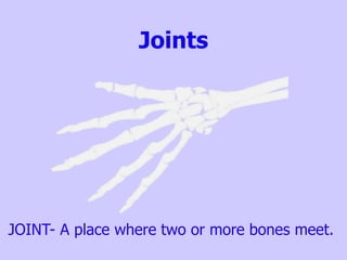
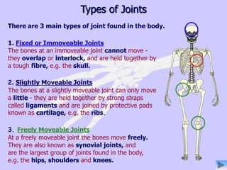
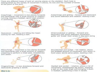
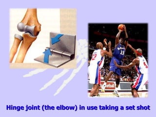
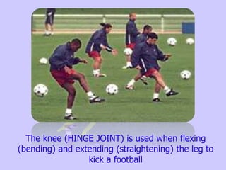
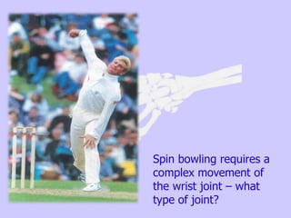
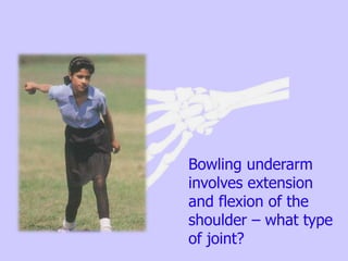
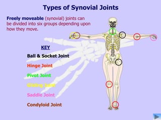
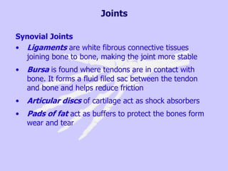
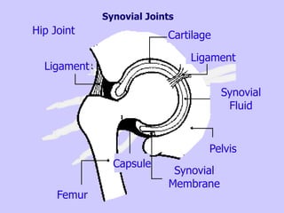
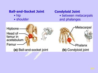
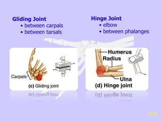
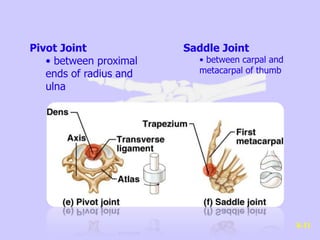
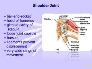
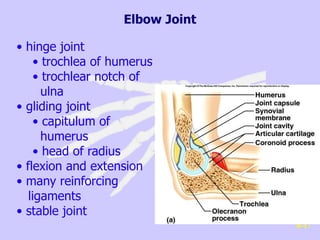
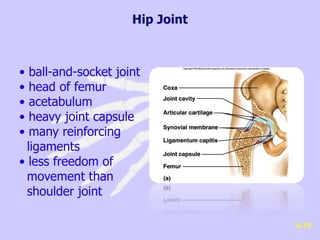
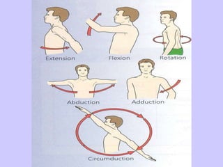
Joints
JOINT- A place where two or more bones meet.
1. Fixed or Immoveable Joints
The bones at an immoveable joint cannot move -
they overlap or interlock, and are held together by
a tough fibre, e.g. the skull.
2. Slightly Moveable Joints
The bones at a slightly moveable joint can only move
a little - they are held together by strong straps
called ligaments and are joined by protective pads
known as cartilage, e.g. the ribs.
3. Freely Moveable Joints
At a freely moveable joint the bones move freely.
They are also known as synovial joints, and
are the largest group of joints found in the body,
e.g. the hips, shoulders and knees.
Types of Joints
There are 3 main types of joint found in the body.
CLASSIFICATION OF JOINT BY
STRUCTURE
Fibrous: Have no cavity and are held together by
fibrous connective tissue – e.g. the sutures of the skull
bones
Cartilaginous: Also have no cavity. There is cartilage
between the bones of the joint. May be found between
the vertebrae of the spine
Synovial: Has a fluid filled cavity surrounded by an
articular capsule. The articulating surfaces are covered in
hyaline cartilage – e.g. the hinge joint of the knee
CLASSIFICATION OF JOINT BY
MOVEMENT
Fibrous or synarthrosis: Does not allow any movement,
which makes more sense when you know where in the
body they occur, as they supply protection – e.g. for the
brain
Cartilaginous or amphiarthrosis: Allows only limited
movement
Synovial or diarthrosis: Is freely moving, as much as
the shape of the articulating surface will allow.
JOINT / MOVEMENT TYPES
Joint type Shape of joint Movement range Body place : articulating bones
Ball + socket ball shaped bone fits 3 axes F/E AB/AD R C hip : femur, acetabulum of pelvis
into cup shaped socket shoulder : scapula, humerus
Hinge convex and concave 1 axis F/E knee : femur, patella, tibia
surfaces fitting together elbow : humerus, radius, ulna
Pivot ring shaped surrounding 1 axis R spine / atlas : odontoid process of axis
a cone (turns head from side to side)
elbow : proximal ends of radius and ulna
Condyloid modified ball and socket 2 axes F/E AB/AD C knuckle joints of fingers : metacarpals,
giving circumduction phalanges
wrist : radius, carpals
Saddle shaped like a saddle 2 axes F/E AB/AD C joint at base of thumb : carpal,
giving circumduction metacarpal
Gliding two flat gliding surfaces a little in all directions centre of chest : clavicle, sternum
wrist : carpals
ankle : tarsals
spine : articulating surfaces of vertebrae
In the picture below the ball and socket joint at the hip allows the player to
get height and then the ball and socket joint at the shoulder allows him to
SLAM DUNK!!
Hinge joint (the elbow) in use taking a set shot
The knee (HINGE JOINT) is used when flexing
(bending) and extending (straightening) the leg to
kick a football
Spin bowling requires a
complex movement of
the wrist joint – what
type of joint?
Bowling underarm
involves extension
and flexion of the
shoulder – what type
of joint?
RELATIONSHIP of MUSCULAR SYSTEM to
SKELETAL SYSTEM
NAMES OF MAJOR MUSCLE GROUPS
Example : biceps
ORIGIN (static end) : coracoid process / glenoid fossa tubercle of humerus
INSERTION (moving end) : tuberosity of radius
FASCIA
connective tissue
example : epimysium
LIGAMENTS
attach bone to bone
example : knee joint
cruciate ligaments
TENDONS
attach muscle to bone
via PERIOSTEUM
example : achilles tendon
PERIOSTEAL
LAYERS
attach tendons to bone
MUSCULAR
ATTACHMENTS
APONEUROSIS
a flattened ribbon
shaped tendon
Cartilage, Tendons, Ligaments: What’s the
difference?
Cartilage Tendons Ligaments
Tough
Attaches bone to
muscle
Attaches bone to
bone
Flexible Sturdy Elastic
At end of bone Non elastic Stabilise
Cushions
Size changes
depending on
muscle
Made of many
fibres
Anchor Strong
Types of Synovial Joints
KEY
Ball & Socket Joint
Hinge Joint
Pivot Joint
Gliding Joint
Saddle Joint
Condyloid Joint
Freely moveable (synovial) joints can
be divided into six groups depending upon
how they move.
Joints
Synovial Joints
• The articular capsule is a fibrous tissue encasing
the joint, forming a capsule
• The synovial membrane acts as a lining to the
joint capsule and secretes synovial fluid
• The articular/hyaline cartilage covers the ends of
the articulating bones
• Synovial fluid fills the joint capsule nourishes and
lubricates the articular cartilage
Joints
Synovial Joints
• Ligaments are white fibrous connective tissues
joining bone to bone, making the joint more stable
• Bursa is found where tendons are in contact with
bone. It forms a fluid filed sac between the tendon
and bone and helps reduce friction
• Articular discs of cartilage act as shock absorbers
• Pads of fat act as buffers to protect the bones form
wear and tear
Synovial Joints
Ligament
Cartilage
Ligament
Synovial
Fluid
Pelvis
Synovial
Membrane
Capsule
Femur
Hip Joint
Ball-and-Socket Joint
• hip
• shoulder
Condyloid Joint
• between metacarpals
and phalanges
8-9
Gliding Joint
• between carpals
• between tarsals
Hinge Joint
• elbow
• between phalanges
8-10
Pivot Joint
• between proximal
ends of radius and
ulna
Saddle Joint
• between carpal and
metacarpal of thumb
8-11
• ball-and-socket
• head of humerus
• glenoid cavity of
scapula
• loose joint capsule
• bursae
• ligaments prevent
displacement
• very wide range of
movement
8-15
Shoulder Joint
• hinge joint
• trochlea of humerus
• trochlear notch of
ulna
• gliding joint
• capitulum of
humerus
• head of radius
• flexion and extension
• many reinforcing
ligaments
• stable joint
8-17
Elbow Joint
• ball-and-socket joint
• head of femur
• acetabulum
• heavy joint capsule
• many reinforcing
ligaments
• less freedom of
movement than
shoulder joint
8-19
Hip Joint
• largest joint
• most complex
• medial and lateral
condyles of distal end of
femur
• medial and lateral
condyles of proximal end
of tibia
• femur articulates
anteriorly with patella
• modified hinge joint
• flexion/extension/little
rotation 8-21
Knee Joint
• strengthened by many
ligaments and tendons
• menisci separate femur
and tibia
• bursae
Joints

Joints

  • 1.
    Joints JOINT- A placewhere two or more bones meet.
  • 2.
    1. Fixed orImmoveable Joints The bones at an immoveable joint cannot move - they overlap or interlock, and are held together by a tough fibre, e.g. the skull. 2. Slightly Moveable Joints The bones at a slightly moveable joint can only move a little - they are held together by strong straps called ligaments and are joined by protective pads known as cartilage, e.g. the ribs. 3. Freely Moveable Joints At a freely moveable joint the bones move freely. They are also known as synovial joints, and are the largest group of joints found in the body, e.g. the hips, shoulders and knees. Types of Joints There are 3 main types of joint found in the body.
  • 3.
    CLASSIFICATION OF JOINTBY STRUCTURE Fibrous: Have no cavity and are held together by fibrous connective tissue – e.g. the sutures of the skull bones Cartilaginous: Also have no cavity. There is cartilage between the bones of the joint. May be found between the vertebrae of the spine Synovial: Has a fluid filled cavity surrounded by an articular capsule. The articulating surfaces are covered in hyaline cartilage – e.g. the hinge joint of the knee
  • 4.
    CLASSIFICATION OF JOINTBY MOVEMENT Fibrous or synarthrosis: Does not allow any movement, which makes more sense when you know where in the body they occur, as they supply protection – e.g. for the brain Cartilaginous or amphiarthrosis: Allows only limited movement Synovial or diarthrosis: Is freely moving, as much as the shape of the articulating surface will allow.
  • 6.
    JOINT / MOVEMENTTYPES Joint type Shape of joint Movement range Body place : articulating bones Ball + socket ball shaped bone fits 3 axes F/E AB/AD R C hip : femur, acetabulum of pelvis into cup shaped socket shoulder : scapula, humerus Hinge convex and concave 1 axis F/E knee : femur, patella, tibia surfaces fitting together elbow : humerus, radius, ulna Pivot ring shaped surrounding 1 axis R spine / atlas : odontoid process of axis a cone (turns head from side to side) elbow : proximal ends of radius and ulna Condyloid modified ball and socket 2 axes F/E AB/AD C knuckle joints of fingers : metacarpals, giving circumduction phalanges wrist : radius, carpals Saddle shaped like a saddle 2 axes F/E AB/AD C joint at base of thumb : carpal, giving circumduction metacarpal Gliding two flat gliding surfaces a little in all directions centre of chest : clavicle, sternum wrist : carpals ankle : tarsals spine : articulating surfaces of vertebrae
  • 7.
    In the picturebelow the ball and socket joint at the hip allows the player to get height and then the ball and socket joint at the shoulder allows him to SLAM DUNK!!
  • 8.
    Hinge joint (theelbow) in use taking a set shot
  • 9.
    The knee (HINGEJOINT) is used when flexing (bending) and extending (straightening) the leg to kick a football
  • 10.
    Spin bowling requiresa complex movement of the wrist joint – what type of joint?
  • 11.
    Bowling underarm involves extension andflexion of the shoulder – what type of joint?
  • 12.
    RELATIONSHIP of MUSCULARSYSTEM to SKELETAL SYSTEM NAMES OF MAJOR MUSCLE GROUPS Example : biceps ORIGIN (static end) : coracoid process / glenoid fossa tubercle of humerus INSERTION (moving end) : tuberosity of radius FASCIA connective tissue example : epimysium LIGAMENTS attach bone to bone example : knee joint cruciate ligaments TENDONS attach muscle to bone via PERIOSTEUM example : achilles tendon PERIOSTEAL LAYERS attach tendons to bone MUSCULAR ATTACHMENTS APONEUROSIS a flattened ribbon shaped tendon
  • 13.
    Cartilage, Tendons, Ligaments:What’s the difference? Cartilage Tendons Ligaments Tough Attaches bone to muscle Attaches bone to bone Flexible Sturdy Elastic At end of bone Non elastic Stabilise Cushions Size changes depending on muscle Made of many fibres Anchor Strong
  • 14.
    Types of SynovialJoints KEY Ball & Socket Joint Hinge Joint Pivot Joint Gliding Joint Saddle Joint Condyloid Joint Freely moveable (synovial) joints can be divided into six groups depending upon how they move.
  • 15.
    Joints Synovial Joints • Thearticular capsule is a fibrous tissue encasing the joint, forming a capsule • The synovial membrane acts as a lining to the joint capsule and secretes synovial fluid • The articular/hyaline cartilage covers the ends of the articulating bones • Synovial fluid fills the joint capsule nourishes and lubricates the articular cartilage
  • 16.
    Joints Synovial Joints • Ligamentsare white fibrous connective tissues joining bone to bone, making the joint more stable • Bursa is found where tendons are in contact with bone. It forms a fluid filed sac between the tendon and bone and helps reduce friction • Articular discs of cartilage act as shock absorbers • Pads of fat act as buffers to protect the bones form wear and tear
  • 17.
  • 18.
    Ball-and-Socket Joint • hip •shoulder Condyloid Joint • between metacarpals and phalanges 8-9
  • 19.
    Gliding Joint • betweencarpals • between tarsals Hinge Joint • elbow • between phalanges 8-10
  • 20.
    Pivot Joint • betweenproximal ends of radius and ulna Saddle Joint • between carpal and metacarpal of thumb 8-11
  • 21.
    • ball-and-socket • headof humerus • glenoid cavity of scapula • loose joint capsule • bursae • ligaments prevent displacement • very wide range of movement 8-15 Shoulder Joint
  • 22.
    • hinge joint •trochlea of humerus • trochlear notch of ulna • gliding joint • capitulum of humerus • head of radius • flexion and extension • many reinforcing ligaments • stable joint 8-17 Elbow Joint
  • 23.
    • ball-and-socket joint •head of femur • acetabulum • heavy joint capsule • many reinforcing ligaments • less freedom of movement than shoulder joint 8-19 Hip Joint
  • 24.
    • largest joint •most complex • medial and lateral condyles of distal end of femur • medial and lateral condyles of proximal end of tibia • femur articulates anteriorly with patella • modified hinge joint • flexion/extension/little rotation 8-21 Knee Joint • strengthened by many ligaments and tendons • menisci separate femur and tibia • bursae

Editor's Notes

  • #3 Task 1 and 2
  • #16  All six differ in movement – as the shape of the articulating bones defines But they are all very similar I structure…. Your example is a knee