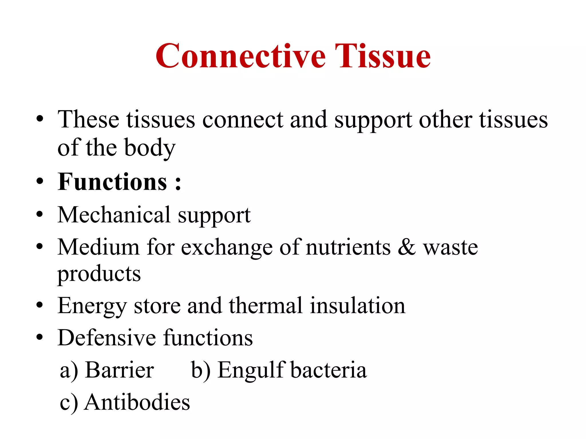
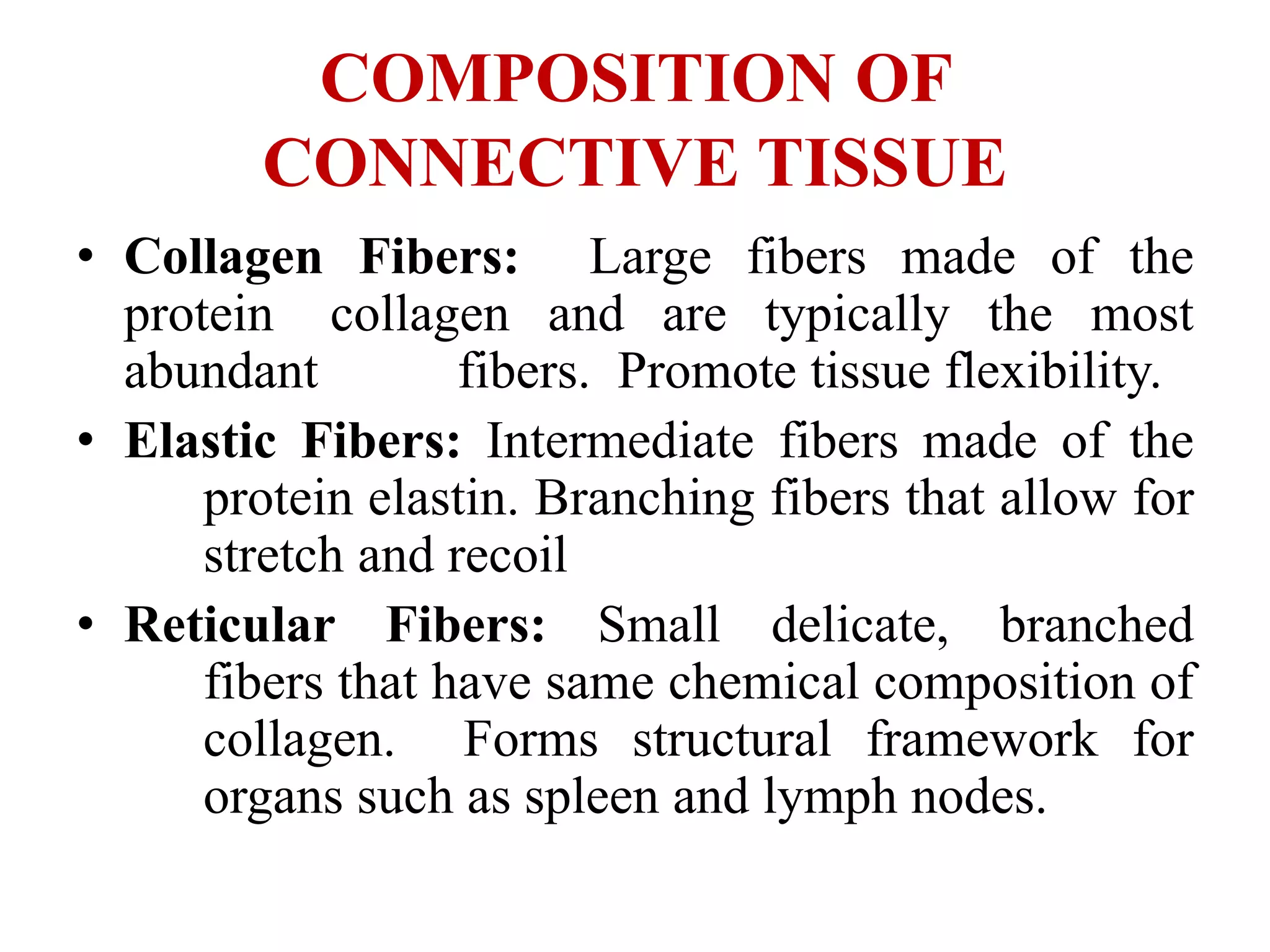
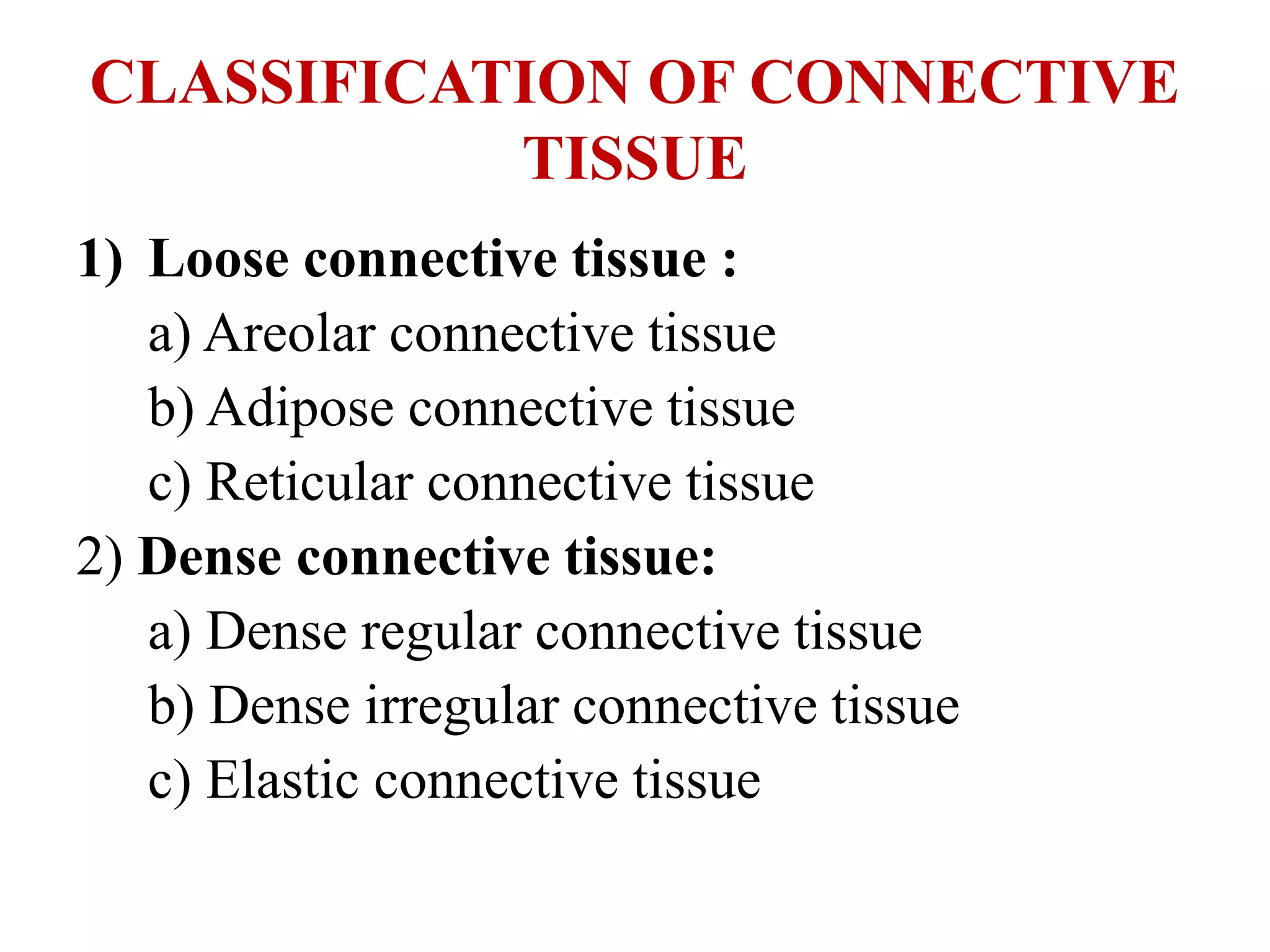
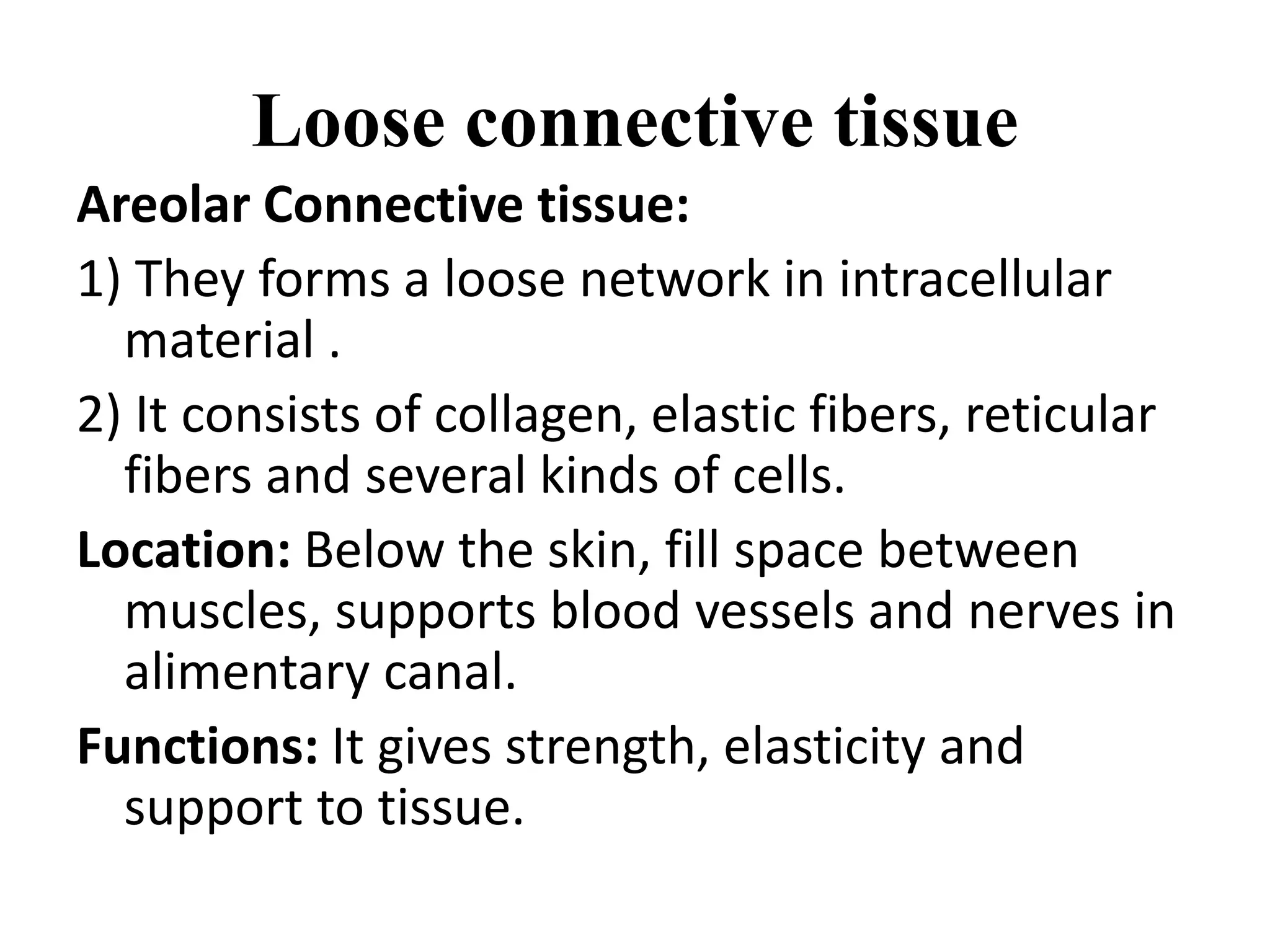
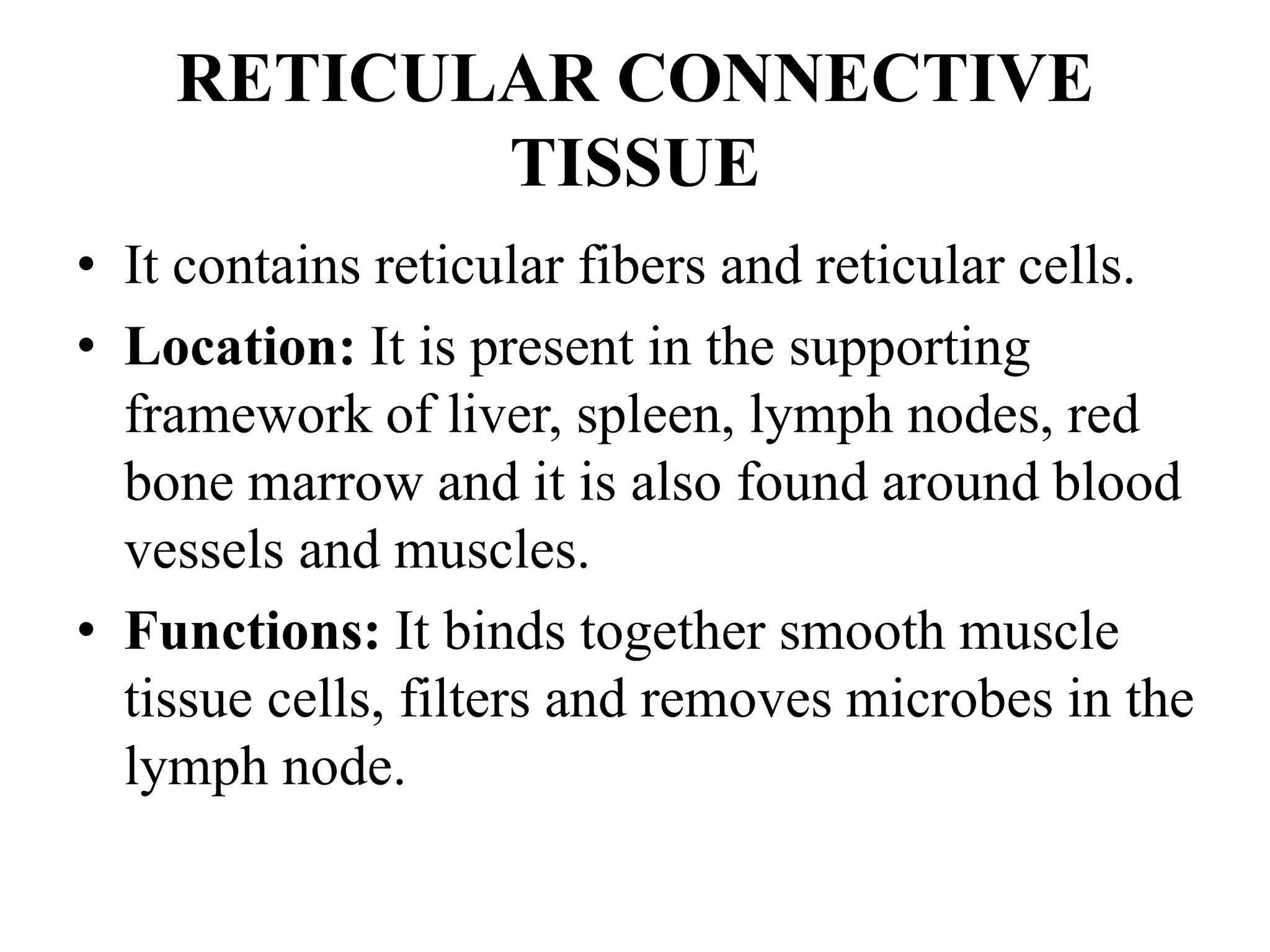
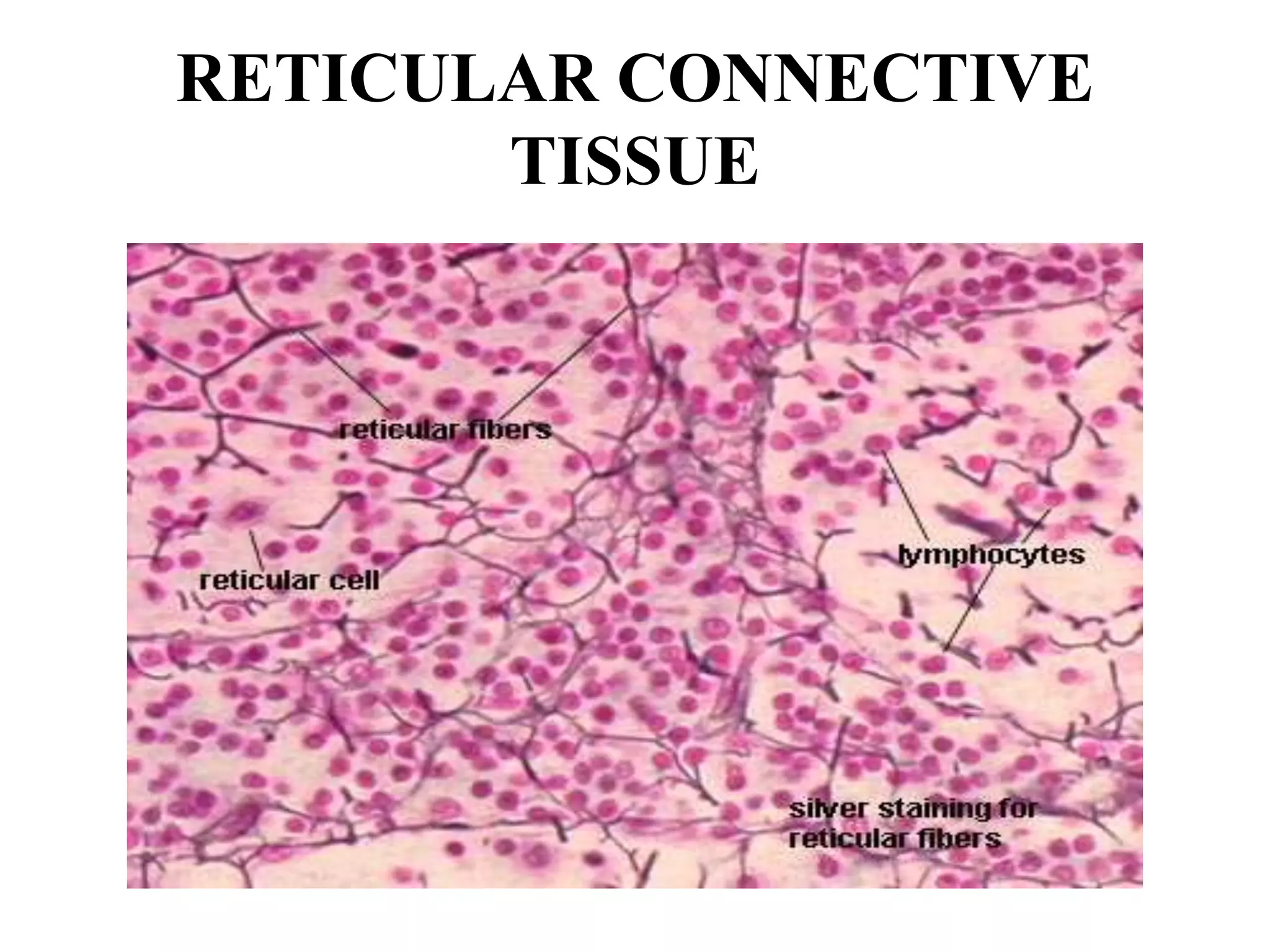
CONNECTIVE TISSUE
Connective Tissue
• These tissues connect and support other tissues
of the body
• Functions :
• Mechanical support
• Medium for exchange of nutrients & waste
products
• Energy store and thermal insulation
• Defensive functions
a) Barrier b) Engulf bacteria
c) Antibodies
COMPOSITION OF
CONNECTIVE TISSUE
• Collagen Fibers: Large fibers made of the
protein collagen and are typically the most
abundant fibers. Promote tissue flexibility.
• Elastic Fibers: Intermediate fibers made of the
protein elastin. Branching fibers that allow for
stretch and recoil
• Reticular Fibers: Small delicate, branched
fibers that have same chemical composition of
collagen. Forms structural framework for
organs such as spleen and lymph nodes.
CLASSIFICATION OF CONNECTIVE
TISSUE
1) Loose connective tissue :
a) Areolar connective tissue
b) Adipose connective tissue
c) Reticular connective tissue
2) Dense connective tissue:
a) Dense regular connective tissue
b) Dense irregular connective tissue
c) Elastic connective tissue
CLASSIFICATION OF
CONNECTIVE TISSUE
3) Cartilage:
a) Hyaline cartilage
b) Fibro cartilage
c) Elastic cartilage
4) Bone tissue
Liquid connective tissue
a) Blood tissue
b) Lymph
Loose connective tissue
Areolar Connective tissue:
1) They forms a loose network in intracellular
material .
2) It consists of collagen, elastic fibers, reticular
fibers and several kinds of cells.
Location: Below the skin, fill space between
muscles, supports blood vessels and nerves in
alimentary canal.
Functions: It gives strength, elasticity and
support to tissue.
Areolar Connective tissue:
ADIPOSE CONNECTIVE TISSUE
• It consists of adipocytes which stores fat.
Location: It is present in subcutaneous layer deep in
the skin, around the heart and kidneys
Functions :
• Prevents heat loose from body.
• Act as reservoir of energy.
• It give shape to the limbs and body.
• It protects underlying organ from injury.
ADIPOSE CONNECTIVE TISSUE
RETICULAR CONNECTIVE
TISSUE
• It contains reticular fibers and reticular cells.
• Location: It is present in the supporting
framework of liver, spleen, lymph nodes, red
bone marrow and it is also found around blood
vessels and muscles.
• Functions: It binds together smooth muscle
tissue cells, filters and removes microbes in the
lymph node.
RETICULAR CONNECTIVE
TISSUE
DENSE CONNECTIVE TISSUE
• In this tissue, fibers are densely packed.
• The fiber content is higher.
• Cell content is lower as compared to loose
connective tissue.
DENSE REGULAR CONNECTIVE
TISSUE
• Bundles of collagen fibers are arranged in
parallel patterns to provide strength to tissue.
• Fibroblast are appear in rows between the
fibers.
• It is tough in nature.
Location: It forms tendons (attach muscle bone)
and ligaments(attach bone to bone).
Functions: It provides strong attachment to
structure.
DENSE REGULAR CONNECTIVE
TISSUE
DENSE IRREGULAR
CONNECTIVE TISSUE
• It contains collagen fibers which are irregular
arranged and a few fibroblasts are appear in
rows between the fibers.
Location: It present in dermis layer of skin,
membrane capsules around kidneys, liver,
testes and lymph node, heart valves.
Functions: It provides strength to different
organs.
DENSE IRREGULAR
CONNECTIVE TISSUE
ELASTIC CONNECTIVE TISSUE
• It consists of freely branching elastic fibers.
• Fibroblast are present in space between fibers.
• It is yellowish in colour.
Location: It is present in tissues , walls of elastic
arteries, trachea, bronchial tubes and vocal
cords.
Functions: It allows stretching of various
organs.
ELASTIC CONNECTIVE TISSUE
CARTILAGE
• It consist of network of closely packed
collagen fibers and elastic fibers.
• The cells of mature cartilage called as
chondrocytes.
HYALINE CARTILAGE
• It is bluish white in color.
• It consists of fine collagen fibers and many
chondrocytes.
Location: It is present at the end of long bones,
anterior ends of ribs, nose and parts of larynx,
trachea, bronchi, bronchial tubes.
Function: It provides small surface for
movement at joints, flexibility and support.
HYALINE CARTILAGE
FIBRO CARTILAGE
• It is strongest form of cartilage.
• The chondrocytes are scattered among the
bundle collagen fibers within the extracellular
matrix.
Location: It is present in inter-verteblar disc.
Functions: It covers and protects bony structures
of body.
FIBRO CARTILAGE
ELASTIC CARTILAGE
• The chondrocytes are located within a
threadlike network of elastic fibers within
extracellular matrix.
Location: It is present in pinna of ear and top of
larynx.
Functions: It provides strength and elasticity
and maintain the shape of certain organs such
as the external ear.
ELASTIC CARTILAGE
THANK YOU

Connective tissue

  • 1.
  • 2.
    Connective Tissue • Thesetissues connect and support other tissues of the body • Functions : • Mechanical support • Medium for exchange of nutrients & waste products • Energy store and thermal insulation • Defensive functions a) Barrier b) Engulf bacteria c) Antibodies
  • 3.
    COMPOSITION OF CONNECTIVE TISSUE •Collagen Fibers: Large fibers made of the protein collagen and are typically the most abundant fibers. Promote tissue flexibility. • Elastic Fibers: Intermediate fibers made of the protein elastin. Branching fibers that allow for stretch and recoil • Reticular Fibers: Small delicate, branched fibers that have same chemical composition of collagen. Forms structural framework for organs such as spleen and lymph nodes.
  • 4.
    CLASSIFICATION OF CONNECTIVE TISSUE 1)Loose connective tissue : a) Areolar connective tissue b) Adipose connective tissue c) Reticular connective tissue 2) Dense connective tissue: a) Dense regular connective tissue b) Dense irregular connective tissue c) Elastic connective tissue
  • 5.
    CLASSIFICATION OF CONNECTIVE TISSUE 3)Cartilage: a) Hyaline cartilage b) Fibro cartilage c) Elastic cartilage 4) Bone tissue Liquid connective tissue a) Blood tissue b) Lymph
  • 6.
    Loose connective tissue AreolarConnective tissue: 1) They forms a loose network in intracellular material . 2) It consists of collagen, elastic fibers, reticular fibers and several kinds of cells. Location: Below the skin, fill space between muscles, supports blood vessels and nerves in alimentary canal. Functions: It gives strength, elasticity and support to tissue.
  • 7.
  • 8.
    ADIPOSE CONNECTIVE TISSUE •It consists of adipocytes which stores fat. Location: It is present in subcutaneous layer deep in the skin, around the heart and kidneys Functions : • Prevents heat loose from body. • Act as reservoir of energy. • It give shape to the limbs and body. • It protects underlying organ from injury.
  • 9.
  • 10.
    RETICULAR CONNECTIVE TISSUE • Itcontains reticular fibers and reticular cells. • Location: It is present in the supporting framework of liver, spleen, lymph nodes, red bone marrow and it is also found around blood vessels and muscles. • Functions: It binds together smooth muscle tissue cells, filters and removes microbes in the lymph node.
  • 11.
  • 12.
    DENSE CONNECTIVE TISSUE •In this tissue, fibers are densely packed. • The fiber content is higher. • Cell content is lower as compared to loose connective tissue.
  • 13.
    DENSE REGULAR CONNECTIVE TISSUE •Bundles of collagen fibers are arranged in parallel patterns to provide strength to tissue. • Fibroblast are appear in rows between the fibers. • It is tough in nature. Location: It forms tendons (attach muscle bone) and ligaments(attach bone to bone). Functions: It provides strong attachment to structure.
  • 14.
  • 15.
    DENSE IRREGULAR CONNECTIVE TISSUE •It contains collagen fibers which are irregular arranged and a few fibroblasts are appear in rows between the fibers. Location: It present in dermis layer of skin, membrane capsules around kidneys, liver, testes and lymph node, heart valves. Functions: It provides strength to different organs.
  • 16.
  • 17.
    ELASTIC CONNECTIVE TISSUE •It consists of freely branching elastic fibers. • Fibroblast are present in space between fibers. • It is yellowish in colour. Location: It is present in tissues , walls of elastic arteries, trachea, bronchial tubes and vocal cords. Functions: It allows stretching of various organs.
  • 18.
  • 19.
    CARTILAGE • It consistof network of closely packed collagen fibers and elastic fibers. • The cells of mature cartilage called as chondrocytes.
  • 20.
    HYALINE CARTILAGE • Itis bluish white in color. • It consists of fine collagen fibers and many chondrocytes. Location: It is present at the end of long bones, anterior ends of ribs, nose and parts of larynx, trachea, bronchi, bronchial tubes. Function: It provides small surface for movement at joints, flexibility and support.
  • 21.
  • 22.
    FIBRO CARTILAGE • Itis strongest form of cartilage. • The chondrocytes are scattered among the bundle collagen fibers within the extracellular matrix. Location: It is present in inter-verteblar disc. Functions: It covers and protects bony structures of body.
  • 23.
  • 24.
    ELASTIC CARTILAGE • Thechondrocytes are located within a threadlike network of elastic fibers within extracellular matrix. Location: It is present in pinna of ear and top of larynx. Functions: It provides strength and elasticity and maintain the shape of certain organs such as the external ear.
  • 25.
  • 26.