Downloaded 472 times
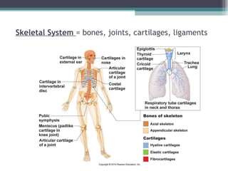
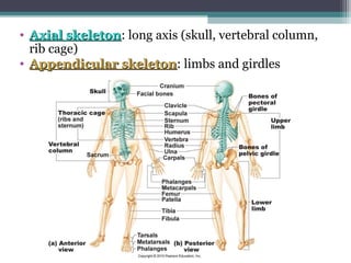
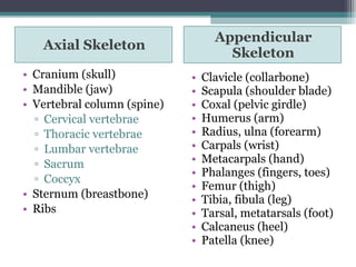
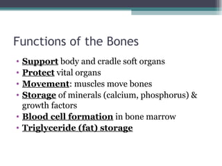
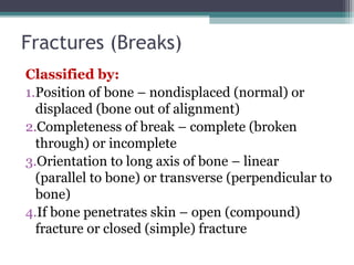
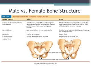
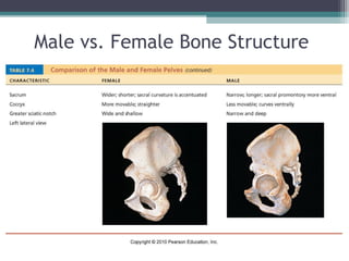
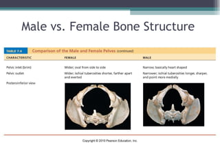
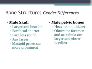
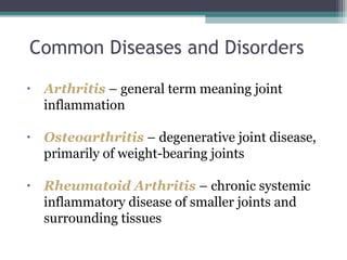
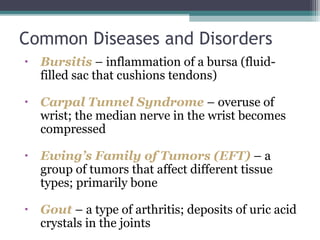
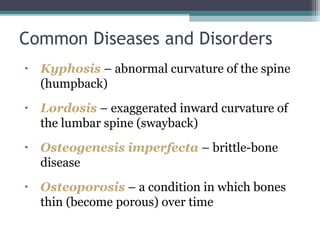

This document provides an overview of the skeletal system, detailing the types and functions of bones, their classifications, and the differences in male and female bone structures. It outlines bone development, the hormonal control involved, and the different types of fractures. Furthermore, it discusses common skeletal diseases and disorders such as arthritis, osteoporosis, and scoliosis.