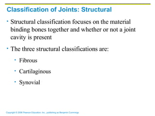
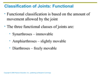
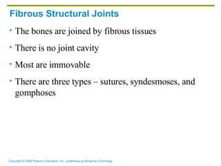
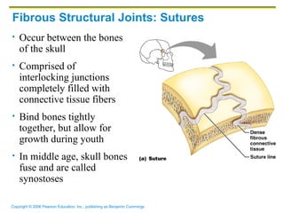
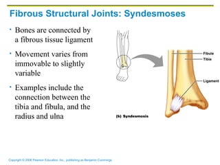
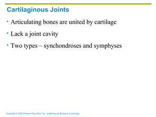
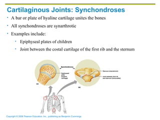
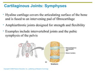
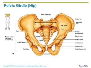
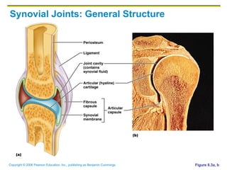
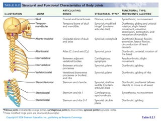
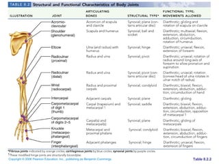
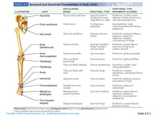
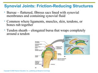
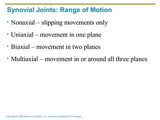
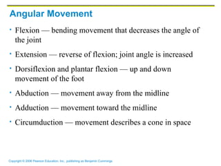
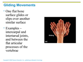
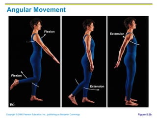
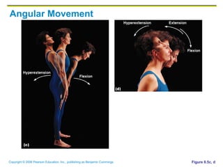
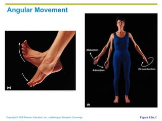
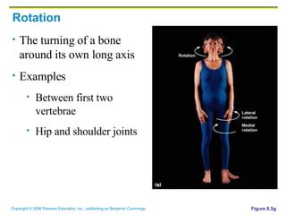
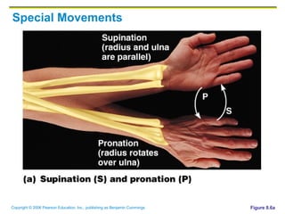
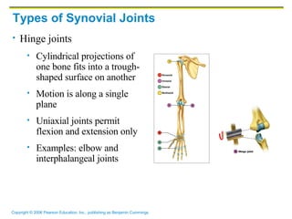
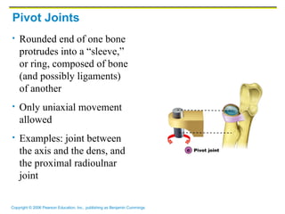
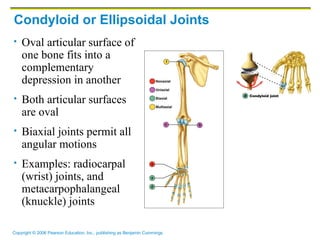
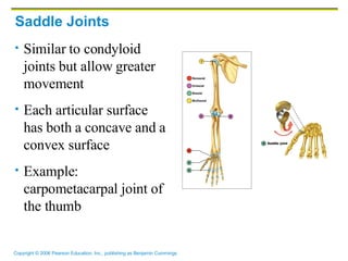
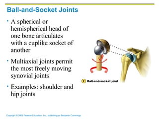
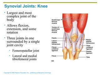
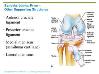
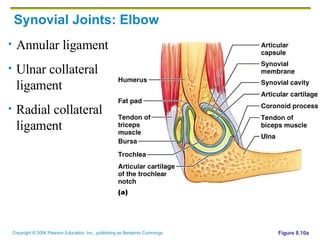
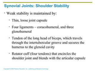
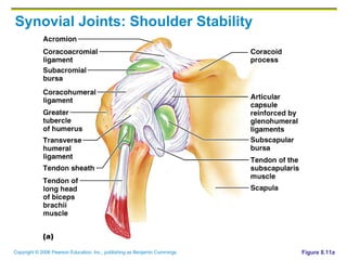
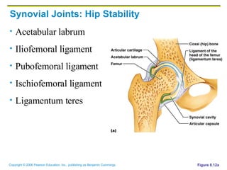
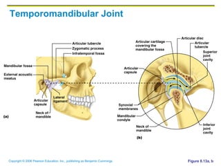
Chapter 8 Joints (Articulations) Weakest parts of the skeleton Articulation – site where two or more bones meet Functions of joints Give the skeleton mobility Hold the skeleton together
Classification of Joints: Structural Structural classification focuses on the material binding bones together and whether or not a joint cavity is present The three structural classifications are: Fibrous Cartilaginous  Synovial
Classification of Joints: Functional Functional classification is based on the amount of movement allowed by the joint The three functional classes of joints are: Synarthroses – immovable  Amphiarthroses – slightly movable  Diarthroses – freely movable
Fibrous Structural Joints The bones are joined by fibrous tissues There is no joint cavity Most are immovable There are three types – sutures, syndesmoses, and gomphoses
Fibrous Structural Joints: Sutures Occur between the bones of the skull Comprised of interlocking junctions completely filled with connective tissue fibers Bind bones tightly together, but allow for growth during youth In middle age, skull bones fuse and are called synostoses
Fibrous Structural Joints: Syndesmoses Bones are connected by a fibrous tissue ligament Movement varies from immovable to slightly variable Examples include the connection between the tibia and fibula, and the radius and ulna
Fibrous Structural Joints: Gomphoses The peg-in-socket fibrous joint between a tooth and its alveolar socket The fibrous connection is the periodontal ligament
Cartilaginous Joints Articulating bones are united by cartilage Lack a joint cavity Two types – synchondroses and symphyses
Cartilaginous Joints: Synchondroses A bar or plate of hyaline cartilage unites the bones All synchondroses are synarthrotic Examples include:  Epiphyseal plates of children Joint between the costal cartilage of the first rib and the sternum
Cartilaginous Joints: Symphyses Hyaline cartilage covers the articulating surface of the bone and is fused to an intervening pad of fibrocartilage Amphiarthrotic joints designed for strength and flexibility Examples include intervertebral joints and the pubic symphysis of the pelvis
Pelvic Girdle (Hip) Figure 7.27a
Synovial Joints Those joints in which the articulating bones are separated by a fluid-containing joint cavity All are freely movable diarthroses Examples – all limb joints, and most joints of the body Synovial joints all have the following Articular cartilage Joint (synovial) cavity Articular capsule Synovial fluid Reinforcing ligaments
Synovial Joints: General Structure Figure 8.3a, b
Table 8.2.1
Table 8.2.2
Table 8.2.3
Synovial Joints: Friction-Reducing Structures Bursae – flattened, fibrous sacs lined with synovial membranes and containing synovial fluid Common where ligaments, muscles, skin, tendons, or bones rub together Tendon sheath – elongated bursa that wraps completely around a tendon
Synovial Joints: Stability Stability is determined by: Articular surfaces – shape determines what movements are possible Ligaments – unite bones and prevent excessive or undesirable motion Muscle tone is accomplished by: Muscle tendons across joints acting as stabilizing factors Tendons that are kept tight at all times by muscle tone
Synovial Joints: Movement The two muscle attachments across a joint are: Origin – attachment to the immovable bone Insertion – attachment to the movable bone Described as movement along transverse, frontal, or sagittal planes
Synovial Joints: Range of Motion Nonaxial – slipping movements only Uniaxial – movement in one plane Biaxial – movement in two planes Multiaxial – movement in or around all three planes
Gliding Movements One flat bone surface glides or slips over another similar surface Examples – intercarpal and intertarsal joints, and between the flat articular processes of the vertebrae
Angular Movement Flexion — bending movement that decreases the angle of the joint Extension — reverse of flexion; joint angle is increased Dorsiflexion and plantar flexion — up and down movement of the foot Abduction — movement away from the midline Adduction — movement toward the midline Circumduction — movement describes a cone in space
Gliding Movements One flat bone surface glides or slips over another similar surface Examples – intercarpal and intertarsal joints, and between the flat articular processes of the vertebrae
Angular Movement Figure 8.5b
Angular Movement Figure 8.5c, d
Angular Movement Figure 8.5e, f
Rotation The turning of a bone around its own long axis Examples Between first two vertebrae Hip and shoulder joints Figure 8.5g
Special Movements Figure 8.6a
Special Movements Figure 8.6b
Special Movements Figure 8.6c
Special Movements Figure 8.6d
Special Movements Figure 8.6e
Plane Joint Plane joints Articular surfaces are essentially flat Allow only slipping or gliding movements Only examples of nonaxial joints Figure 8.7a
Types of Synovial Joints Hinge joints Cylindrical projections of one bone fits into a trough-shaped surface on another Motion is along a single plane Uniaxial joints permit flexion and extension only Examples: elbow and interphalangeal joints
Pivot Joints Rounded end of one bone protrudes into a “sleeve,” or ring, composed of bone (and possibly ligaments) of another Only uniaxial movement allowed Examples: joint between the axis and the dens, and the proximal radioulnar joint
Condyloid or Ellipsoidal Joints Oval articular surface of one bone fits into a complementary depression in another Both articular surfaces are oval Biaxial joints permit all angular motions Examples: radiocarpal (wrist) joints, and metacarpophalangeal (knuckle) joints
Saddle Joints Similar to condyloid joints but allow greater movement Each articular surface has both a concave and a convex surface Example: carpometacarpal joint of the thumb
Ball-and-Socket Joints A spherical or hemispherical head of one bone articulates with a cuplike socket of another Multiaxial joints permit the most freely moving synovial joints Examples: shoulder and hip joints
Synovial Joints: Knee Largest and most complex joint of the body Allows flexion, extension, and some rotation Three joints in one surrounded by a single joint cavity Femoropatellar joint Lateral and medial tibiofemoral joints
Synovial Joints: Knee –  Other Supporting Structures Anterior cruciate ligament Posterior cruciate ligament Medial meniscus (semilunar cartilage) Lateral meniscus
Synovial Joints: Shoulder Stability Figure 8.11a
Synovial Joints: Elbow Annular ligament Ulnar collateral ligament Radial collateral ligament Figure 8.10a
Synovial Joints: Shoulder Stability Weak stability is maintained by: Thin, loose joint capsule Four ligaments – coracohumeral, and three glenohumeral Tendon of the long head of biceps, which travels through the intertubercular groove and secures the humerus to the glenoid cavity Rotator cuff (four tendons) that encircles the shoulder joint and blends with the articular capsule
Synovial Joints: Shoulder Stability Figure 8.11a
Synovial Joints: Hip Stability Acetabular labrum Iliofemoral ligament Pubofemoral ligament Ischiofemoral ligament Ligamentum teres Figure 8.12a
Temporomandibular Joint Figure 8.13a, b
Sprains The ligaments reinforcing a joint are stretched or torn Partially torn ligaments slowly repair themselves Completely torn ligaments require prompt surgical repair Cartilage Injuries The snap and pop of overstressed cartilage Common aerobics injury Repaired with arthroscopic surgery
Dislocations Occur when bones are forced out of alignment Usually accompanied by sprains, inflammation, and joint immobilization Caused by serious falls and are common sports injuries Subluxation – partial dislocation of a joint
Inflammatory and Degenerative Conditions Bursitis An inflammation of a bursa, usually caused by a blow or friction Symptoms are pain and swelling Treated with anti-inflammatory drugs; excessive fluid may be aspirated Tendonitis Inflammation of tendon sheaths typically caused by overuse Symptoms and treatment are similar to bursitis
Arthritis More than 100 different types of inflammatory or degenerative diseases that damage the joints Most widespread crippling disease in the U.S. Symptoms – pain, stiffness, and swelling of a joint Acute forms are caused by bacteria and are treated with antibiotics Chronic forms include osteoarthritis, rheumatoid arthritis, and gouty arthritis
Osteoarthritis (OA) Most common chronic arthritis; often called “wear-and-tear” arthritis Affects women more than men 85% of all Americans develop OA More prevalent in the aged, and is probably related to the normal aging process OA reflects the years of abrasion and compression causing increased production of metalloproteinase enzymes that break down cartilage As one ages, cartilage is destroyed more quickly than it is replaced The exposed bone ends thicken, enlarge, form bone spurs, and restrict movement Joints most affected are the cervical and lumbar spine, fingers, knuckles, knees, and hips
Osteoarthritis: Treatments OA is slow and irreversible Treatments include: Mild pain relievers, along with moderate activity Magnetic therapy  Glucosamine sulfate decreases pain and inflammation
Rheumatoid Arthritis (RA) Chronic, inflammatory, autoimmune disease of unknown cause, with an insidious onset Usually arises between the ages of 40 to 50, but may occur at any age Signs and symptoms include joint tenderness, anemia, osteoporosis, muscle atrophy, and cardiovascular problems The course of RA is marked with exacerbations and remissions RA begins with synovitis of the affected joint Inflammatory chemicals are inappropriately released Inflammatory blood cells migrate to the joint, causing swelling Inflamed synovial membrane thickens into a pannus Pannus erodes cartilage, scar tissue forms, articulating bone ends connect The end result, ankylosis, produces bent, deformed fingers
Rheumatoid Arthritis: Treatment Conservative therapy – aspirin, long-term use of antibiotics, and physical therapy Progressive treatment – anti-inflammatory drugs or immunosuppressants  The drug Enbrel, a biological response modifier, neutralizes the harmful properties of inflammatory chemicals
Gouty Arthritis Deposition of uric acid crystals in joints and soft tissues, followed by an inflammation response Typically, gouty arthritis affects the joint at the base of the great toe In untreated gouty arthritis, the bone ends fuse and immobilize the joint Treatment – colchicine, nonsteroidal anti-inflammatory drugs, and glucocorticoids
Developmental Aspects of Joints By embryonic week 8, synovial joints resemble adult joints Few problems occur until late middle age Advancing years take their toll on joints: Ligaments and tendons shorten and weaken Intervertebral discs become more likely to herniate Most people in their 70s have some degree of OA Prudent exercise (especially swimming) that coaxes joints through their full range of motion is key to postponing joint problems

Chapter 8

  • 1.
    Chapter 8 Joints(Articulations) Weakest parts of the skeleton Articulation – site where two or more bones meet Functions of joints Give the skeleton mobility Hold the skeleton together
  • 2.
    Classification of Joints:Structural Structural classification focuses on the material binding bones together and whether or not a joint cavity is present The three structural classifications are: Fibrous Cartilaginous Synovial
  • 3.
    Classification of Joints:Functional Functional classification is based on the amount of movement allowed by the joint The three functional classes of joints are: Synarthroses – immovable Amphiarthroses – slightly movable Diarthroses – freely movable
  • 4.
    Fibrous Structural JointsThe bones are joined by fibrous tissues There is no joint cavity Most are immovable There are three types – sutures, syndesmoses, and gomphoses
  • 5.
    Fibrous Structural Joints:Sutures Occur between the bones of the skull Comprised of interlocking junctions completely filled with connective tissue fibers Bind bones tightly together, but allow for growth during youth In middle age, skull bones fuse and are called synostoses
  • 6.
    Fibrous Structural Joints:Syndesmoses Bones are connected by a fibrous tissue ligament Movement varies from immovable to slightly variable Examples include the connection between the tibia and fibula, and the radius and ulna
  • 7.
    Fibrous Structural Joints:Gomphoses The peg-in-socket fibrous joint between a tooth and its alveolar socket The fibrous connection is the periodontal ligament
  • 8.
    Cartilaginous Joints Articulatingbones are united by cartilage Lack a joint cavity Two types – synchondroses and symphyses
  • 9.
    Cartilaginous Joints: SynchondrosesA bar or plate of hyaline cartilage unites the bones All synchondroses are synarthrotic Examples include: Epiphyseal plates of children Joint between the costal cartilage of the first rib and the sternum
  • 10.
    Cartilaginous Joints: SymphysesHyaline cartilage covers the articulating surface of the bone and is fused to an intervening pad of fibrocartilage Amphiarthrotic joints designed for strength and flexibility Examples include intervertebral joints and the pubic symphysis of the pelvis
  • 11.
    Pelvic Girdle (Hip)Figure 7.27a
  • 12.
    Synovial Joints Thosejoints in which the articulating bones are separated by a fluid-containing joint cavity All are freely movable diarthroses Examples – all limb joints, and most joints of the body Synovial joints all have the following Articular cartilage Joint (synovial) cavity Articular capsule Synovial fluid Reinforcing ligaments
  • 13.
    Synovial Joints: GeneralStructure Figure 8.3a, b
  • 14.
  • 15.
  • 16.
  • 17.
    Synovial Joints: Friction-ReducingStructures Bursae – flattened, fibrous sacs lined with synovial membranes and containing synovial fluid Common where ligaments, muscles, skin, tendons, or bones rub together Tendon sheath – elongated bursa that wraps completely around a tendon
  • 18.
    Synovial Joints: StabilityStability is determined by: Articular surfaces – shape determines what movements are possible Ligaments – unite bones and prevent excessive or undesirable motion Muscle tone is accomplished by: Muscle tendons across joints acting as stabilizing factors Tendons that are kept tight at all times by muscle tone
  • 19.
    Synovial Joints: MovementThe two muscle attachments across a joint are: Origin – attachment to the immovable bone Insertion – attachment to the movable bone Described as movement along transverse, frontal, or sagittal planes
  • 20.
    Synovial Joints: Rangeof Motion Nonaxial – slipping movements only Uniaxial – movement in one plane Biaxial – movement in two planes Multiaxial – movement in or around all three planes
  • 21.
    Gliding Movements Oneflat bone surface glides or slips over another similar surface Examples – intercarpal and intertarsal joints, and between the flat articular processes of the vertebrae
  • 22.
    Angular Movement Flexion— bending movement that decreases the angle of the joint Extension — reverse of flexion; joint angle is increased Dorsiflexion and plantar flexion — up and down movement of the foot Abduction — movement away from the midline Adduction — movement toward the midline Circumduction — movement describes a cone in space
  • 23.
    Gliding Movements Oneflat bone surface glides or slips over another similar surface Examples – intercarpal and intertarsal joints, and between the flat articular processes of the vertebrae
  • 24.
  • 25.
  • 26.
  • 27.
    Rotation The turningof a bone around its own long axis Examples Between first two vertebrae Hip and shoulder joints Figure 8.5g
  • 28.
  • 29.
  • 30.
  • 31.
  • 32.
  • 33.
    Plane Joint Planejoints Articular surfaces are essentially flat Allow only slipping or gliding movements Only examples of nonaxial joints Figure 8.7a
  • 34.
    Types of SynovialJoints Hinge joints Cylindrical projections of one bone fits into a trough-shaped surface on another Motion is along a single plane Uniaxial joints permit flexion and extension only Examples: elbow and interphalangeal joints
  • 35.
    Pivot Joints Roundedend of one bone protrudes into a “sleeve,” or ring, composed of bone (and possibly ligaments) of another Only uniaxial movement allowed Examples: joint between the axis and the dens, and the proximal radioulnar joint
  • 36.
    Condyloid or EllipsoidalJoints Oval articular surface of one bone fits into a complementary depression in another Both articular surfaces are oval Biaxial joints permit all angular motions Examples: radiocarpal (wrist) joints, and metacarpophalangeal (knuckle) joints
  • 37.
    Saddle Joints Similarto condyloid joints but allow greater movement Each articular surface has both a concave and a convex surface Example: carpometacarpal joint of the thumb
  • 38.
    Ball-and-Socket Joints Aspherical or hemispherical head of one bone articulates with a cuplike socket of another Multiaxial joints permit the most freely moving synovial joints Examples: shoulder and hip joints
  • 39.
    Synovial Joints: KneeLargest and most complex joint of the body Allows flexion, extension, and some rotation Three joints in one surrounded by a single joint cavity Femoropatellar joint Lateral and medial tibiofemoral joints
  • 40.
    Synovial Joints: Knee– Other Supporting Structures Anterior cruciate ligament Posterior cruciate ligament Medial meniscus (semilunar cartilage) Lateral meniscus
  • 41.
    Synovial Joints: ShoulderStability Figure 8.11a
  • 42.
    Synovial Joints: ElbowAnnular ligament Ulnar collateral ligament Radial collateral ligament Figure 8.10a
  • 43.
    Synovial Joints: ShoulderStability Weak stability is maintained by: Thin, loose joint capsule Four ligaments – coracohumeral, and three glenohumeral Tendon of the long head of biceps, which travels through the intertubercular groove and secures the humerus to the glenoid cavity Rotator cuff (four tendons) that encircles the shoulder joint and blends with the articular capsule
  • 44.
    Synovial Joints: ShoulderStability Figure 8.11a
  • 45.
    Synovial Joints: HipStability Acetabular labrum Iliofemoral ligament Pubofemoral ligament Ischiofemoral ligament Ligamentum teres Figure 8.12a
  • 46.
  • 47.
    Sprains The ligamentsreinforcing a joint are stretched or torn Partially torn ligaments slowly repair themselves Completely torn ligaments require prompt surgical repair Cartilage Injuries The snap and pop of overstressed cartilage Common aerobics injury Repaired with arthroscopic surgery
  • 48.
    Dislocations Occur whenbones are forced out of alignment Usually accompanied by sprains, inflammation, and joint immobilization Caused by serious falls and are common sports injuries Subluxation – partial dislocation of a joint
  • 49.
    Inflammatory and DegenerativeConditions Bursitis An inflammation of a bursa, usually caused by a blow or friction Symptoms are pain and swelling Treated with anti-inflammatory drugs; excessive fluid may be aspirated Tendonitis Inflammation of tendon sheaths typically caused by overuse Symptoms and treatment are similar to bursitis
  • 50.
    Arthritis More than100 different types of inflammatory or degenerative diseases that damage the joints Most widespread crippling disease in the U.S. Symptoms – pain, stiffness, and swelling of a joint Acute forms are caused by bacteria and are treated with antibiotics Chronic forms include osteoarthritis, rheumatoid arthritis, and gouty arthritis
  • 51.
    Osteoarthritis (OA) Mostcommon chronic arthritis; often called “wear-and-tear” arthritis Affects women more than men 85% of all Americans develop OA More prevalent in the aged, and is probably related to the normal aging process OA reflects the years of abrasion and compression causing increased production of metalloproteinase enzymes that break down cartilage As one ages, cartilage is destroyed more quickly than it is replaced The exposed bone ends thicken, enlarge, form bone spurs, and restrict movement Joints most affected are the cervical and lumbar spine, fingers, knuckles, knees, and hips
  • 52.
    Osteoarthritis: Treatments OAis slow and irreversible Treatments include: Mild pain relievers, along with moderate activity Magnetic therapy Glucosamine sulfate decreases pain and inflammation
  • 53.
    Rheumatoid Arthritis (RA)Chronic, inflammatory, autoimmune disease of unknown cause, with an insidious onset Usually arises between the ages of 40 to 50, but may occur at any age Signs and symptoms include joint tenderness, anemia, osteoporosis, muscle atrophy, and cardiovascular problems The course of RA is marked with exacerbations and remissions RA begins with synovitis of the affected joint Inflammatory chemicals are inappropriately released Inflammatory blood cells migrate to the joint, causing swelling Inflamed synovial membrane thickens into a pannus Pannus erodes cartilage, scar tissue forms, articulating bone ends connect The end result, ankylosis, produces bent, deformed fingers
  • 54.
    Rheumatoid Arthritis: TreatmentConservative therapy – aspirin, long-term use of antibiotics, and physical therapy Progressive treatment – anti-inflammatory drugs or immunosuppressants The drug Enbrel, a biological response modifier, neutralizes the harmful properties of inflammatory chemicals
  • 55.
    Gouty Arthritis Depositionof uric acid crystals in joints and soft tissues, followed by an inflammation response Typically, gouty arthritis affects the joint at the base of the great toe In untreated gouty arthritis, the bone ends fuse and immobilize the joint Treatment – colchicine, nonsteroidal anti-inflammatory drugs, and glucocorticoids
  • 56.
    Developmental Aspects ofJoints By embryonic week 8, synovial joints resemble adult joints Few problems occur until late middle age Advancing years take their toll on joints: Ligaments and tendons shorten and weaken Intervertebral discs become more likely to herniate Most people in their 70s have some degree of OA Prudent exercise (especially swimming) that coaxes joints through their full range of motion is key to postponing joint problems