
Download as PPSX, PPTX


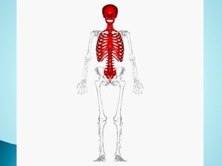
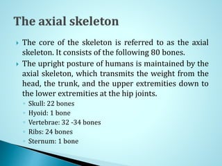
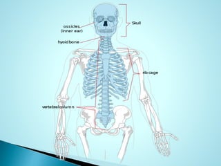

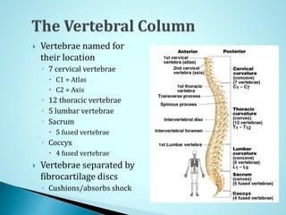

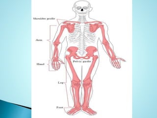
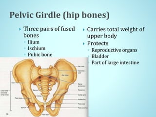
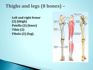
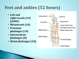


The human skeleton can be divided into two parts: the axial skeleton and the appendicular skeleton. The axial skeleton consists of 80 bones including the skull, vertebral column, ribs, and sternum, and forms the core of the body providing support and protecting organs. The appendicular skeleton is made up of 126 bones organized into the upper and lower limbs, including shoulders, pelvis, arms, forearms, hands, thighs, legs, feet, and ankles. Together the skeleton provides structure, movement, protection, storage, and production of blood cells to the body.


