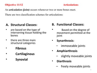
SlideShare a Scribd company logo
Objective 11
• Define “articulation” and discuss the
classification schemes for the joints of the
body: functional classes (synarthrosis,
amphiarthrosis, diarthrosis) and structural
classes (fibrous, cartilaginous, synovial).
1
Objective 11/12 Articulations
An articulation (joint) occurs whenever two or more bones meet.
There are two classification schemes for articulations:
A. Structural Classes:
• are based on the type of
intervening tissue holding the
bones
• there are three main
structural categories
• Fibrous
• Cartilaginous
• Synovial
B. Functional Classes:
• Based on the degree of
movement permitted at the
joint
• Synarthrosis:
– immoveable joints
• Amphiarthrosis
– slightly moveable joints
• Diarthrosis
– freely moveable joints
2
1. Fibrous Joints
- the bones are joined by fibrous connective tissue
- the amount of movement permitted depends on the length of the fibers holding
the bones together
1a. Sutures
- short fibers hold the bones together
- fontanels in infants
- these joints ossify (form a syntosis) in adults
- are immoveable joints
What type of bone formation?
3
Intramembraneous Ossification
via fibrous membrane
4
Fibrous Joints:
Sutures
• Rigid, interlocking joints
• Immovable joints for protection of brain
• Contain short connective tissue fibers
• Allow for growth during youth
• In middle age, sutures ossify and fuse
– Called Synostoses
1b. Syndesmosis: longer bands of fibers connect the bones; these
bands are called ligaments or
interosseous membranes
Slightly moveable-Amphiarthrotic
Immoveable-SynarthroticDistal radius and ulna
Distal tibia/fibula
6
1c. Gomphosis: the periodontal ligament holds the tooth in
its alveolar socket
Immoveable
7
© 2013 Pearson Education, Inc.
Synchondroses
Bones united by hyaline cartilage
Epiphyseal
plate (temporary
hyaline cartilage
joint)
Sternum
(manubrium)
Joint between first
rib and sternum
(immovable)
2. Cartilaginous Joints
- held together by hyaline or fibro cartilage; immoveable (syn-) or slightly moveable
(amphi-)
Symphyses
Bones united by fibrocartilage
Body of vertebra
Fibrocartilaginous
intervertebral disc
(sandwiched between
hyaline cartilage)
Pubic symphysis
most of these joints eventually ossify (form a syntosis)
Articular cartilage: hyaline cartilage that covers
the ends of the
articulating bones
Joint cavity: potential space that lies
(synovial) between the articulating
bones; this space is filled with
synovial fluid
Articular capsule: has two portions:
fibrous capsule: dense irregular
connective tissue that holds the
bones together
synovial membrane: inner
structure that secretes
synovial fluid
1
4
3
2
9
Ligament
Joint cavity
Articular (hyaline)
cartilage
Fibrous
layer
Synovial
membrane
(secretes
synovial
fluid)
Articular
capsule
Periosteum
1
4
3
2
3. Synovial Joints
- highly innervated, complicated structure; all
moveable (diarthrotic)
Articular cartilage: hyaline cartilage that
covers the ends of articulating bones
Joint (synovial) cavity: space between
articulating bones; this space is filled
with synovial fluid
Articular capsule: has two portions:
fibrous capsule: dense irregular CT that
holds bones together
synovial membrane: inner structure that
secretes synovial fluid
Reinforcing Ligaments
Joint Cavity space between articulating
bones; filled with synovial fluid
Articular (hyaline) cartilage
Fibrous
layer
Synovial
membrane
(secretes
synovial
fluid)
Articular Capsule::
FC: dense irregular CT that holds bones
together
SM: inner structure that secretes synovial fluid
Periosteum
1
4
3
2
3. Synovial Joints
- highly innervated, complicated structure; all
moveable (diarthrotic)
Synovial fluid: viscous fluid (yolk-like) that is found
in the joint cavity; functions
include:
1. Lubricate the joint surfaces
and reduce friction
2. Helps to nourish articular
cartilage
3. Phagocytic cells keep
the joint free of debris
Reinforcing dense, regular connective tissue
Ligaments: that connects the articulating
bones
intrinsic (capsular) ligaments
are parts of the joint capsule
extrinsic (extracapsular) ligaments
lie external to the joint capsule
intracapsular ligaments are deep to
the joint capsule
12
Objective 13 Additional Synovial Structures
Bursae/Tendon Sheaths/Menisci
Bursae: flattened fibrous sac lined with synovial membrane and filled with
synovial fluid
are inserted at strategic points to protect soft tissues from being
damaged while rubbing over bone
Tendon Sheaths: function like bursae, but are elongated and are wrapped around
tendons
13
© 2013 Pearson Education, Inc.
Figure 8.4b Bursae and tendon sheaths.
Bursa rolls
and lessens
friction.
Humerus head
rolls medially as
arm abducts.
Humerus moving
Enlargement of (a), showing how
a bursa eliminates friction where
a ligament (or other structure) would
rub against a bone
15
Menisci:
fibrocartilaginous pads that are found within
the joint cavity of some joints (knee)
Function:
1. Help protect the articular surfaces
2. Help to improve the fit of articular
surfaces that are not
congruent
Labrum: similar to the meniscus but located
in the shoulder or hip joint
Additional Structures:
Objectives 15 and 16 Joint Range of Motion
A. Range of motion:
movement at a joint ; can
occur in a variety of
directions or planes; and is
determined by:
1. The shapes of the articulating
surface
2. The pressure of ligaments and
tendons
3. The presence of adjacent tissue
B. Terms for range of motion:
Non-axial: non-directional movement;
slipping movements only
Uniaxial: movement in one plane
Biaxial: movement in two planes
Multiaxial: movement in three planes
16
C. Specific Movements
• Gliding:
- When flat or nearly flat articular
surfaces slip or glide over each
other
- Examples are intertarsal,
intercarpal, intervertbral (at
articular processes) and sterno-
clavicular joints
• Protraction/Retraction:
17
• Elevation/Depression:
Angular Movements
-the angle between two bones increases or decreases
Flexion: movement in a sagittal plane so that the angle between two
bones is decreased
Extension: movement into anatomic position from a flexed position
(increases ventral angle)
Hyperextension: extension beyond anatomic position
18
19
20
• Six types, based on shape of articular surfaces:
– Plane
– Hinge
– Pivot
– Condylar
– Saddle
– Ball-and-socket
Objective 17
Classes of Synovial Joints
Gliding
Plane joint Nonaxial movement
Ex: Intercarpal/intertarsal joints, articularr suface of vertebral joints
Hinge joint Uniaxial movement
Ex: Elbow & interphalangeal joints
Flexion and extension
Pivot joint Uniaxial movement
Rotation
Ex: Proximal radioulnar joints, atlantoaxial joint
Condylar joint Biaxial movement
Ex: Metacarpophalangeal (knuckle) & wrist joints
Flexion and
extension
Adduction and
abduction
Flexion and
extension
Adduction and
abduction
Ex: Carpometacarpal joints of the thumbs
Saddle joint Biaxial movement
Ex: Shoulder joints and hip joints
Flexion
extension
Adduction
abduction Rotation
Ball-and-socket joint Multiaxial movement
Types of Synovial Joints
There are six classes of synovial joints:
Class Movement Permitted Structure
Gliding (Plane. Non-axial flat or slightly curved articular
Arthrodia) surface
Examples include intercarpal and intertarsal joints; joints between the vertebral
articular processes; sternoclavicular joints
23
Class Movement Permitted Structure
Hinge (ginglymus) uniaxial; flexion/extension convex or cylindrical
projection of one bone
fits into a concave or
cylindrical groove of
another bone
Examples include the elbow, knee and interphlangeal joints
24
Class Movement Permitted Structure
Pivot (trochoid) uniaxial; rotation only a rounded or conical surface
of one bone fits into a
“sleeve” or ring of bone or
ligaments
Examples include the joint between the atlas and axis; proximal radius and
ulna
C1 Atlas C2 Axis
25
Class Movement Permitted Structure
Ellipsoidal biaxial; circumduction the oval articular surface of
(condyloid) one bone fits into a complex
concavity of another
Examples include the radiocarpal joint; metacarophalangeal joint;
atlanto-occipital joint
26
Class Movement Permitted Structure
Saddle (sellaris) biaxial; circumduction but a structured like an ellipsoidal
greater degree of movement is joint
permitted than in an ellipsoidal
joint
Example is the carpometacarpal joint of the thumb
27
Class Movement Permitted Structure
Ball and Socket multiaxial a spherical head or
(Spheroid) hemispherical head of a bone
fits into the concave socket of
another bone
28
Objective 16 Selected Synovial Joints
A. Shoulder Joint:
- a ball and socket with great range of motion
Bones: arm bone (humerus),
shoulder blade (scapula), and
collarbone (clavicle).
29
Rotator Cuff Muscles:
SITS
Supraspinatus
Infraspinatus
Teres minor
Subscapularis
The rotator cuff muscles and triceps brachii (long head)
provide support
?
30
31
B. The Hip Joint
- ball and socket with less range of motion, limited by the deep
socket of the acetabulum and strong ligaments
- Weight bearing joint
The acetabular labrum makes
the acetabulum deeper!
Reinforcing ligaments:
ligamentum teres
ischiofemoral ligament
pubofemoral ligament
ilio femoral ligament
32
C. The Elbow Joint
- A hinge joint with substantial anterior/posterior movement
- Radial and ulnar collateral ligaments limit side to side motion
- Bones: humerus, radius, ulnar
33
D. The Knee Joint
- a modified hinge joint that allows extension and some rotation
- An incomplete articular capsule is found on the lateral and posterior
surfaces
- Extracapsular ligaments include the fibular and tibial collateral
ligaments, oblique popliteal ligament and arcuate popliteal ligament
- Intracapsular ligaments are the ACL and the PCL
http://www.youtube.com/watch?v=uuoj_HFG5Z0
34
YouTube - Knee Ligament Anatomy Animation
35
Objective 18 Joint Disorders
Disorder Cause(s) Characteristics
Rheumatoid arthritis autoimmune inflammation of the synovial
membranes causes them to thicken
and accumulate fluid leading to
pain
often involves interphalangeal
wrist and knee joints
mild cases result in a reduced
range of motion because of
inflammation
severe cases may cause the joint
to fuse (ankylosis) and become
immoveable
36
Rheumatoid Arthritis
37
Disorder Cause Characteristics
Osteoarthritis disease of cartilage progressive deterioration of articular
cartilage in weight bearing joints
such as the knee, the spine and hips
bone spurs can grow that inflame
surrounding tissues
associated with aging; may be
exacerbated by obesity, athletics
                                                                                                                                                    
38
Disorder Cause Characteristics
Gouty arthritis sodium ureate crystals irritation of articular cartilage causing
inflammation, swelling and pain
likely to occur in the joint at the base
of the big toe, the elbow, knee, hand
foot, ankle, arm or shoulder
effects primarily men of any age
precipitated by diet, diuretics, stress
and some antibiotics
                                                                                                                                                    
39
Disorder Cause(s) Characteristics
Bursitis joint injury or infection inflammation of bursae causing swelling,
pain, tenderness and a reduced range of
motion
most frequent in bursae near the shoulders,
hips, pelvis and knees
Dislocation traumatic joint injury, displacement of a bone from its joint
congenital joint causing swelling, pain and temporary
malformation paralysis of the joint
most common in fingers, and fairly connon
at the mandible, elbow, hip and knee
Tendinitis traumatic injury, poor painful inflammation of a tendon
causing posture and some tenderness and restricted movement
musculoskeletal disorders common sites are the shoulder, elbow and
achilles tendon
Sprain overuse or stress of a injury to ligaments resulting in tearing and
ligament stretching causes pain, swelling and
immobility common in wrists, ankles 40
Bursitis Images
41
                                                                                                                                                          
Dislocation
Images
42

More Related Content

What's hot

Temporo mandibular joint
Temporo mandibular jointTemporo mandibular joint
Temporo mandibular joint
Martin Bush
 

What's hot (20)

Periodontal ligament (PDL)
Periodontal ligament (PDL)Periodontal ligament (PDL)
Periodontal ligament (PDL)
 
Malunion - Principals and Management - Dr Chintan N. Patel
Malunion - Principals and Management - Dr Chintan N. PatelMalunion - Principals and Management - Dr Chintan N. Patel
Malunion - Principals and Management - Dr Chintan N. Patel
 
Perioontal ligament lecture
Perioontal ligament lecturePerioontal ligament lecture
Perioontal ligament lecture
 
Knee Problems and Knee Injuries Overview
Knee Problems and Knee Injuries OverviewKnee Problems and Knee Injuries Overview
Knee Problems and Knee Injuries Overview
 
Periodontal ligament
Periodontal ligamentPeriodontal ligament
Periodontal ligament
 
Perthes disease ADOLESCENT COXA VARA
Perthes disease ADOLESCENT COXA VARAPerthes disease ADOLESCENT COXA VARA
Perthes disease ADOLESCENT COXA VARA
 
Cementum
CementumCementum
Cementum
 
Lumbar Disc Replacement
Lumbar Disc ReplacementLumbar Disc Replacement
Lumbar Disc Replacement
 
Human bones
Human bonesHuman bones
Human bones
 
Fibrousdysplasia
Fibrousdysplasia Fibrousdysplasia
Fibrousdysplasia
 
Vol 25 arthritis
Vol 25 arthritisVol 25 arthritis
Vol 25 arthritis
 
Osteology of head n neck ppt 1
Osteology of head n neck ppt 1Osteology of head n neck ppt 1
Osteology of head n neck ppt 1
 
Cubitus valgus varus
Cubitus valgus varusCubitus valgus varus
Cubitus valgus varus
 
Temporo mandibular joint
Temporo mandibular jointTemporo mandibular joint
Temporo mandibular joint
 
Comprehensive Vertebral Column Anatomy, MDIRT Nchanji Nkeh Keneth
Comprehensive Vertebral Column Anatomy, MDIRT Nchanji Nkeh KenethComprehensive Vertebral Column Anatomy, MDIRT Nchanji Nkeh Keneth
Comprehensive Vertebral Column Anatomy, MDIRT Nchanji Nkeh Keneth
 
Bone tumors
Bone tumors   Bone tumors
Bone tumors
 
Legg calve perthes
Legg calve perthesLegg calve perthes
Legg calve perthes
 
Bone : Basics
Bone : BasicsBone : Basics
Bone : Basics
 
Cleidocranial dysplasia
Cleidocranial dysplasiaCleidocranial dysplasia
Cleidocranial dysplasia
 
Perthes Disease LCPD
Perthes Disease LCPDPerthes Disease LCPD
Perthes Disease LCPD
 

Similar to Topic 3 obj 11 17

4. Joints,,Articulations.pptx
4. Joints,,Articulations.pptx4. Joints,,Articulations.pptx
4. Joints,,Articulations.pptx
AnwarTawaam
 
Skeletal system axil & appendicular skeleton powerpoint (chapter 6)
Skeletal system axil & appendicular skeleton powerpoint (chapter 6)Skeletal system axil & appendicular skeleton powerpoint (chapter 6)
Skeletal system axil & appendicular skeleton powerpoint (chapter 6)
Chitranjan kumar
 

Similar to Topic 3 obj 11 17 (20)

1 точно ушул .pdf
1 точно ушул .pdf1 точно ушул .pdf
1 точно ушул .pdf
 
anatomy of joints
anatomy of jointsanatomy of joints
anatomy of joints
 
Joints
JointsJoints
Joints
 
Joints
JointsJoints
Joints
 
Joints (1).ppt
Joints (1).pptJoints (1).ppt
Joints (1).ppt
 
4. Joints,,Articulations.pptx
4. Joints,,Articulations.pptx4. Joints,,Articulations.pptx
4. Joints,,Articulations.pptx
 
JOINTS.pptx
JOINTS.pptxJOINTS.pptx
JOINTS.pptx
 
The Joints
The JointsThe Joints
The Joints
 
joints and its classification different types of joints .pptx
joints and its classification   different types of joints .pptxjoints and its classification   different types of joints .pptx
joints and its classification different types of joints .pptx
 
Chapter 9
Chapter 9Chapter 9
Chapter 9
 
mbbs ims msu
mbbs ims msumbbs ims msu
mbbs ims msu
 
JOINt.pptx
JOINt.pptxJOINt.pptx
JOINt.pptx
 
Skeletal system axil & appendicular skeleton powerpoint (chapter 6)
Skeletal system axil & appendicular skeleton powerpoint (chapter 6)Skeletal system axil & appendicular skeleton powerpoint (chapter 6)
Skeletal system axil & appendicular skeleton powerpoint (chapter 6)
 
2-chap-1-bones-joints-typesmuscless.pptx
2-chap-1-bones-joints-typesmuscless.pptx2-chap-1-bones-joints-typesmuscless.pptx
2-chap-1-bones-joints-typesmuscless.pptx
 
Kinesiology types of joints
Kinesiology types of jointsKinesiology types of joints
Kinesiology types of joints
 
Kinesiology types of joints
Kinesiology types of jointsKinesiology types of joints
Kinesiology types of joints
 
Share_784_Joints.pdf
Share_784_Joints.pdfShare_784_Joints.pdf
Share_784_Joints.pdf
 
joints-150716123754-lva1-app6891.pptx
joints-150716123754-lva1-app6891.pptxjoints-150716123754-lva1-app6891.pptx
joints-150716123754-lva1-app6891.pptx
 
Joints and its classification
Joints and its classification Joints and its classification
Joints and its classification
 
ABHI (TYPES OF JOINTS 2).docx
ABHI (TYPES OF  JOINTS 2).docxABHI (TYPES OF  JOINTS 2).docx
ABHI (TYPES OF JOINTS 2).docx
 

Recently uploaded

Industrial Training Report- AKTU Industrial Training Report
Industrial Training Report- AKTU Industrial Training ReportIndustrial Training Report- AKTU Industrial Training Report
Industrial Training Report- AKTU Industrial Training Report
Avinash Rai
 
Additional Benefits for Employee Website.pdf
Additional Benefits for Employee Website.pdfAdditional Benefits for Employee Website.pdf
Additional Benefits for Employee Website.pdf
joachimlavalley1
 

Recently uploaded (20)

The Art Pastor's Guide to Sabbath | Steve Thomason
The Art Pastor's Guide to Sabbath | Steve ThomasonThe Art Pastor's Guide to Sabbath | Steve Thomason
The Art Pastor's Guide to Sabbath | Steve Thomason
 
Sha'Carri Richardson Presentation 202345
Sha'Carri Richardson Presentation 202345Sha'Carri Richardson Presentation 202345
Sha'Carri Richardson Presentation 202345
 
Students, digital devices and success - Andreas Schleicher - 27 May 2024..pptx
Students, digital devices and success - Andreas Schleicher - 27 May 2024..pptxStudents, digital devices and success - Andreas Schleicher - 27 May 2024..pptx
Students, digital devices and success - Andreas Schleicher - 27 May 2024..pptx
 
Sectors of the Indian Economy - Class 10 Study Notes pdf
Sectors of the Indian Economy - Class 10 Study Notes pdfSectors of the Indian Economy - Class 10 Study Notes pdf
Sectors of the Indian Economy - Class 10 Study Notes pdf
 
Basic_QTL_Marker-assisted_Selection_Sourabh.ppt
Basic_QTL_Marker-assisted_Selection_Sourabh.pptBasic_QTL_Marker-assisted_Selection_Sourabh.ppt
Basic_QTL_Marker-assisted_Selection_Sourabh.ppt
 
50 ĐỀ LUYỆN THI IOE LỚP 9 - NĂM HỌC 2022-2023 (CÓ LINK HÌNH, FILE AUDIO VÀ ĐÁ...
50 ĐỀ LUYỆN THI IOE LỚP 9 - NĂM HỌC 2022-2023 (CÓ LINK HÌNH, FILE AUDIO VÀ ĐÁ...50 ĐỀ LUYỆN THI IOE LỚP 9 - NĂM HỌC 2022-2023 (CÓ LINK HÌNH, FILE AUDIO VÀ ĐÁ...
50 ĐỀ LUYỆN THI IOE LỚP 9 - NĂM HỌC 2022-2023 (CÓ LINK HÌNH, FILE AUDIO VÀ ĐÁ...
 
Industrial Training Report- AKTU Industrial Training Report
Industrial Training Report- AKTU Industrial Training ReportIndustrial Training Report- AKTU Industrial Training Report
Industrial Training Report- AKTU Industrial Training Report
 
[GDSC YCCE] Build with AI Online Presentation
[GDSC YCCE] Build with AI Online Presentation[GDSC YCCE] Build with AI Online Presentation
[GDSC YCCE] Build with AI Online Presentation
 
PART A. Introduction to Costumer Service
PART A. Introduction to Costumer ServicePART A. Introduction to Costumer Service
PART A. Introduction to Costumer Service
 
NCERT Solutions Power Sharing Class 10 Notes pdf
NCERT Solutions Power Sharing Class 10 Notes pdfNCERT Solutions Power Sharing Class 10 Notes pdf
NCERT Solutions Power Sharing Class 10 Notes pdf
 
Open Educational Resources Primer PowerPoint
Open Educational Resources Primer PowerPointOpen Educational Resources Primer PowerPoint
Open Educational Resources Primer PowerPoint
 
How to the fix Attribute Error in odoo 17
How to the fix Attribute Error in odoo 17How to the fix Attribute Error in odoo 17
How to the fix Attribute Error in odoo 17
 
Keeping Your Information Safe with Centralized Security Services
Keeping Your Information Safe with Centralized Security ServicesKeeping Your Information Safe with Centralized Security Services
Keeping Your Information Safe with Centralized Security Services
 
B.ed spl. HI pdusu exam paper-2023-24.pdf
B.ed spl. HI pdusu exam paper-2023-24.pdfB.ed spl. HI pdusu exam paper-2023-24.pdf
B.ed spl. HI pdusu exam paper-2023-24.pdf
 
Additional Benefits for Employee Website.pdf
Additional Benefits for Employee Website.pdfAdditional Benefits for Employee Website.pdf
Additional Benefits for Employee Website.pdf
 
Pragya Champions Chalice 2024 Prelims & Finals Q/A set, General Quiz
Pragya Champions Chalice 2024 Prelims & Finals Q/A set, General QuizPragya Champions Chalice 2024 Prelims & Finals Q/A set, General Quiz
Pragya Champions Chalice 2024 Prelims & Finals Q/A set, General Quiz
 
The impact of social media on mental health and well-being has been a topic o...
The impact of social media on mental health and well-being has been a topic o...The impact of social media on mental health and well-being has been a topic o...
The impact of social media on mental health and well-being has been a topic o...
 
UNIT – IV_PCI Complaints: Complaints and evaluation of complaints, Handling o...
UNIT – IV_PCI Complaints: Complaints and evaluation of complaints, Handling o...UNIT – IV_PCI Complaints: Complaints and evaluation of complaints, Handling o...
UNIT – IV_PCI Complaints: Complaints and evaluation of complaints, Handling o...
 
Jose-Rizal-and-Philippine-Nationalism-National-Symbol-2.pptx
Jose-Rizal-and-Philippine-Nationalism-National-Symbol-2.pptxJose-Rizal-and-Philippine-Nationalism-National-Symbol-2.pptx
Jose-Rizal-and-Philippine-Nationalism-National-Symbol-2.pptx
 
Mattingly "AI & Prompt Design: Limitations and Solutions with LLMs"
Mattingly "AI & Prompt Design: Limitations and Solutions with LLMs"Mattingly "AI & Prompt Design: Limitations and Solutions with LLMs"
Mattingly "AI & Prompt Design: Limitations and Solutions with LLMs"
 

Topic 3 obj 11 17

  • 1. Objective 11 • Define “articulation” and discuss the classification schemes for the joints of the body: functional classes (synarthrosis, amphiarthrosis, diarthrosis) and structural classes (fibrous, cartilaginous, synovial). 1
  • 2. Objective 11/12 Articulations An articulation (joint) occurs whenever two or more bones meet. There are two classification schemes for articulations: A. Structural Classes: • are based on the type of intervening tissue holding the bones • there are three main structural categories • Fibrous • Cartilaginous • Synovial B. Functional Classes: • Based on the degree of movement permitted at the joint • Synarthrosis: – immoveable joints • Amphiarthrosis – slightly moveable joints • Diarthrosis – freely moveable joints 2
  • 3. 1. Fibrous Joints - the bones are joined by fibrous connective tissue - the amount of movement permitted depends on the length of the fibers holding the bones together 1a. Sutures - short fibers hold the bones together - fontanels in infants - these joints ossify (form a syntosis) in adults - are immoveable joints What type of bone formation? 3
  • 5. Fibrous Joints: Sutures • Rigid, interlocking joints • Immovable joints for protection of brain • Contain short connective tissue fibers • Allow for growth during youth • In middle age, sutures ossify and fuse – Called Synostoses
  • 6. 1b. Syndesmosis: longer bands of fibers connect the bones; these bands are called ligaments or interosseous membranes Slightly moveable-Amphiarthrotic Immoveable-SynarthroticDistal radius and ulna Distal tibia/fibula 6
  • 7. 1c. Gomphosis: the periodontal ligament holds the tooth in its alveolar socket Immoveable 7
  • 8. © 2013 Pearson Education, Inc. Synchondroses Bones united by hyaline cartilage Epiphyseal plate (temporary hyaline cartilage joint) Sternum (manubrium) Joint between first rib and sternum (immovable) 2. Cartilaginous Joints - held together by hyaline or fibro cartilage; immoveable (syn-) or slightly moveable (amphi-) Symphyses Bones united by fibrocartilage Body of vertebra Fibrocartilaginous intervertebral disc (sandwiched between hyaline cartilage) Pubic symphysis most of these joints eventually ossify (form a syntosis)
  • 9. Articular cartilage: hyaline cartilage that covers the ends of the articulating bones Joint cavity: potential space that lies (synovial) between the articulating bones; this space is filled with synovial fluid Articular capsule: has two portions: fibrous capsule: dense irregular connective tissue that holds the bones together synovial membrane: inner structure that secretes synovial fluid 1 4 3 2 9
  • 10. Ligament Joint cavity Articular (hyaline) cartilage Fibrous layer Synovial membrane (secretes synovial fluid) Articular capsule Periosteum 1 4 3 2 3. Synovial Joints - highly innervated, complicated structure; all moveable (diarthrotic) Articular cartilage: hyaline cartilage that covers the ends of articulating bones Joint (synovial) cavity: space between articulating bones; this space is filled with synovial fluid Articular capsule: has two portions: fibrous capsule: dense irregular CT that holds bones together synovial membrane: inner structure that secretes synovial fluid
  • 11. Reinforcing Ligaments Joint Cavity space between articulating bones; filled with synovial fluid Articular (hyaline) cartilage Fibrous layer Synovial membrane (secretes synovial fluid) Articular Capsule:: FC: dense irregular CT that holds bones together SM: inner structure that secretes synovial fluid Periosteum 1 4 3 2 3. Synovial Joints - highly innervated, complicated structure; all moveable (diarthrotic)
  • 12. Synovial fluid: viscous fluid (yolk-like) that is found in the joint cavity; functions include: 1. Lubricate the joint surfaces and reduce friction 2. Helps to nourish articular cartilage 3. Phagocytic cells keep the joint free of debris Reinforcing dense, regular connective tissue Ligaments: that connects the articulating bones intrinsic (capsular) ligaments are parts of the joint capsule extrinsic (extracapsular) ligaments lie external to the joint capsule intracapsular ligaments are deep to the joint capsule 12
  • 13. Objective 13 Additional Synovial Structures Bursae/Tendon Sheaths/Menisci Bursae: flattened fibrous sac lined with synovial membrane and filled with synovial fluid are inserted at strategic points to protect soft tissues from being damaged while rubbing over bone Tendon Sheaths: function like bursae, but are elongated and are wrapped around tendons 13
  • 14. © 2013 Pearson Education, Inc. Figure 8.4b Bursae and tendon sheaths. Bursa rolls and lessens friction. Humerus head rolls medially as arm abducts. Humerus moving Enlargement of (a), showing how a bursa eliminates friction where a ligament (or other structure) would rub against a bone
  • 15. 15 Menisci: fibrocartilaginous pads that are found within the joint cavity of some joints (knee) Function: 1. Help protect the articular surfaces 2. Help to improve the fit of articular surfaces that are not congruent Labrum: similar to the meniscus but located in the shoulder or hip joint Additional Structures:
  • 16. Objectives 15 and 16 Joint Range of Motion A. Range of motion: movement at a joint ; can occur in a variety of directions or planes; and is determined by: 1. The shapes of the articulating surface 2. The pressure of ligaments and tendons 3. The presence of adjacent tissue B. Terms for range of motion: Non-axial: non-directional movement; slipping movements only Uniaxial: movement in one plane Biaxial: movement in two planes Multiaxial: movement in three planes 16
  • 17. C. Specific Movements • Gliding: - When flat or nearly flat articular surfaces slip or glide over each other - Examples are intertarsal, intercarpal, intervertbral (at articular processes) and sterno- clavicular joints • Protraction/Retraction: 17 • Elevation/Depression:
  • 18. Angular Movements -the angle between two bones increases or decreases Flexion: movement in a sagittal plane so that the angle between two bones is decreased Extension: movement into anatomic position from a flexed position (increases ventral angle) Hyperextension: extension beyond anatomic position 18
  • 19. 19
  • 20. 20
  • 21. • Six types, based on shape of articular surfaces: – Plane – Hinge – Pivot – Condylar – Saddle – Ball-and-socket Objective 17 Classes of Synovial Joints
  • 22. Gliding Plane joint Nonaxial movement Ex: Intercarpal/intertarsal joints, articularr suface of vertebral joints Hinge joint Uniaxial movement Ex: Elbow & interphalangeal joints Flexion and extension Pivot joint Uniaxial movement Rotation Ex: Proximal radioulnar joints, atlantoaxial joint Condylar joint Biaxial movement Ex: Metacarpophalangeal (knuckle) & wrist joints Flexion and extension Adduction and abduction Flexion and extension Adduction and abduction Ex: Carpometacarpal joints of the thumbs Saddle joint Biaxial movement Ex: Shoulder joints and hip joints Flexion extension Adduction abduction Rotation Ball-and-socket joint Multiaxial movement Types of Synovial Joints
  • 23. There are six classes of synovial joints: Class Movement Permitted Structure Gliding (Plane. Non-axial flat or slightly curved articular Arthrodia) surface Examples include intercarpal and intertarsal joints; joints between the vertebral articular processes; sternoclavicular joints 23
  • 24. Class Movement Permitted Structure Hinge (ginglymus) uniaxial; flexion/extension convex or cylindrical projection of one bone fits into a concave or cylindrical groove of another bone Examples include the elbow, knee and interphlangeal joints 24
  • 25. Class Movement Permitted Structure Pivot (trochoid) uniaxial; rotation only a rounded or conical surface of one bone fits into a “sleeve” or ring of bone or ligaments Examples include the joint between the atlas and axis; proximal radius and ulna C1 Atlas C2 Axis 25
  • 26. Class Movement Permitted Structure Ellipsoidal biaxial; circumduction the oval articular surface of (condyloid) one bone fits into a complex concavity of another Examples include the radiocarpal joint; metacarophalangeal joint; atlanto-occipital joint 26
  • 27. Class Movement Permitted Structure Saddle (sellaris) biaxial; circumduction but a structured like an ellipsoidal greater degree of movement is joint permitted than in an ellipsoidal joint Example is the carpometacarpal joint of the thumb 27
  • 28. Class Movement Permitted Structure Ball and Socket multiaxial a spherical head or (Spheroid) hemispherical head of a bone fits into the concave socket of another bone 28
  • 29. Objective 16 Selected Synovial Joints A. Shoulder Joint: - a ball and socket with great range of motion Bones: arm bone (humerus), shoulder blade (scapula), and collarbone (clavicle). 29
  • 30. Rotator Cuff Muscles: SITS Supraspinatus Infraspinatus Teres minor Subscapularis The rotator cuff muscles and triceps brachii (long head) provide support ? 30
  • 31. 31
  • 32. B. The Hip Joint - ball and socket with less range of motion, limited by the deep socket of the acetabulum and strong ligaments - Weight bearing joint The acetabular labrum makes the acetabulum deeper! Reinforcing ligaments: ligamentum teres ischiofemoral ligament pubofemoral ligament ilio femoral ligament 32
  • 33. C. The Elbow Joint - A hinge joint with substantial anterior/posterior movement - Radial and ulnar collateral ligaments limit side to side motion - Bones: humerus, radius, ulnar 33
  • 34. D. The Knee Joint - a modified hinge joint that allows extension and some rotation - An incomplete articular capsule is found on the lateral and posterior surfaces - Extracapsular ligaments include the fibular and tibial collateral ligaments, oblique popliteal ligament and arcuate popliteal ligament - Intracapsular ligaments are the ACL and the PCL http://www.youtube.com/watch?v=uuoj_HFG5Z0 34
  • 35. YouTube - Knee Ligament Anatomy Animation 35
  • 36. Objective 18 Joint Disorders Disorder Cause(s) Characteristics Rheumatoid arthritis autoimmune inflammation of the synovial membranes causes them to thicken and accumulate fluid leading to pain often involves interphalangeal wrist and knee joints mild cases result in a reduced range of motion because of inflammation severe cases may cause the joint to fuse (ankylosis) and become immoveable 36
  • 38. Disorder Cause Characteristics Osteoarthritis disease of cartilage progressive deterioration of articular cartilage in weight bearing joints such as the knee, the spine and hips bone spurs can grow that inflame surrounding tissues associated with aging; may be exacerbated by obesity, athletics                                                                                                                                                      38
  • 39. Disorder Cause Characteristics Gouty arthritis sodium ureate crystals irritation of articular cartilage causing inflammation, swelling and pain likely to occur in the joint at the base of the big toe, the elbow, knee, hand foot, ankle, arm or shoulder effects primarily men of any age precipitated by diet, diuretics, stress and some antibiotics                                                                                                                                                      39
  • 40. Disorder Cause(s) Characteristics Bursitis joint injury or infection inflammation of bursae causing swelling, pain, tenderness and a reduced range of motion most frequent in bursae near the shoulders, hips, pelvis and knees Dislocation traumatic joint injury, displacement of a bone from its joint congenital joint causing swelling, pain and temporary malformation paralysis of the joint most common in fingers, and fairly connon at the mandible, elbow, hip and knee Tendinitis traumatic injury, poor painful inflammation of a tendon causing posture and some tenderness and restricted movement musculoskeletal disorders common sites are the shoulder, elbow and achilles tendon Sprain overuse or stress of a injury to ligaments resulting in tearing and ligament stretching causes pain, swelling and immobility common in wrists, ankles 40

Editor's Notes

  1. Synovial fluid is a viscous, non-Newtonian fluid found in the cavities ofsynovial joints. With its yolk-like consistency ("synovial" partially derives from ovum, Latin for egg), the principal role of synovial fluid is to reduce friction between the articular cartilage of synovial joints during movement.